Apparatus and methods of brain shift compensation and applications of the same

a brain shift and apparatus technology, applied in the field of image-guided surgery, can solve the problems of limited surface alignment techniques, scalps lacking geometric specificity, skin surface deformation,

Active Publication Date: 2005-05-12
VANDERBILT UNIV
View PDF5 Cites 70 Cited by
  • Summary
  • Abstract
  • Description
  • Claims
  • Application Information

AI Technical Summary

Benefits of technology

[0011] The present invention provides apparatus and methods of compensation for intra-operative brain shift of a living subject, being

Problems solved by technology
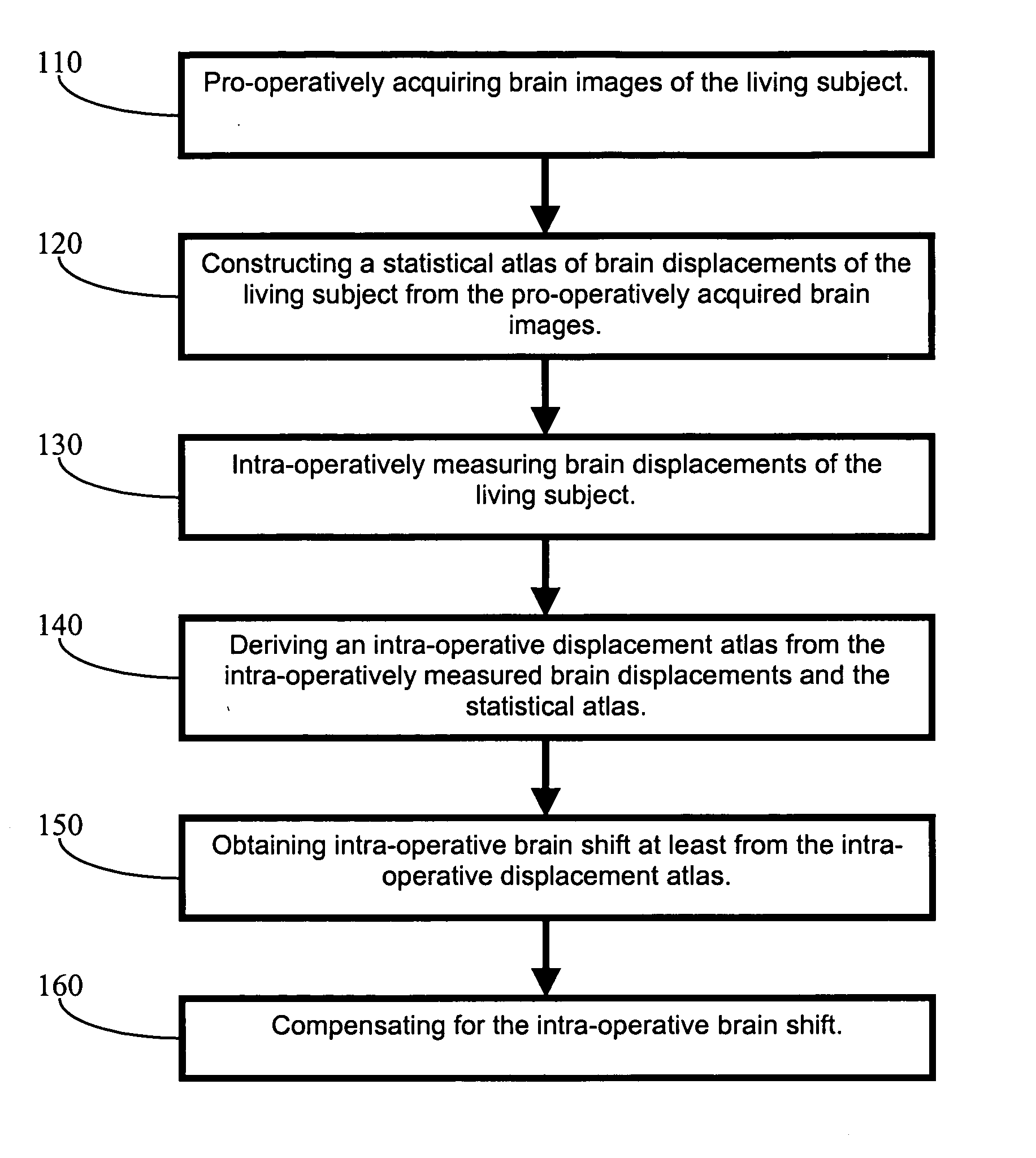
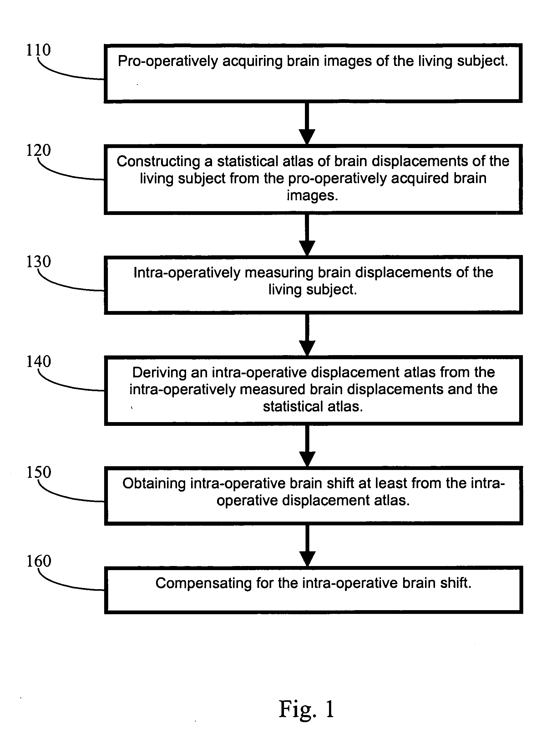
However, utilization of the PRG method requires a preliminary surgery for implantation of the fiducial markers to predetermined positions in a patient's anatomy.
However, the surface based alignment techniques are limited with facts, for example, scalps lack geometric specificity, and skin surfaces may deform due to intra-operative drugs or procedural retraction [9].
However, recent studies have shown limitations in accuracy with current image-guided procedures.
Specifically, with neurosurgery, registration is provided by markers attached to the skull of a patient or on the skin surrounding the skull of a patient, where soft-tissue deformations of the brain during surgery may result in significant errors in aligning a pre-operative image space to an actual physical space.
However, dose considerations of repeatedly using computed tomography (hereinafter “CT”) scanning in the OR have hindered adoption of the iCT technique [14].
Although conceptually appealing, the exorbit

Method used


Image

  • Apparatus and methods of brain shift compensation and applications of the same
  • Apparatus and methods of brain shift compensation and applications of the same
  • Apparatus and methods of brain shift compensation and applications of the same

Examples


example 1

Animal Experiment

[0052] To evaluate the accuracy and effectiveness of the computational model for predicting of brain deformations, in vivo experiments were conducted with a porcine. In these experiments, a porcine brain was implanted with small CT-visible markers in a grid-like fashion using a needle. Following implantation, a series of gradual deformations consistent with neurosurgical loading rates were applied and recorded using the CT. By recording the bead positions in each image volume, a complete subsurface trajectory for all deformation stages was determined. Following the experiment, subject-specific finite element models of all test subjects were constructed from the pre-operative MR and registered to the CT data set. Deformations applied in the OR were now applied in simulation and bead trajectories were compared to ascertain model-predictive fidelity. In addition to uni-axial deformations, validation with realistic loading conditions such as from the retraction of tiss...

example 2

Clinical Trials

[0054] In one embodiment of the present invention, four patients who undergo brain surgery were chosen to gather data for evaluating the invented method. The four patients were employed merely as an example to acquire data for practicing the present invention, and the use of the four patients should not limit the scope of the present invention. Each patient was assigned a number from Patent 1 to Patient 4 as his or her identification. Additionally, prior to clinical data acquisition, the surgical procedures for human patients were approved by the Vanderbilt University Institutional Review Board (hereinafter “VUIRB”) and patient consent was acquired for all clinical data.

[0055] In four clinical cases studying the effects of gravity-induced brain sag, features on the cortical surface (i.e. blood vessel bifurcations) were digitized and tracked in the OR in the direction of gravity. The cases involved 4 supine cases with 2 patients having the head in the neutral positio...


PUM


Abstract

A method of compensation for intra-operative brain shift of a living subject. In one embodiment, the method includes the steps of pro-operatively acquiring brain images of the living subject, constructing a statistical atlas of brain displacements of the living subject from the pro-operatively acquired brain images, intra-operatively measuring brain displacements of the living subject, deriving an intra-operative displacement atlas from the intra-operatively measured brain displacements and the statistical atlas, obtaining intra-operative brain shift at least from the intra-operative displacement atlas, and compensating for the intra-operative brain shift.

Description

CROSS-REFERENCE TO RELATED PATENT APPLICATION [0001] This application is a continuation-in-part of U.S. patent application Ser. No. 10 / 936,339, filed Sep. 8, 2004, entitled “APPARATUS AND METHODS OF CORTICAL SURFACE REGISTRATION AND DEFORMATION TRACKING FOR PATIENT-TO-IMAGE ALIGNMENT IN RELATION TO IMAGE-GUIDED SURGERY,” by Michael I. Miga, Benoit M. Dawant and Tuhin K. Sinha, the disclosure of which is hereby incorporated herein by reference in its entirety, which itself claims the benefit, pursuant to 35 U.S.C. § 119(e), of provisional U.S. patent application Ser. No. 60 / 501,514, filed Sep. 8, 2003, entitled “APPARATUS AND METHODS OF CORTICAL SURFACE REGISTRATION AND DEFORMATION TRACKING FOR PATIENT-TO-IMAGE ALIGNMENT DURING IMAGE-GUIDED SURGERY,” by Michael I. Miga, Benoit M. Dawant and Tuhin K. Sinha, which is incorporated herein by reference in its entirety. [0002] Some references, which may include patents, patent applications and various publications, are cited and discussed ...

Claims


Application Information

Patent Timeline
IPC IPC(8): A61BA61B5/05A61B19/00A61N5/02G06K9/00G06T7/00
CPCA61B19/50G06T7/0038A61B19/5244A61B2017/00694A61B2019/505A61B2019/507A61B2019/5231A61B2019/5255A61B2019/5265A61B2019/5276A61B2019/5289A61B2019/566G06K9/00214G06K9/6211G06K9/6212G06K2209/051G06T7/0034A61B19/52A61B90/36A61B2090/373A61B2034/2055A61B2034/256A61B34/20A61B2090/364A61B34/10A61B2034/105A61B2034/107A61B2034/2065A61B2090/378G06T7/35G06T7/38G06V20/653G06V10/758G06V10/757G06V2201/031
Inventor MIGA, MICHAEL I.DUMPURI, PRASHANTHCHEN, CHUN-CHENG R.
Owner VANDERBILT UNIV
Why Eureka
Try Eureka