
Signal-adaptive noise reduction in digital radiographic images

a digital radiographic image and signal-adaptive technology, applied in the field of radiographic imaging, can solve the problems of reducing the perceived quality of final images, reducing the perceived image quality, and reducing the contrast at lowly attenuated regions, so as to reduce the perceived noise in digital x-ray images, improve the displayed image quality of digital x-ray images, and reduce noise

Inactive Publication Date: 2005-06-09
GE MEDICAL SYST GLOBAL TECH CO LLC
View PDF4 Cites 7 Cited by
  • Summary
  • Abstract
  • Description
  • Claims
  • Application Information

AI Technical Summary

Benefits of technology

[0004] Accordingly, the above-identified shortcomings of existing noise reduction systems and methods for digital x-ray imaging are overcome by embodiments of the present invention, which relates to novel systems and methods for reducing the perceived noise in digital x-ray images. These systems and methods improve the displayed image quality of digital x-ray images by reducing noise at highly attenuated regions without affecting the image contrast at relatively lowly attenuated regions. This is accomplished by taking the detected signal properties into account in a noise reduction framework, where the framework is adaptive to the detected signal.
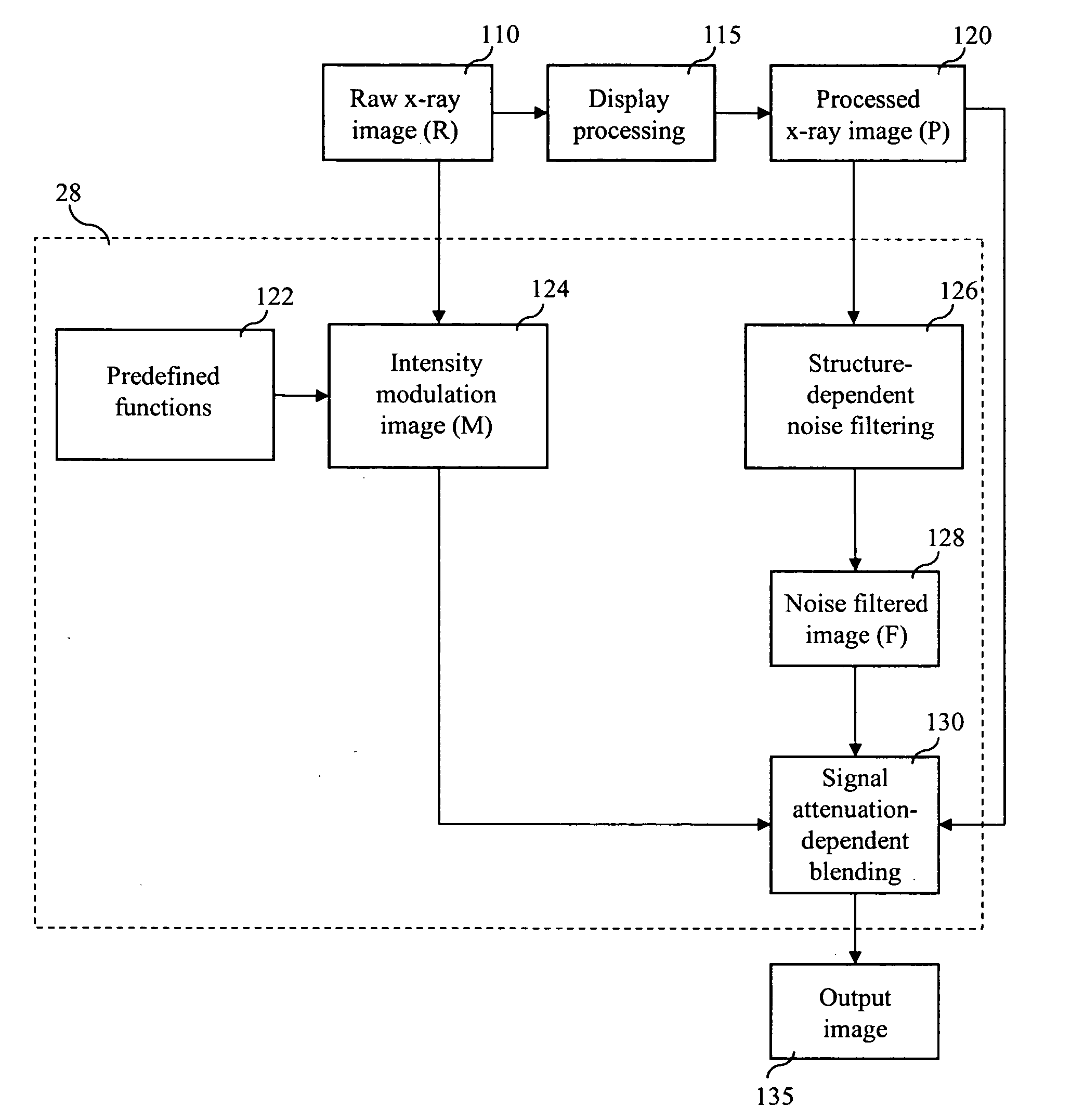
[0007] Yet other embodiments of these methods comprise the steps of: obtaining raw x-ray image data of an imaged object; processing the raw x-ray image data to create processed x-ray image data; inputting the raw x-ray image data and the processed x-ray image data to an image processor; developing a first intensity modulation image from the raw x-ray image data; developing a second intensity modulation image from the processed x-ray image data; deriving a structure-dependent noise filtered image using the processed x-ray image data; performing signal attenuation-dependent blending; and creating a noise-reduced digital x-ray image therefrom.

Problems solved by technology

During the display processing of images from digital radiographic imaging systems, the image noise at highly attenuated regions in the images becomes more noticeable, and can therefore decrease the perceived quality of the final images.
This perceived image degradation is mainly due to the contrast enhancement that occurs in the highly attenuated regions of the image.
However, that comes at the cost of having decreased contrast at lowly attenuated regions in the images, such as the lung parenchyma.

Method used


Image

  • Signal-adaptive noise reduction in digital radiographic images
  • Signal-adaptive noise reduction in digital radiographic images
  • Signal-adaptive noise reduction in digital radiographic images

Examples


Embodiment Construction

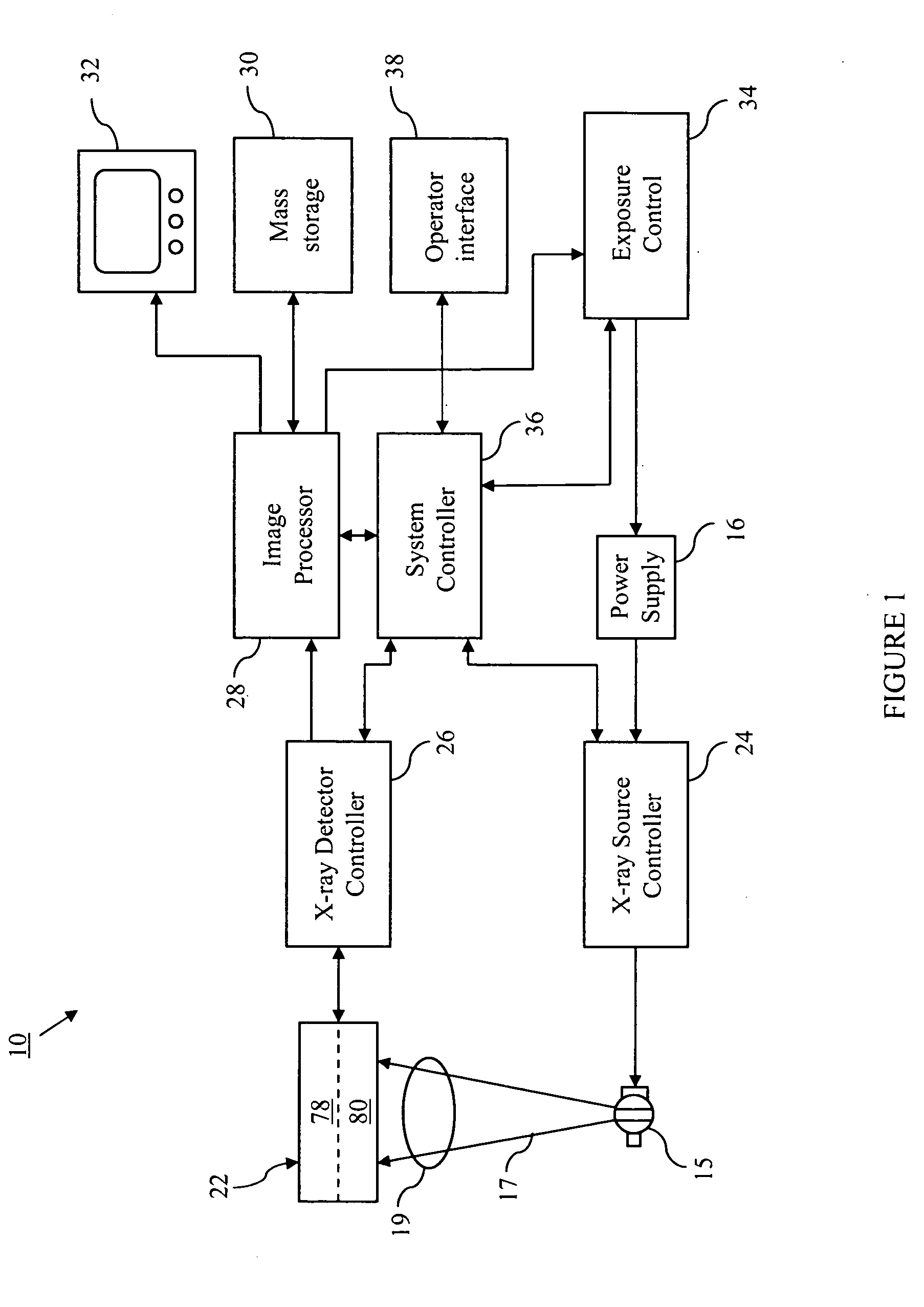
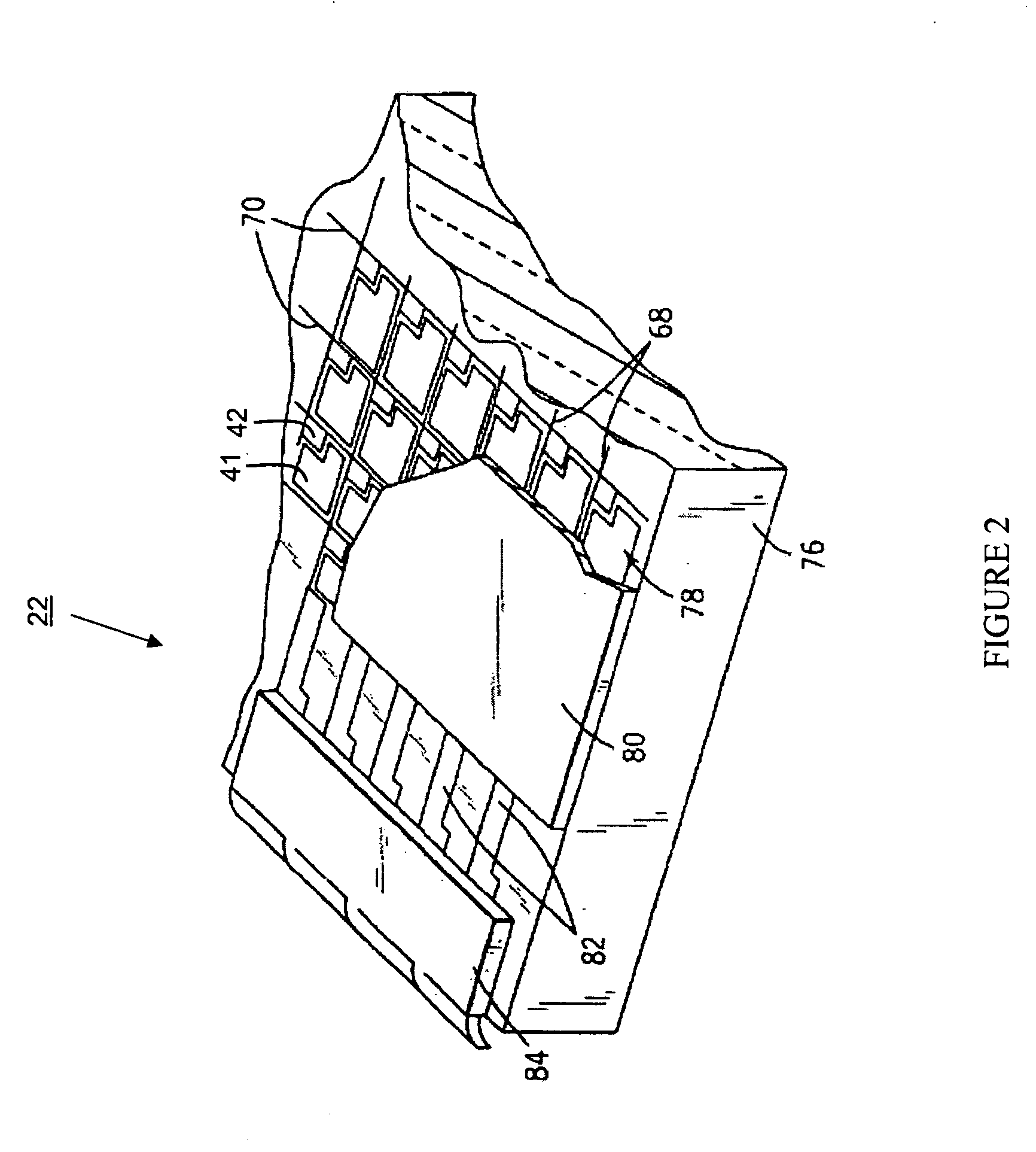
[0021] For the purposes of promoting an understanding of the invention, reference will now be made to some preferred embodiments of the present invention as illustrated in FIGS. 1-7 and specific language used to describe the same. The terminology used herein is for the purpose of description, not limitation. Specific structural and functional details disclosed herein are not to be interpreted as limiting, but merely as a basis for the claims as a representative basis for teaching one skilled in the art to variously employ the present invention. Any modifications or variations in the depicted structures and methods, and such further applications of the principles of the invention as illustrated herein, as would normally occur to one skilled in the art, are considered to be within the spirit and scope of this invention.

[0022] This invention relates to systems and methods for reducing the perceived noise in digital x-ray images. These systems and methods improve the displayed image qu...


PUM


Abstract

Signal-adaptive noise reduction in digital radiographic images is described herein. Embodiments comprise methods for signal-adaptive noise reduction in digital radiographic images, comprising the steps of: obtaining raw x-ray image data of an imaged object; processing the raw x-ray image data to create processed x-ray image data; inputting at least one of the raw x-ray image data and the processed x-ray image data to an image processor; developing at least one of a first intensity modulation image from the raw x-ray image data and a second intensity modulation image from the processed x-ray image data; deriving a structure-dependent noise filtered image using the processed x-ray image data; performing signal attenuation-dependent blending; and creating a noise-reduced digital x-ray image therefrom. Computer-readable mediums encoded with programming for facilitating signal-adaptive noise reduction in digital radiographic images, and digital radiographic imaging systems comprising such programming, are also described.

Description

FIELD OF THE INVENTION [0001] The present invention relates generally to radiation imaging. More specifically, the present invention relates to systems and methods for signal-adaptive noise reduction in digital radiographic images, where the detected signal properties are taken into account in a noise reduction framework, and where the noise reduction framework is adaptive to the detected signal. BACKGROUND OF THE INVENTION [0002] With applications ranging from diagnostic procedures to radiation therapy, the importance of high-performance medical imaging is immeasurable. As such, new advanced medical imaging technologies continue to be developed. Digital medical imaging techniques represent the future of medical imaging. Digital imaging systems produce far more accurate and detailed images of an object than conventional film-based imaging systems, and also allow further enhancements of the images to be made once an object is scanned. [0003] During the display processing of images fr...

Claims


Application Information

Patent Timeline
Patent Type & Authority Applications(United States)
IPC IPC(8): G06K9/00G06K9/40G06T5/00H04N5/32
CPCH04N5/217H04N5/32G06T2207/30004G06T2207/10116G06T2207/20012G06T5/002H04N23/81G06T5/70H04N23/30
Inventor AVINASH, GOPAL B.JABRI, KADRI NIZARSEAMANS, JOHN L.
Owner GE MEDICAL SYST GLOBAL TECH CO LLC
Learn More