

Methods for determining binding affinities
a technology of binding affinity and method, applied in the direction of fluorescence/phosphorescence, biochemistry apparatus and processes, instruments, etc., can solve the problems of ksub>a, time-consuming and expensive current methods, and inability to determine the kinetic association rate constant of screening methods in complex solutions
- Summary
- Abstract
- Description
- Claims
- Application Information
AI Technical Summary
Problems solved by technology
Method used
Image

Examples
example 1
Preparation of Biorecognition Surfaces
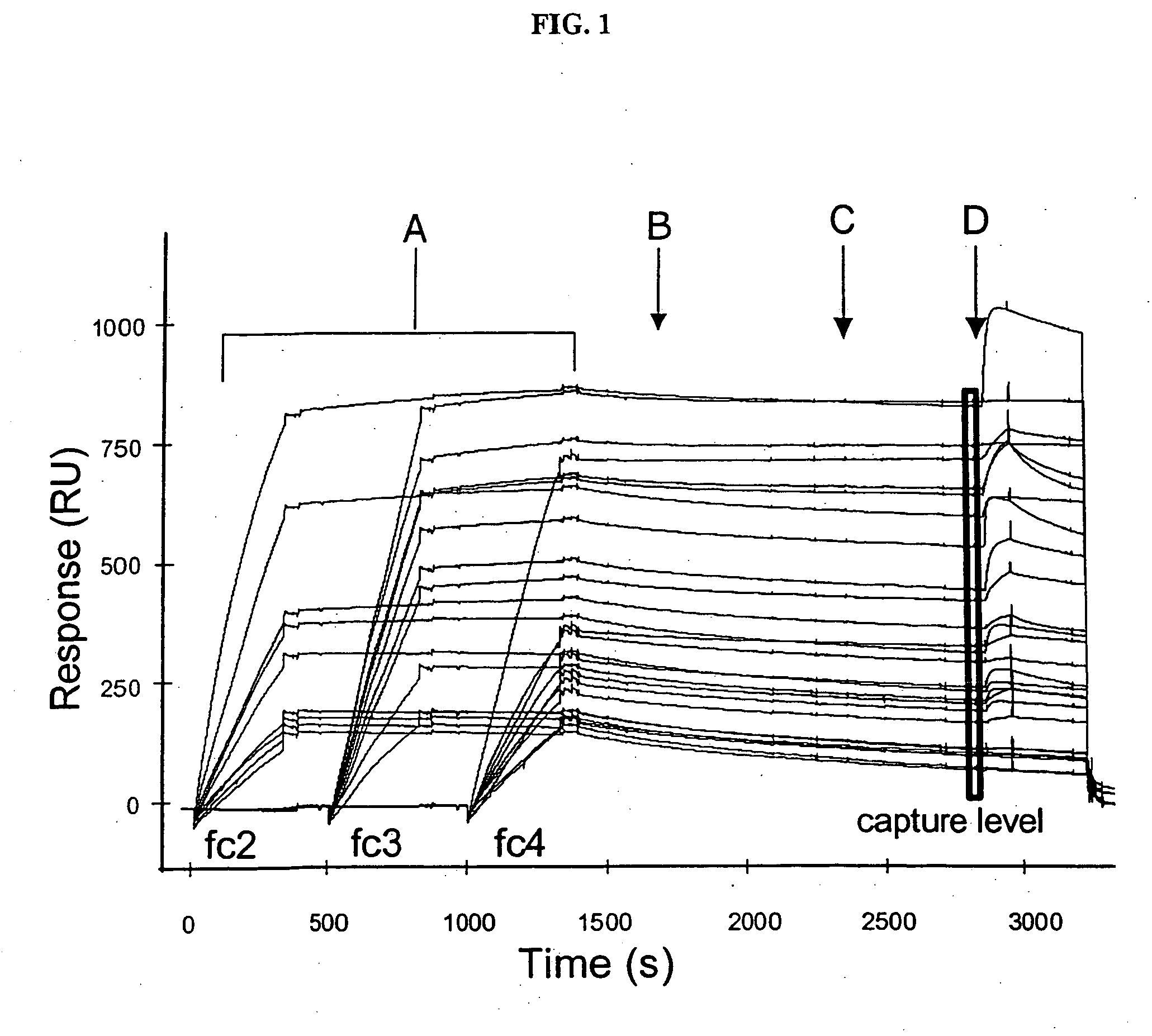
[0062] We prepared a biorecognition surface by first immobilizing Immunopure® Protein A (Pierce, Rockford, Ill.) to CM5 sensor chips (BIACORE AB, Uppsala, Sweden) in a BIACORE 2000 or 3000 instrument (FIG. 1) as follows.
[0063] In the BIACORE instrument, the sensor chips were pre-conditioned in water at a flow rate of 100 μL / min by applying two consecutive 20-μL pulses of 50 mM NaOH, 0.1% HCl (v / v) and 0.1% SDS. The individual flow cells were equilibrated with 10 mM HEPES buffer containing 150 mM NaCl and 0.005% P-20 Surfactant (BIACORE AB), pH 7.4 (“HBSP running buffer”) at a flow rate of 20 μL / min. Next, a solution of 70 μL of 50 mM N-hydroxysuccinimide (“NHS;” BIACORE AB) and 70 μL of 200 mM 1-(3-dimethylaminopropyl)-ethylcarbodiimide hydrochloride (“EDC;” BIACORE AB) was injected over the flow cells to activate the CM-dextran. Johnsson et al., Anal Biochem, 198, pp. 268-277 (1991). A solution of protein A (reconstituted in water to 5 mg / mL ...
example 2
Ligand Capture
[0065] We used the protein A or goat anti-IgG (Fc sp.) immobilized on the sensor chip surface to capture monoclonal antibodies from hybridoma supernatants (FIG. 1A) as follows. Myszka, J. Mol. Recognit., 12, pp. 279-284 (1999) and Svensson et al., Eur. J. Biochem., 258, pp. 890-896 (1998). Buffer (10 mM HEPES, 150 mM NaCl, 0.005% polysorbate-20, pH 7.4) containing 12 mg / mL of each BSA (Sigma, St. Louis, Mo.) and soluble carboxymethyl-dextran (“CM-dextran;” Fluka BioChemika) was flowed across the individual flow cells at a flow rate of 100 μL / min. Three hybridoma supernatant solutions containing antibodies of interest were diluted 1 / 25 in the same buffer and then separately injected over three flow cells for 5 min at a flow rate of 50 μL / min. The fourth flow cell was not exposed to an antibody solution and therefore, served as a control. The antibody-captured protein A surface was then washed for 10 min at a flow rate of 50 μL / min to remove any non-specific components ...
example 3
Screening of Binding Partner
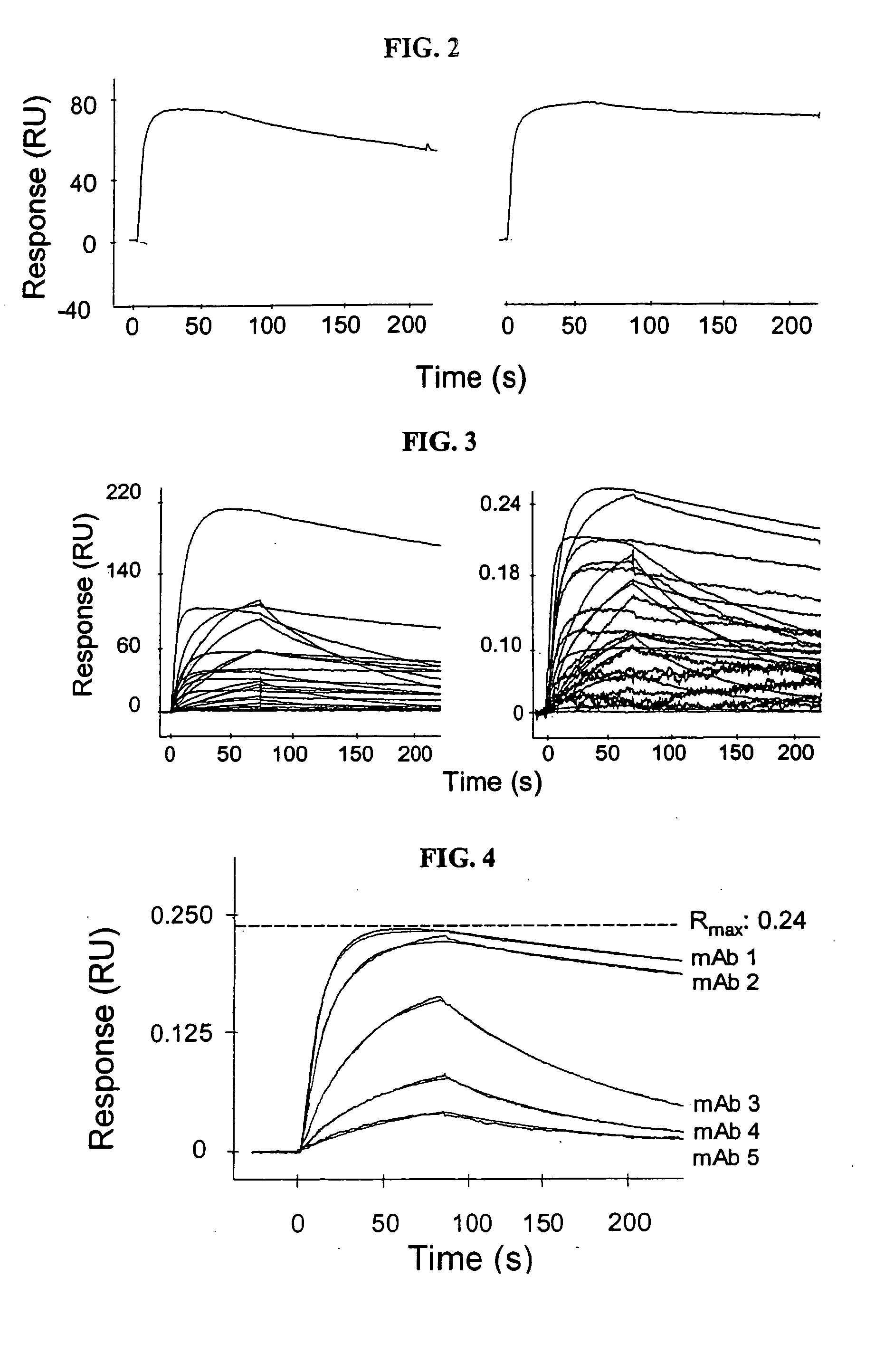
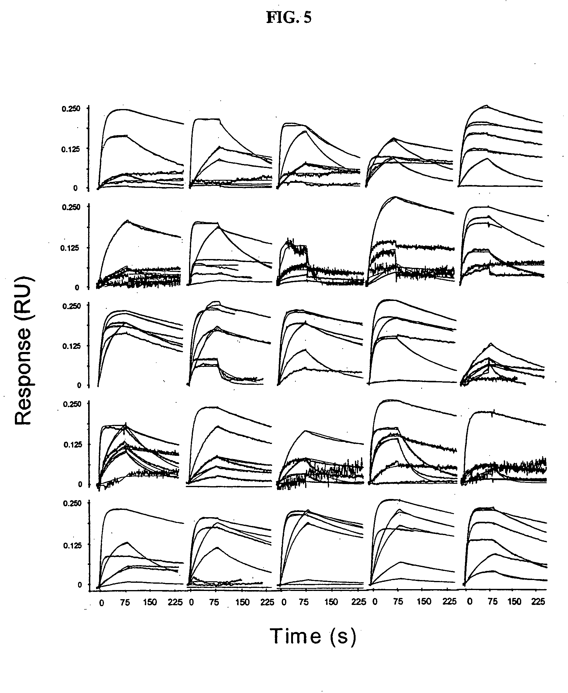
[0066] We screened the antibody-captured protein A surfaces for binding to antigen (FIG. 1D) as follows. Prior to antigen injection, a solution of buffer was injected to determine baseline drift caused by the decay of the antibody-captured protein A surface (FIG. 1C). Antigen binding was measured by flowing an antigen at a predetermined concentration across the individual flow cells for 1 min at a flow rate of 100 μL / min and then reintroducing the buffer for 5 min to initiate dissociation. After dissociation, the protein A surface was regenerated by injecting 10 μL of 100 mM H3PO4 for 12 sec. After regeneration, we repeated the antibody capture procedure described in Example 2 using three supernatants from the panel at a time until the entire panel was screened.
[0067] To determine the concentration of antigen to be used in the antigen-binding step, we first assessed the quality of antigen binding to the antibody-captured protein A surfaces using three d...
PUM
| Property | Measurement | Unit |
|---|---|---|
| flow rate | aaaaa | aaaaa |
| pH | aaaaa | aaaaa |
| flow rate | aaaaa | aaaaa |
Abstract
Description
Claims
Application Information