
Polypeptide display libraries and methods of making and using thereof

a polypeptide and display library technology, applied in the field of bacteria polypeptide display libraries, can solve the problems of clonal loss, phage display presents a few undesirable properties, and overall low quality results

Active Publication Date: 2005-09-08
RGT UNIV OF CALIFORNIA
View PDF4 Cites 39 Cited by
  • Summary
  • Abstract
  • Description
  • Claims
  • Application Information

AI Technical Summary

Benefits of technology

[0082]FIG. 34 shows flow cytometric analysis of cells displaying of peptides in an Omp encoding gene modified to possess no leucine codons, thus enabling display of peptides that incorporate a variety of synthetic leucine analogs. Display of a non-canonical amino acid is exemplified using trifluoroleucine (Tfl) and OmpX. This figure also shows a comparison of the level of display of the T7tag peptide (that does not contain any leucine residues) using either unmodified wild-type (top) or NoLeu-OmpX (bottom) scaffolds in the presence of (left) 19 amino acids (deficient in Leu), (middle) 19 amino acids+trifluoroleucine (No Leu), or (right) 20 standard amino acids. These were labeled with an anti-T7tag biotinylated antibody, washed once in PBS, and labeled with streptavidin-phycoerythrin (Molecular Probes, Eugene, Oreg.). The increase in green fluorescence for the 19+Tfl samples (middle) from about 5.6 to about 63.7 allows for the screening of bacterial display libraries for peptides that incorporate Tfl.

Problems solved by technology

Unfortunately, phage display presents a few undesirable properties.
Phage are also difficult to elute or recover from an immobilized target ligand, thereby resulting in clonal loss.
Phage display also requires an infection step wherein viruses that do not bind and enter a cell are lost early in the process, thereby resulting in lower quality results overall, e.g., affinity of isolated binding molecules.
Further, phage display selections are time consuming requiring typically about two to about three weeks for the isolation of phage display polypeptides that bind a given target.
Second, phage display methods can lead to Darwinian outgrowth of particular clones owing to their relative infectivity, assembly efficiency, and toxicity to the host cell.
Third, the rate at which desired binding clones can be enriched is slowed by relatively low enrichment ratios.
Some of these prior art display systems have been tested for library screening without success for isolation of high affinity protein binding peptides.
Prior art expression vectors for polypeptide display libraries using host cells suffer from a variety of problems.
The problems of the prior art methods include (1) only small peptides may be expressed, (2) large libraries cannot be selected, (3) the polypeptides are not expressed on the outer membrane surface, but are instead expressed in the periplasmic space between the inner and the outer membranes, (4) polypeptides that are displayed on the outer membrane surface do not properly bind or interact with large molecules and certain targets, and (5) analyzing expression on fimbrial or flagella results in loss of some desired polypeptides due to mechanical shearing.
Though several different bacterial display systems have been reported, their usefulness has been restricted by technical limitations including accessibility on the cell surface, inability to display highly diverse sequences, and adverse effects on cell growth and viability.
Consequently, these techniques do not enable isolation of high affinity peptide ligands.
Additionally, these techniques do not provide peptide exposure on the cell surface suitable for binding to analytes including antibodies, proteins, viruses, cells, macromolecules.
Thus, these display formats are not compatible with certain isolation methods, since the peptides produced do not bind to large molecules and other surfaces, e.g., magnetic particles.
The prior art process also reduces cell viability and alters membrane permeability, thereby reducing process efficiency.
Thus far, routine isolation of high affinity peptide ligands for arbitrary protein targets has not been demonstrated.
However, the C and N-termini of these “carrier” proteins are not naturally located on the cell surface which precludes the ability to display polypeptides as terminal fusions.
As a result, proteins which are not capable of folding in the insertional fusion context, when their C and N termini are fused to the “carrier” protein sequence, as well as those for which the C and N termini are physically separated in space, e.g., single chain Fv antibody fragments, cannot be displayed effectively as insertions.
Similarly, the restriction to the use of insertional fusions, interferes with the display of a large number of proteins encoded by cDNA libraries on the cell surface.
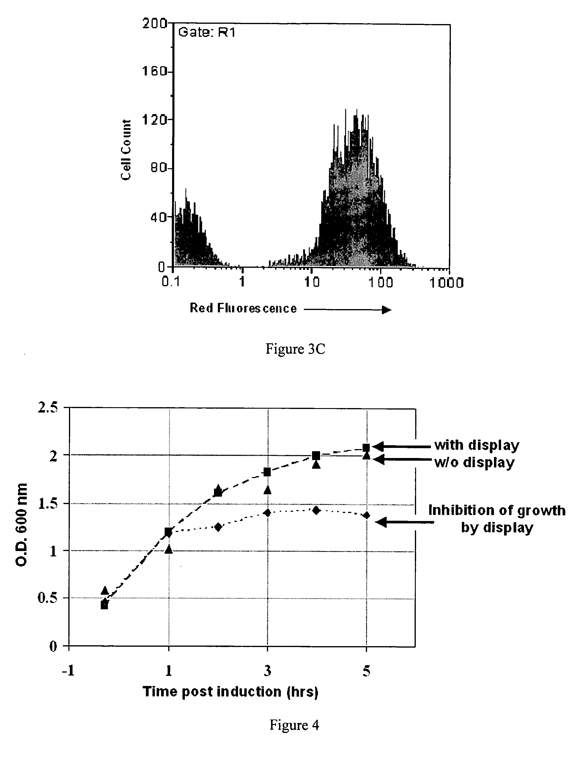
Unfortunately, the performance of LppOmpA expression vector as a general process for isolating and expressing polypeptides from large libraries is significantly restricted by i) the reduced structural stability of the modified OmpA protein, ii) intolerance to expression at high temperatures, iii) reduced viability, and iv) most importantly, its inability to display polypeptides on the cell surface in a manner compatible with binding to large proteins without compromising viability and / or growth rate See Christman, A.et al., 1999.
In addition, expression vectors in the prior art are problematic because (1) the polypeptides produced by the expression vectors are not capable of binding externally added proteins, cells, or surfaces to the host cells, (2) the expression vectors does not allow surface presentation of large polypeptides, and (3) the expressed polypeptides are only expressed in the periplasmic region (between the inner and outer membrane) and not on the outer surface of the host cell, and therefore any expressed protein can only interact with small molecules that pass through the outer membrane and into the periplasmic space.
These problems have prevented the application of this technology as a general process for isolating high affinity binding polypeptides.

Method used


Image

  • Polypeptide display libraries and methods of making and using thereof
  • Polypeptide display libraries and methods of making and using thereof
  • Polypeptide display libraries and methods of making and using thereof

Examples


example 1

OmpA Loop 1 Expression Vector

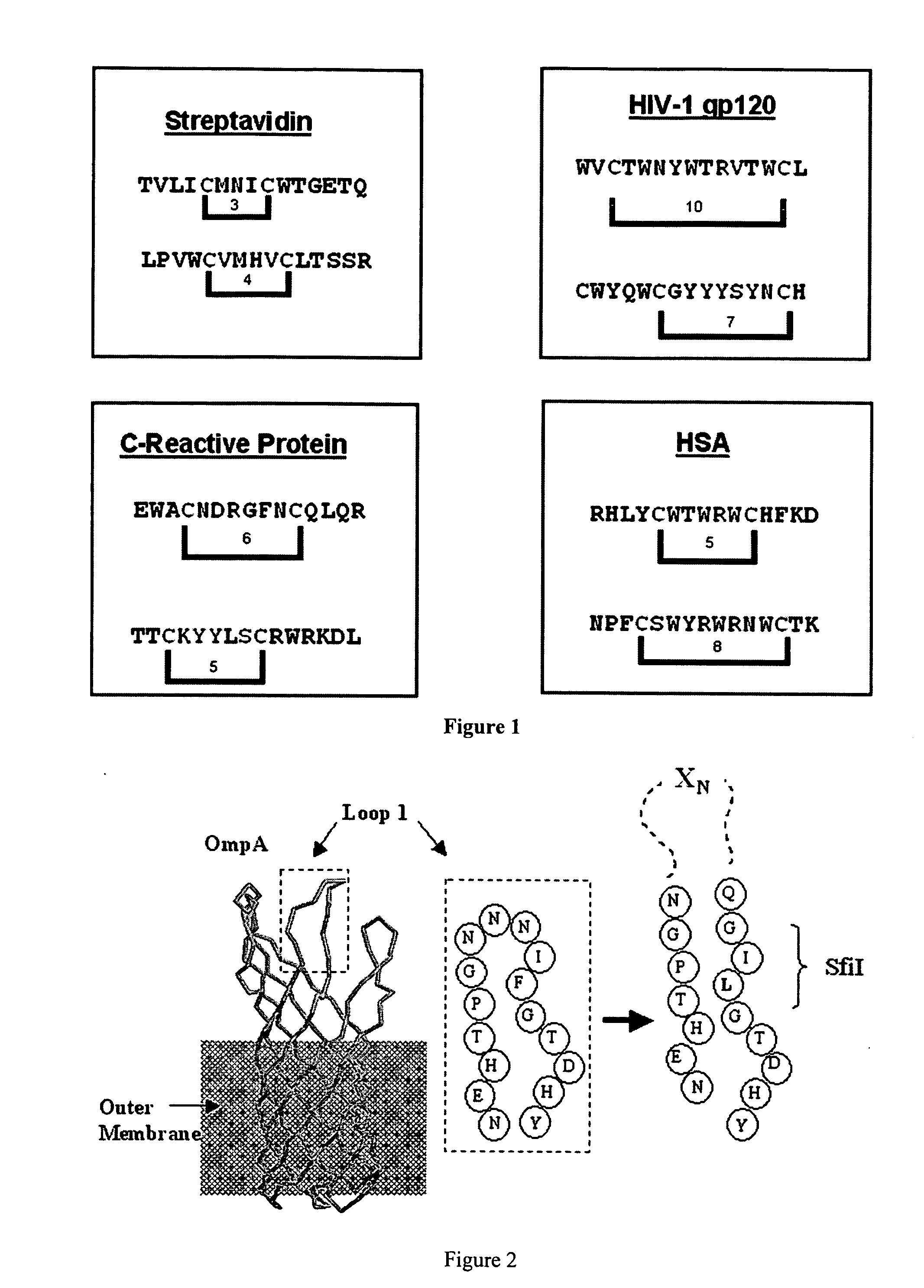
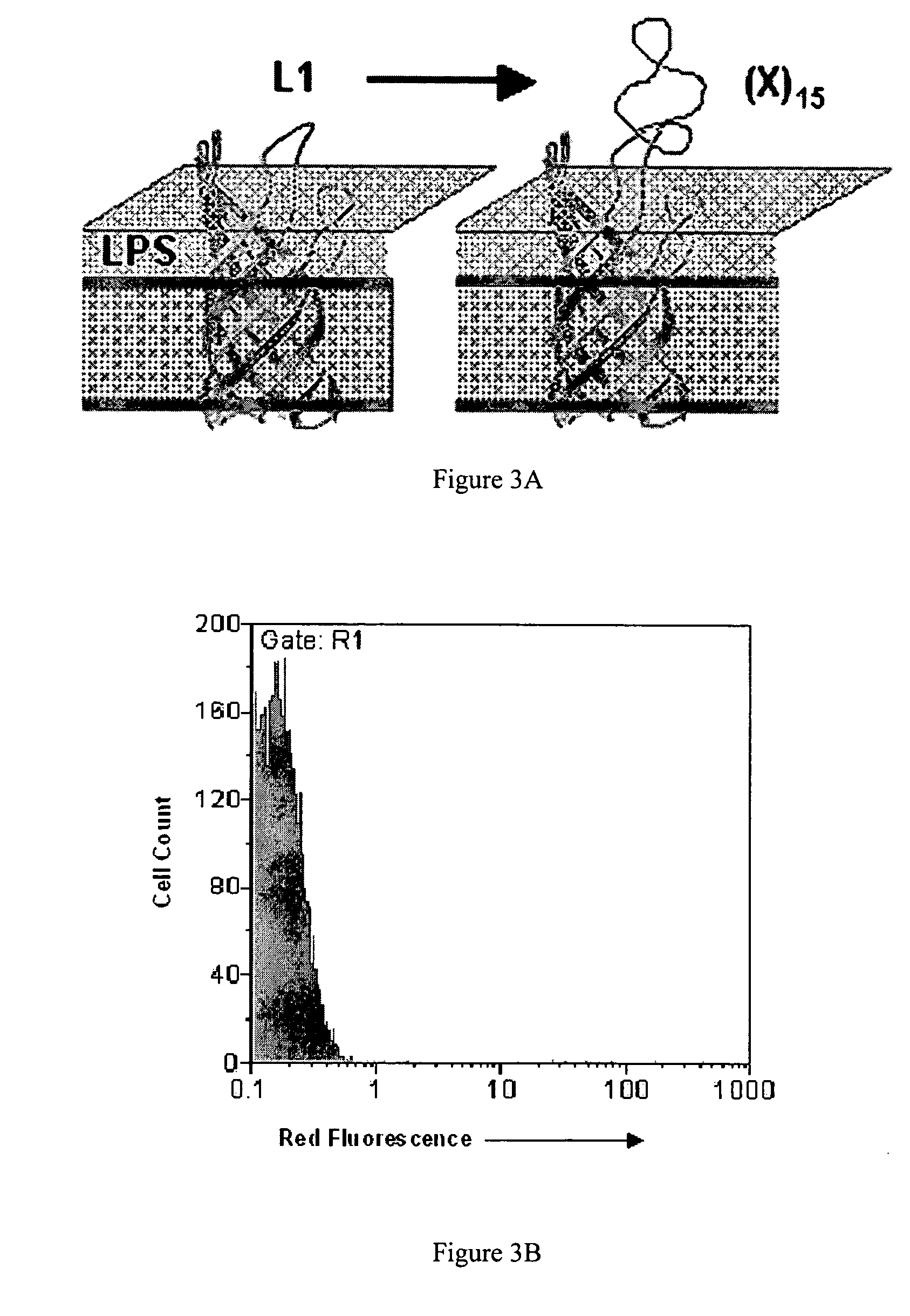
[0182] A 15-mer random insert sequence which provides a balance between sequence complexity and maintenance of the stability and folding and export of OmpA was selected. See FIG. 2. It should be noted that longer length insert, e.g., 15 mer, libraries provide more copies of short sequences while allowing for possible longer cell binding motifs requiring 10 or more amino acids. Although an engineered disulphide bridge may be used for stabilization, such was not used as cystein oxidation in the E. coli periplasm could lead to aggregation and reduced export and disulfides could potentially emerge by chance. Moreover, the membrane spanning domain of OmpA already provides a rigid structural anchor for the peptide inserts into the more flexible loops.

[0183] After optimizing the library construction process through the use of the pBAB33L1, construction of a high quality library of about 4.5×10¹⁰ independent transformants was found to be possible. This library...

example 2

OmpX Loop 2 and OmpX Loop 3 Expression Vectors

[0214] While the following protocol specifically describes the construction of vectors for the display of polypeptides and polypeptide libraries in loop 2 of OmpX, this procedure may be readily applied to loop 3 of OmpX, by consideration of the non-conserved regions in loop 3 as described in Table 3, by one skilled in the art. In loop 3, peptide insertions are preferred between residues 94-99, and preferably between residues 95-97, with Pro96 removed. The wild-type OmpX gene from E. coli MC1061:

atgaaaaaaattgcatgtctttcagcactggccgcagttctggctttcaccgcaggtacttccgta(SEQ ID NO:102)gctgcgacttctactgtaactggcggttacgcacagagcgacgctcagggccaaatgaacaaaatgggcggtttcaacctgaaataccgctatgaagaagacaacagcccgctgggtgtgatcggttctttcacttacaccgagaaaagccgtactgcaagc / tctggtgactacaacaaaaaccagtactacggcatcactgctggtccggcttaccgcattaacgactgggcaagcatctacggtgtagtgggtgtgggttatggtaaattccagaccactgaatac / ccg / acctacaaacacgacaccagcgactacggtttctcctacggtgcgggtctgcagttcaacccgatggaaaacg...

example 3

Circularly Permuted OmpX (CPX)

[0217] Display and expression of passenger polypeptides as N or C terminal fusions is accomplished by topological permutation of an Omp as shown in FIG. 14. Sequence rearrangement of an outer membrane protein, in this case OmpX, was accomplished using an overlap extension PCR methods known in the art in order to create either N or C terminal fusion constructs. See Ho, et al. (1989) Gene 77(1):51-59, which is herein incorporated by reference; and see FIG. 14, FIG. 27, FIG. 28, FIG. 30, and FIG. 32. Polypeptide passenger insertion points are chosen to occur within non-conserved, surface exposed loop sequences of surface exposed proteins, such as monomeric Omps (including OmpA, OmpX, OmpT, and the like) using methods known in the art.

[0218] The DNA sequence of the N / C terminal fusion expression vector provides the following contiguous components fused or linked in linear order from N to C terminus (See FIG. 14): [0219] 1. A DNA sequence encoding an N-ter...


PUM

PropertyMeasurementUnit
timeaaaaaaaaaa
concentrationsaaaaaaaaaa
volumeaaaaaaaaaa

Abstract

Disclosed herein are expression vectors which display a passenger polypeptide on the outer surface of a biological entity. As disclosed herein the displayed passenger polypeptide is capable of interacting or binding with a given ligand. Also disclosed are methods of making and using the expression vectors. N / C terminal fusion expression vectors and methods of making and using are also disclosed.

Description

CROSS REFERENCE TO RELATED APPLICATIONS [0001] This application claims the benefit of U.S. Provisional Patent Application No. 60 / 495,698, filed Aug. 18, 2003, listing Patrick S. Daugherty, Paul H. Bessette, and Jeffrey Rice, as joint inventors, which is herein incorporated by reference in its entirety.BACKGROUND OF THE INVENTION [0002] 1. Field of the Invention [0003] The present invention generally relates to bacterial polypeptide display libraries and methods of making and using thereof. [0004] 2. Description of the Related Art [0005] Polypeptide display technologies have substantially impacted basic and applied research applications ranging from drug discovery to materials synthesis. See Clackson, T. and J. A. Wells (1994) Trends In Biotech. 12(5):173-184; and Shusta, E. V., et al. (1999) Curr. Opin. Biotechnol. 10(2):117-122; and Kodadek, T., (2001) Chem. Biol. 8(2):105-158; Lee, S. W., et al. (2002) Science 296 (5569):892-859; and Nixon, A. E. (2002) Curr. Pharm. Biotechnol. 3(...

Claims


Application Information

Patent Timeline
Patent Type & Authority Applications(United States)
IPC IPC(8): A61K39/02C07H21/04C07K14/195C12NC12N1/21C12N15/10C12N15/74C12Q1/68
CPCC12N15/1044C12N15/1037G01N33/554C07K14/245C40B40/10C40B30/04
Inventor DAUGHERTY, PATRICKBESSETTE, PAULRICE, JEFFREY
Owner RGT UNIV OF CALIFORNIA
Learn More