Magnetic resonance method and apparatus for determining the position and/or orientation of the image plane of slice image exposures of a vessel region in a contrast agent bolus examination

a magnetic resonance system and image plane technology, applied in the direction of instruments, magnetic variable regulation, application, etc., can solve the problems of insufficient use and plagued errors

Inactive Publication Date: 2005-09-15
SIEMENS HEALTHCARE GMBH
View PDF16 Cites 13 Cited by
  • Summary
  • Abstract
  • Description
  • Claims
  • Application Information

AI Technical Summary

Benefits of technology

[0013] An object of the present invention to provide a method that enables as optimal a determin...

Problems solved by technology

First, the determination ensues in a computer-controlled manner, meaning that the conventional manual...

Method used


Image

  • Magnetic resonance method and apparatus for determining the position and/or orientation of the image plane of slice image exposures of a vessel region in a contrast agent bolus examination
  • Magnetic resonance method and apparatus for determining the position and/or orientation of the image plane of slice image exposures of a vessel region in a contrast agent bolus examination
  • Magnetic resonance method and apparatus for determining the position and/or orientation of the image plane of slice image exposures of a vessel region in a contrast agent bolus examination

Examples


Embodiment Construction

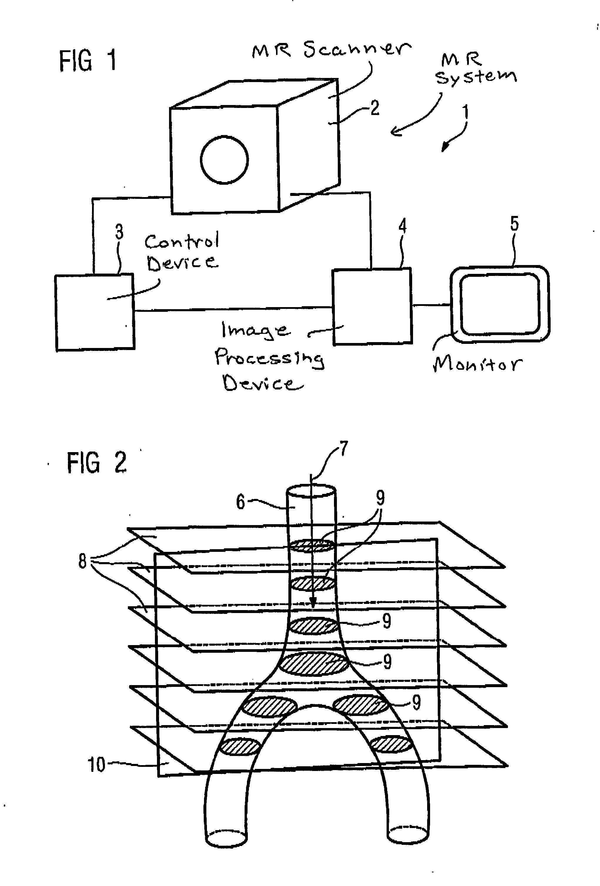
[0025]FIG. 1 shows an inventive magnetic resonance system having a scanner 1 in which a patient is positioned, a control device 3 for controlling the image acquisition operation as well as an image-processing device 4 for generation and output of the slice images using the acquired image signals on a monitor 5.

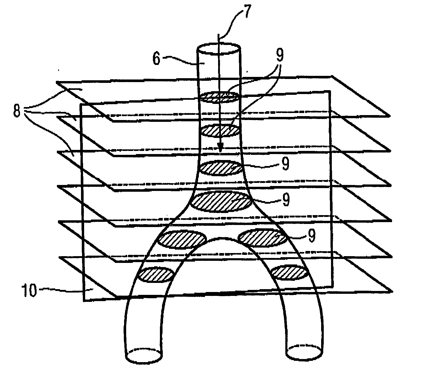
[0026] The actual image acquisition is initiated via the control device 3. A contrast agent bolus examination is to be implemented, with the bolus being present in a vessel region 6, for example the aorta bifurcation as shown in FIG. 2. The flow direction of the blood, and inevitably also of the contrast agent bolus to be detected, is indicated by the arrow 7. A rough position / orientation determination with regard to the position of the vessel region 6 is initially implemented using one or more localizer slice images (not shown in detail in FIG. 2). As a consequence, the position and orientation of the transversal planes serving for the subsequent image plane determination, i...


PUM


Abstract

In a method and a magnetic resonance system for determining the position and/or orientation of the image plane of slice image exposures of a vessel region in a contrast agent bolus examination, in particular in the region of the aorta bifurcation or the aorta arch, wherein the image plane traverses the vessel region in the longitudinal section, initially a group of slice image exposures disposed essentially orthogonal to the vessel is automatically acquired from different planes of the vessel region lying parallel to one another, and the image regions showing the vessel region are automatically determined within the orthogonal slice image exposures in an image-processing device, and using the determined image regions the spatial position of the vessel region is determined. The position and/or orientation of the image plane disposed essentially orthogonal to the planes of the slice image exposures and traversing the vessel region in the longitudinal section is determined automatically using the position information, such that the image plane lies in the center of the vessel region.

Description

BACKGROUND OF THE INVENTION [0001] 1. Field of the Invention [0002] The present invention concerns a method for determination with a magnetic resonance system of the position and / or orientation of the image plane of slice image exposures of a vessel region in a contrast agent bolus examination, in particular in the region of the aorta bifurcation or the aorta arch, wherein the image plane traverses the vessel region in the longitudinal section, and wherein a group of slice image exposures lying essentially orthogonal to the vessel is acquired from different planes of the vessel region, [0003] 2. Description of the Prior Art [0004] A contrast agent bolus examination, frequently also called CareBolus measurement, serves for the determination of the flow-through time of the contrast agent in the framework of a magnetic resonance examination, for example in peripheral angiography. For an angiographic examination, the injection of a contrast agent is necessary in order to be able to depi...

Claims


Application Information

Patent Timeline
IPC IPC(8): A61B5/05A61B5/055G01R33/28G01R33/563
CPCG01R33/563G01R33/281
Inventor HARDER, MARTIN
Owner SIEMENS HEALTHCARE GMBH
Why Eureka
Try Eureka