Methods and systems for ultrasound imaging of the heart from the pericardium

a technology of pericardium and ultrasound imaging, applied in the field of system for examining the heart, can solve the problems of poor and discoordinated contraction, difficult to advance the electrode to the proper spot, and capture may not be the best parameter to use, so as to reduce the cost of an ultrasound imaging catheter

Inactive Publication Date: 2005-09-15
EP MEDSYST
View PDF89 Cites 224 Cited by
  • Summary
  • Abstract
  • Description
  • Claims
  • Application Information

AI Technical Summary

Benefits of technology

[0012] Embodiments reduce the cost of an ultrasound imaging catheter by providing

Problems solved by technology

This leads to poor and discoordinated contraction, and in many cases, an outward movement of the heart muscle, so that blood sloshes around rather than being squeezed out of the ventricle.
The problem facing physicians today is to locate the optimal spot for the permanent fixation of the pacing electrode.
Capture may not be the best parameter to use.
Furthermore, advancing the electrode to the proper spot may not be easy.
However, imaging through the ch

Method used


Image

  • Methods and systems for ultrasound imaging of the heart from the pericardium
  • Methods and systems for ultrasound imaging of the heart from the pericardium
  • Methods and systems for ultrasound imaging of the heart from the pericardium

Examples


Embodiment Construction

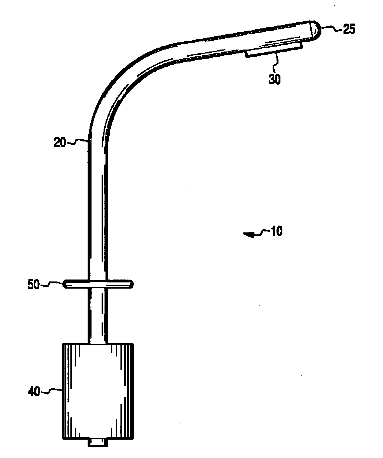
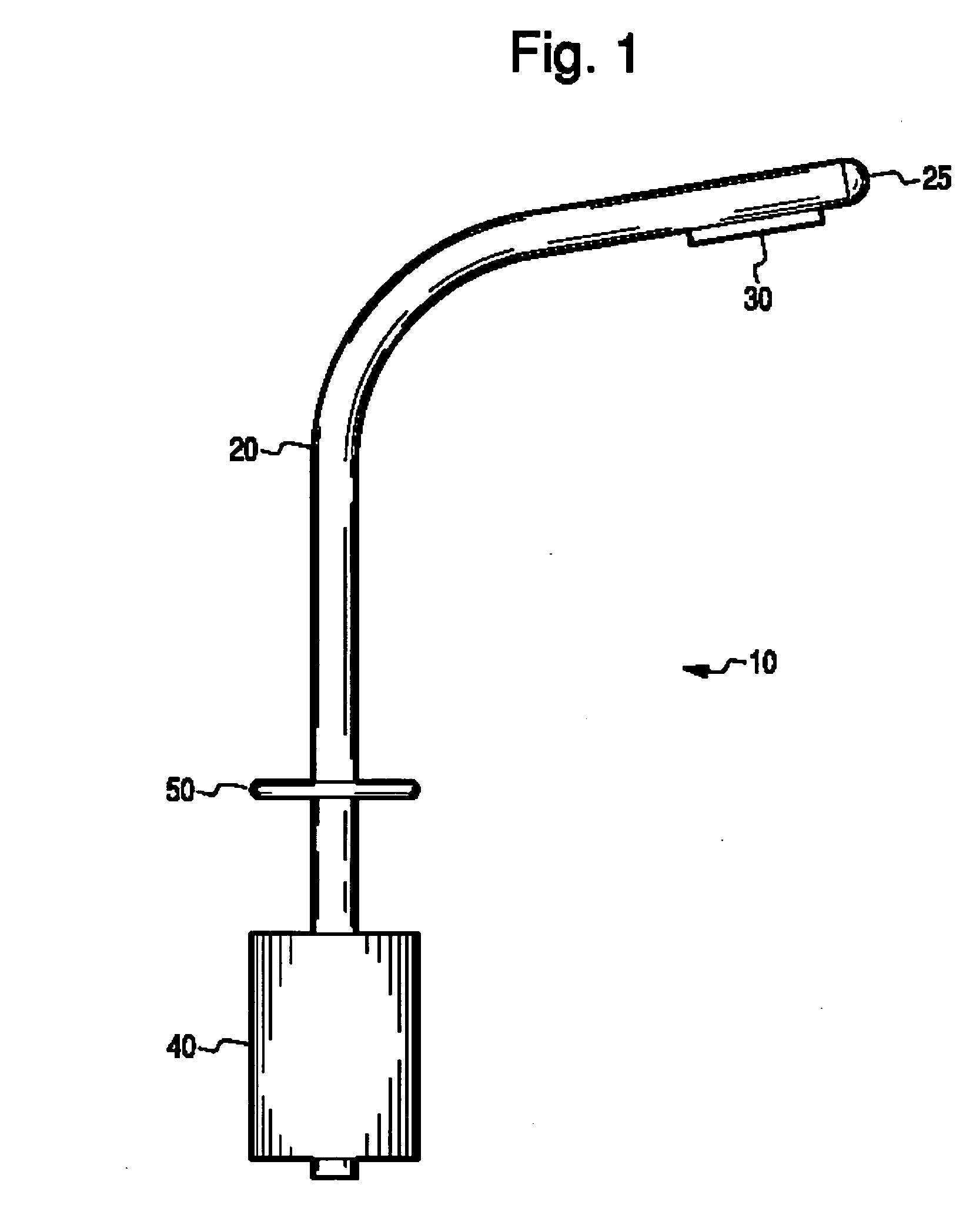
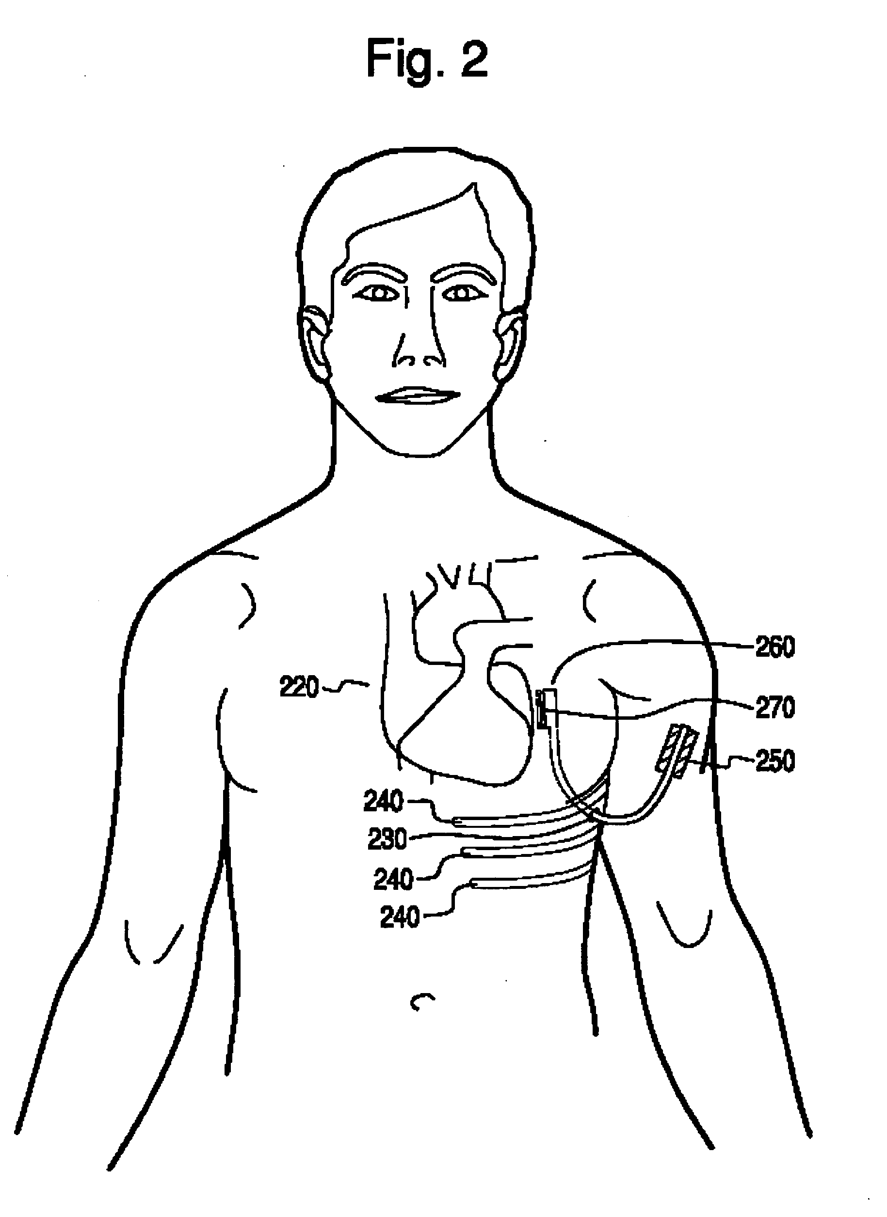
[0027] The various embodiments of the present invention include a much shortened percutaneous catheter system that is configured to be inserted into the chest cavity through a small opening and thence manipulated to the pericardium. An array of ultrasonic transducers and / or ECG sensors on the catheter desirably are positioned on the outside surface of or inside the pericardium in the vicinity of the heart by means of a cannula inserted in the chest. Positioning advantageously is carried out manually or by robotic or semi-robotic manipulators. Once positioned near the heart, the sensors send signals to other externally positioned electronic equipment attached or otherwise in communication with the catheter.

[0028] For example, an array of piezoelectric transducers may be energized to pulse ultrasonic energy and, acting as receivers, detect reflected ultrasound energy, converting received ultrasound into electrical signals (“detected signals.”). The detected signals are conducted to c...


PUM


Abstract

A peritoneal ultrasound imager includes an elongated body less than about 20 inches in length that is adapted to be inserted through a cannula into or near the pericardium space, and an ultrasound transducer array at one end of the body that is suitable for ultrasound echocardiography. The cannula and ultrasound imager may be of a single piece construction. A method for imaging the heart includes introducing a cannula into the wall of a patient's chest, inserting the elongated body into the cannula, moving the inserted elongated body to a position near the heart, and imaging the heart with ultrasound echo.

Description

RELATED APPLICATIONS [0001] The present application claims benefit of and priority to U.S. Provisional Application No. 60 / 548,102 entitled METHODS AND SYSTEMS FOR ULTRASOUND IMAGING OF THE HEART FROM THE PERICARDIUM, filed Feb. 27, 2004, which is hereby incorporated by reference in its entirety.BACKGROUND OF THE INVENTION [0002] 1. Field of the Invention [0003] The present invention is directed at systems for examining a heart, and more particularly to a method and apparatus for imaging the heart using an ultrasound imaging catheter. [0004] 2. Description of the Related Art [0005] Cardiac monitoring and cardiac intervention are important procedures in modern medicine. Information intensive procedures such as cardiac imaging generally requires placing one or more sensors at or in the heart itself, requiring some degree of invasiveness. High resolution heart imaging, for example, often is done by inserting an ultrasound imaging catheter into the heart via the femoral artery. [0006] On...

Claims


Application Information

Patent Timeline
IPC IPC(8): A61B8/00A61B8/02A61B8/12A61B8/14
CPCA61B8/02A61B8/0883A61B8/4488A61B8/12A61B8/0891
Inventor JENKINS, DAVID A.
Owner EP MEDSYST
Why Eureka
Try Eureka