Ultrasonic endoscope

a technology of endoscope and ultrasonic, applied in the field of ultrasonic endoscope, can solve the problems of increasing unable to reduce the diameter of the hard distal portion, and unable to achieve the effect of facilitating the maintenance performan

Active Publication Date: 2005-10-13
FUJI PHOTO OPTICAL CO LTD
View PDF16 Cites 29 Cited by
  • Summary
  • Abstract
  • Description
  • Claims
  • Application Information

AI Technical Summary

Benefits of technology

[0013] To resolve the shortcomings, it is one objective of the present invention to facilitate

Problems solved by technology

When, as is described above, the elevator used for a treatment equipment is attached to the hard distal portion, in addition to the ultrasonic test unit and the endoscopic observation unit, the structure of the hard distal portion becomes extremely complicated, and depending on how the components are arranged, the diameter of the hard distal portion will be increased.
Therefore, unless these components are arranged logically, the diameter of the hard distal portion can not be reduced.
Thus, during the operation performed to remove the constituents of the endoscopic observation unit from the hard distal portion, the enormous number of wires constitutes an obstacle that can interrupt the removal of a member to be repaired, and can result in the br

Method used


Image

  • Ultrasonic endoscope
  • Ultrasonic endoscope
  • Ultrasonic endoscope

Examples


Embodiment Construction

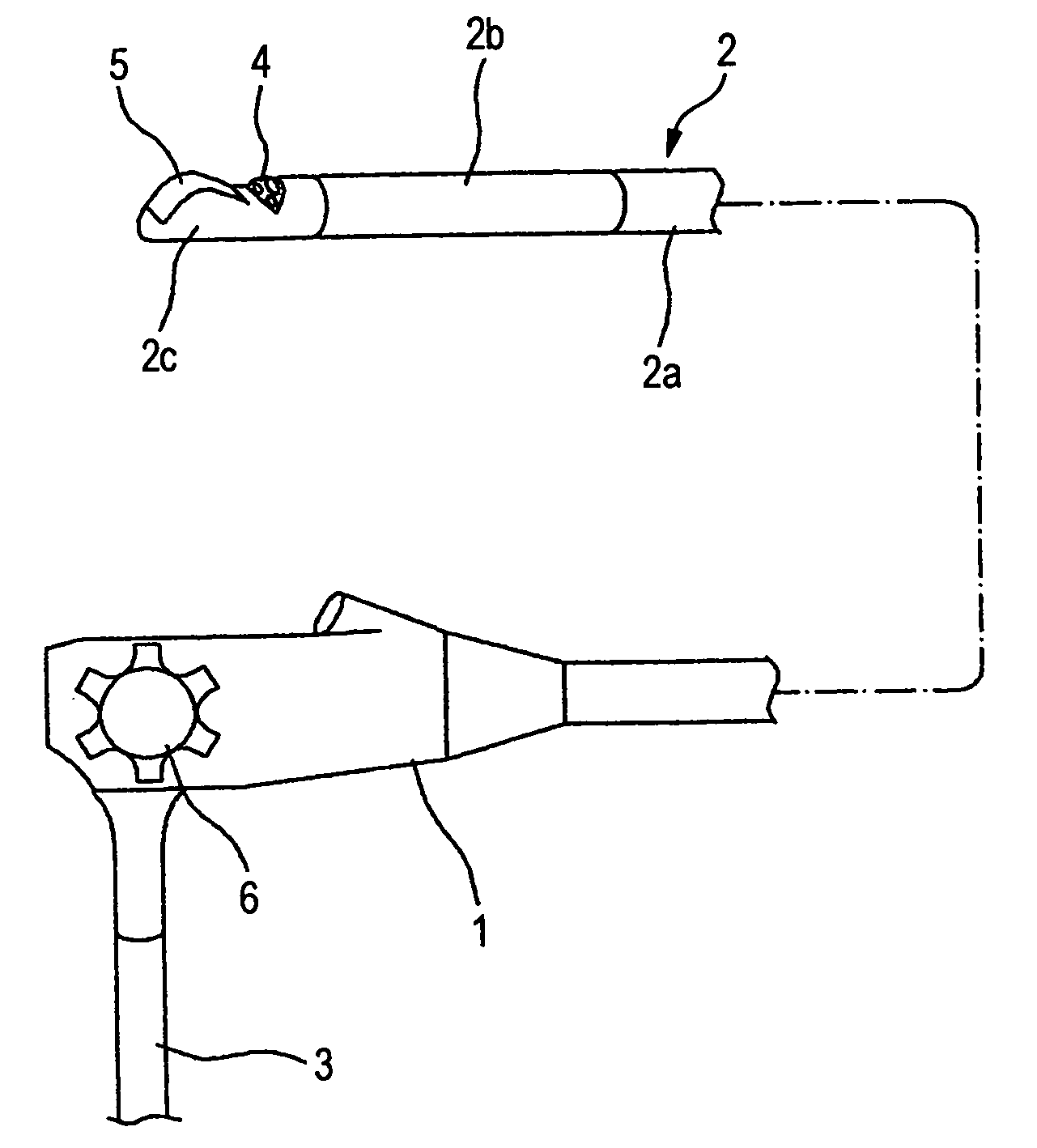
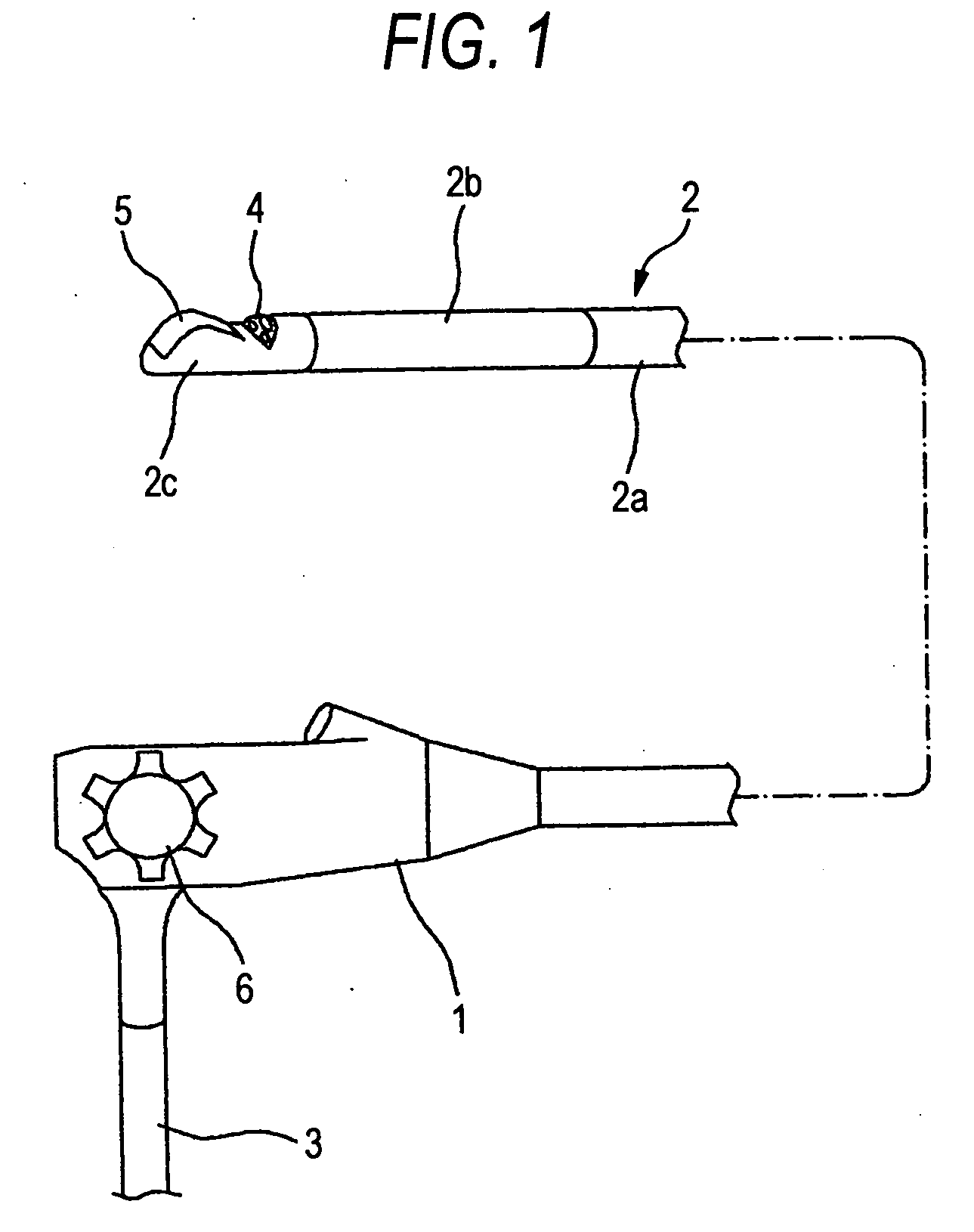
[0037] One embodiment of the present invention will now be explained while referring to the drawings. First, the schematic structure of an ultrasonic endoscope is shown in FIG. 1. In FIG. 1, the ultrasonic endoscope comprises: a main control body 1; an insertion portion 2 to be inserted into a coelom; and a universal cord 3. The insertion portion 2 is constituted by a flexible portion 2a, an angle portion 2b and a hard distal portion 2c, arranged in order from a base end. The flexible portion 2a is flexible so that it can be bent in an arbitrary direction along an insertion path within a coelom. An endoscopic observation unit 4 and an ultrasonic test unit 5 are attached to the hard distal portion 2c, and the angle portion 2b is used to turn the hard distal portion 2c in an arbitrary direction. The angle portion 2b is operated by an angle control portion 6 provided for the main control portion 1.

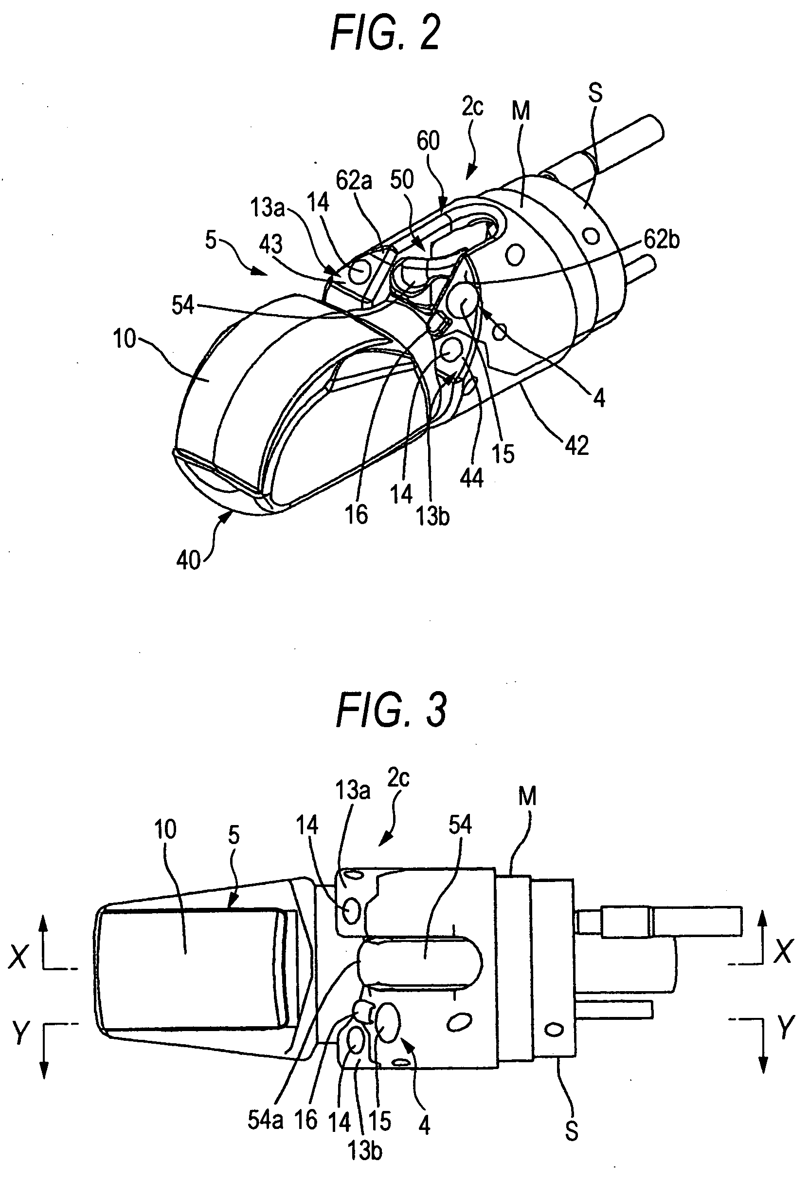
[0038] The external appearance of the hard distal portion 2c when separated from the ang...


PUM


Abstract

An ultrasonic endoscope comprises an insertion portion including: an angle portion; a hard distal portion; an endoscopic observation unit comprising a lighting portion and an observation portion; and an ultrasonic test unit comprising an ultrasonic transducer constituting an ultrasonic test unit, wherein the hard distal portion includes: a distal end main body including an entire portion whereat the ultrasonic transducer is attached; an observation portion block that includes a portion whereat the observation portion is attached and that is separably coupled with the distal end main body; and an elevator block that is securely held between the distal end main body and the observation portion block by engaging the distal end main body and the observation portion block, and that comprises the elevator, and wherein the distal end main body, the observation portion block and the elevator block are assembled so as to be capable of being disassembled.

Description

BACKGROUND OF THE INVENTION [0001] 1. Field of the Invention [0002] The present invention relates to an ultrasonic endoscope that is inserted into a coelom or other body cavity to conduct an endoscopy and an ultrasonic examination. [0003] 2. Description of the Related Art [0004] Generally, an ultrasonic endoscope is so designed that an endoscopic observation unit, constituted by a lighting portion and an observation portion, and an ultrasonic test unit, which includes an ultrasonic transducer, are provided at the distal end of an insertion portion to be inserted in to a coelom. A coelom examination is performed by introducing the insertion portion into the coelom and using the endoscopic observation unit. Then, if during the endoscopy an abnormality is observed, additional information concerning the state of the body tissue can be obtained using the ultrasonic test unit. Further, by taking into account all the results produced by the endoscopy and the ultrasonic examination, therape...

Claims


Application Information

Patent Timeline
IPC IPC(8): A61B1/00A61B8/12A61B1/018A61B1/04A61B1/06A61B8/14
CPCA61B1/00098A61B8/14A61B1/018A61B1/00165
Inventor KOHNO, SHINICHI
Owner FUJI PHOTO OPTICAL CO LTD
Why Eureka
Try Eureka