Method for the early detection of renal injury

a renal injury and early detection technology, applied in the field of early detection of renal injury, can solve the problems of delayed initiation, and persistent high mortality and morbidity

Inactive Publication Date: 2005-12-08
CHILDRENS HOSPITAL MEDICAL CENT CINCINNATI +1
View PDF48 Cites 105 Cited by
  • Summary
  • Abstract
  • Description
  • Claims
  • Application Information

AI Technical Summary

Problems solved by technology

Acute renal failure (ARF) secondary to a renal tubular cell injury, including an ischemic injury or a nephrotoxic injury remains a common and potentially devastating problem in clinical medicine and nephrology, with a persistently high rate of mortality and morbidity despite significant advances in supportive care.
While these studies have suggested possible therapeutic approaches in animal models, translational research efforts in humans have yielded disa...

Method used


Image

  • Method for the early detection of renal injury
  • Method for the early detection of renal injury
  • Method for the early detection of renal injury

Examples


example 1

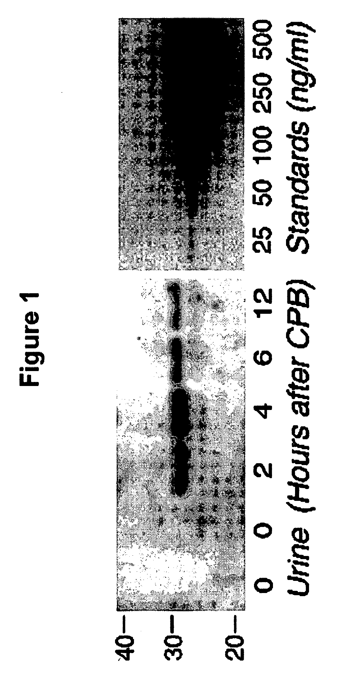
[0067] Western Analysis For NGAL Expression And Quantitation: Equal aliquots (30 μl) of each urine sample were boiled for 10 min in denaturing buffer and subjected to standard Western Blot analysis with an affinity purified goat polyclonal antibody raised against human NGAL (F-19, Santa Cruz Biotechnology). Simultaneous blots were prepared under identical conditions of transfer and exposure with known quantities of recombinant human NGAL, as standards for quantitation of urine NGAL as previously described by Mishra et al. in Am J Nephrol 2004;24:307-315. The laboratory investigators were blinded to the sample sources and clinical outcomes until the end of the study.

[0068] Urine NGAL Measurements—Western Analysis: NGAL was virtually undetectable in the urine of all patients prior to surgery, and in healthy volunteers (n=10). FIG. 1 shows a Western Blot typical of that for a patient undergoing CPB. NGAL is not detected at 0 hours, or before CPB, but rapidly appears in the urine by 2 ...

example 2

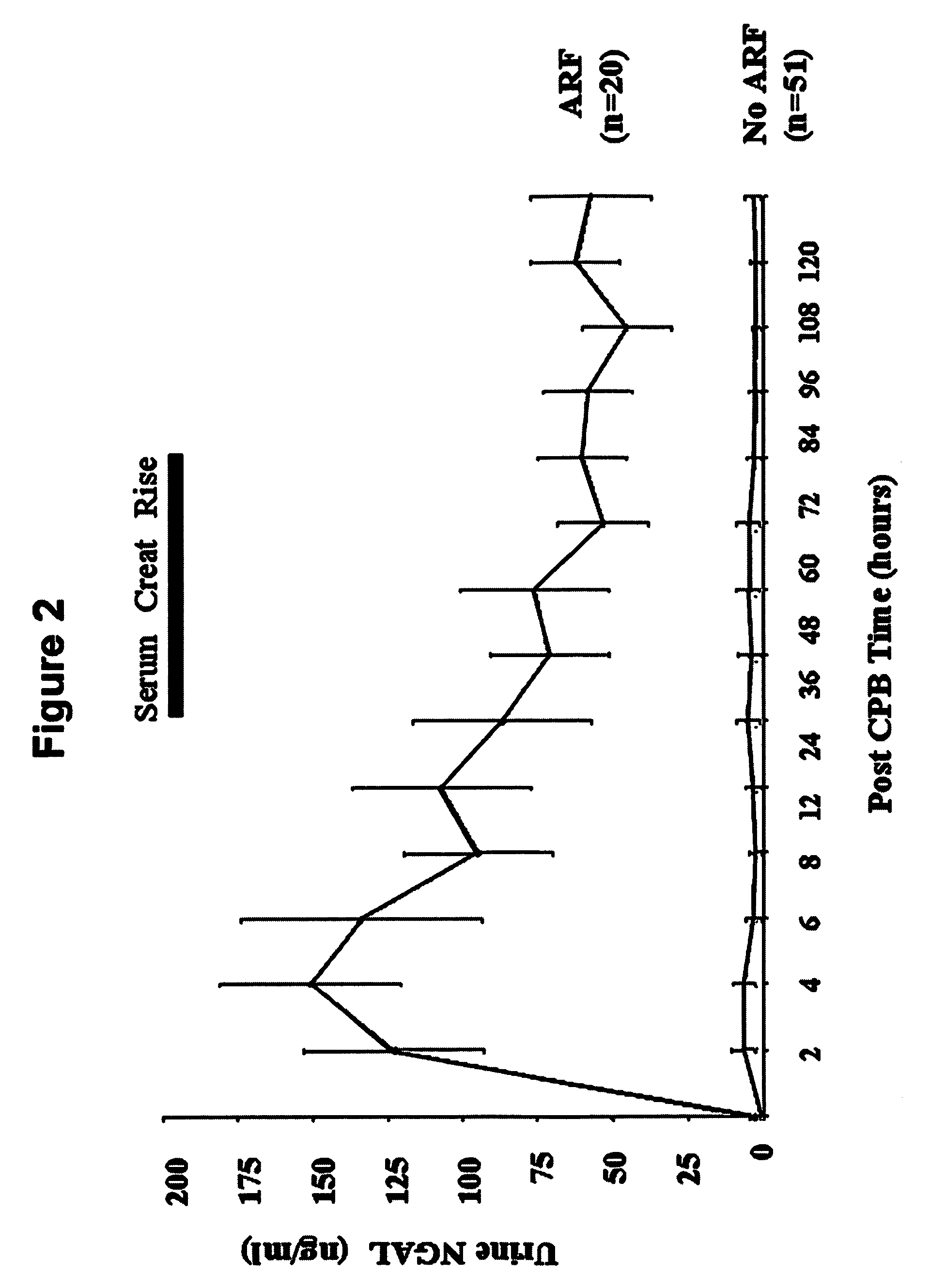
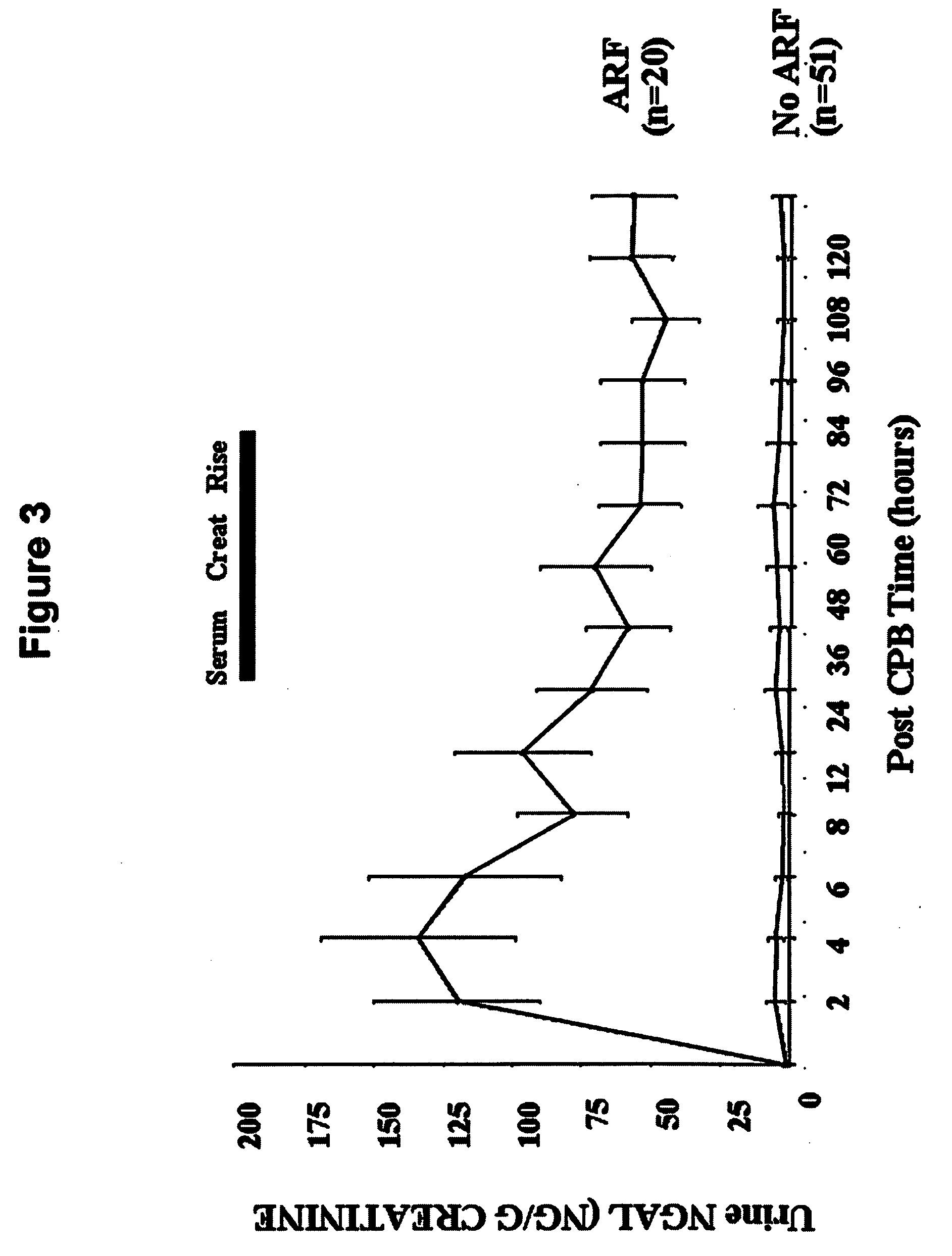
[0070] In patients who never developed acute renal injury, there was a small but statistically significant increase in urinary NGAL at 2 hours or the first available sample post CPB (4.9±1.5 ng / ml versus 0.9±0.3 ng / ml at baseline, p<0.05) and 4 hours post CPB (4.9±1.2 ng / ml, p<0.05 ver baseline). In marked contrast, patients who subsequently developed acute renal injury displayed a dramatic increase in urinary NGAL at all time points examined, as shown in FIG. 2. The pattern of urinary NGAL excretion was characterized by a peak very early after the precipitating event (2-6 hours following CPB), followed by a lesser but sustained increase over the entire duration of the study. This overall pattern remained unchanged when urinary NGAL concentration was normalized for urinary creatinine excretion (FIG. 3).

Example 4

[0071] Urine NGAL levels were consistently low in healthy volunteers (2.2±0.5 ng / ml, n=10) and at baseline in all subjects (1.6±0.3 ng / ml, n=71). In patients who never deve...

example 4

[0072] Serum NGAL Measurements—ELISA: Serum NGAL is a novel early biomarker of ischemic renal injury, similar to troponins in myocardial ischemia, and detection of serum NGAL is an example of the invention. Serum NGAL levels were consistently low in normal healthy volunteers (2.5±0.8, n=6) and all study subjects prior to surgery (3.2±0.5 ng / ml, n=71). Patients who never developed acute renal injury showed a small but statistically significant increase in serum NGAL at 2 hours or the first available sample post CPB (7.0±1.1 ng / ml, p<0.05 versus baseline) and 12 hours post CPB (5.2±0.8 ng / ml, p<0.05 versus baseline). Patients who subsequently developed acute renal injury displayed a striking increase in serum NGAL at all time points examined, as shown in FIG. 7. Similar to urine NGAL, the serum NGAL peaked very early after CPB, followed by a lesser but sustained increase over the entire duration of the study. Serum NGAL levels were 61±10 ng / ml at 2 hours, 54.7±7.9 ng / ml at 12 hours, a...


PUM


Abstract

A method and kit for detecting the immediate or early onset of renal disease and injury, including renal tubular cell injury, utilizing NGAL as an immediate or early on-set biomarker in a sample of blood serum. NGAL is a small secreted polypeptide that is protease resistant and consequently readily detected in the blood serum following renal tubule cell injury. NGAL protein expression is detected predominantly in proximal tubule cells, in a punctuate cytoplasmic distribution reminiscent of a secreted protein. The appearance NGAL in the serum is related to the dose and duration of renal ischemia and nephrotoxemia, and is diagnostic of renal tubule cell injury and renal failure. NGAL detection is also a useful marker for monitoring the nephrotoxic side effects of drugs or other therapeutic agents.

Description

BACKGROUND OF THE INVENTION [0001] Acute renal failure (ARF) secondary to a renal tubular cell injury, including an ischemic injury or a nephrotoxic injury remains a common and potentially devastating problem in clinical medicine and nephrology, with a persistently high rate of mortality and morbidity despite significant advances in supportive care. Pioneering studies over several decades have illuminated the roles of persistent vasoconstriction, tubular obstruction, cellular structural and metabolic alterations, and the inflammatory response in the pathogenesis of ARF. While these studies have suggested possible therapeutic approaches in animal models, translational research efforts in humans have yielded disappointing results. The reasons for this may include the multifaceted response of the kidney to ischemic injury and nephrotoxins, and a paucity of early biomarkers for ARF with a resultant delay in initiating therapy. [0002] An individual is considered to have acute renal failu...

Claims


Application Information

Patent Timeline
IPC IPC(8): G01N33/53G01N33/542G01N33/68
CPCG01N33/6893G01N2333/82G01N33/573G01N2800/52G01N2800/347
Inventor DEVARAJAN, PRASADBARASCH, JONATHAN M.
Owner CHILDRENS HOSPITAL MEDICAL CENT CINCINNATI
Why Eureka
Try Eureka