Shunt and method treatment of glaucoma

a glaucoma and shunt technology, applied in the field of shunt and method treatment of glaucoma, can solve the problems of eye damage, eye pain, eye irritation, etc., and achieve the effects of reducing the risk of glaucoma, and improving the quality of li

Inactive Publication Date: 2005-12-08
UNIVERSITY OF SASKATCHEWAN
View PDF20 Cites 184 Cited by
  • Summary
  • Abstract
  • Description
  • Claims
  • Application Information

AI Technical Summary

Benefits of technology

[0011] Broadly, the invention provides a shunt for implantation between the anterior chamber of the eye and the epithelial-lined space through the frontal sinus bone of a patient for the treatment of glaucoma. The shunt includes a tube having a length sufficient to span the distance between the anterior chamber of the eye and the epithelial-lined space of the patient, the tube having an open anterior chamber end and a closed epithelial-lined space end, and a seal device associated with the tube between the anterior chamber and epithelial-lined space ends, for sealing a hole in the frontal sinus bone, and for anchoring the tube against movement from the frontal sinus bone. The shunt also includes a fluid pressure openable valve in the tube, located at or near the epithelial-lined space sinus end, allowing for controlled flow of aqueous humor through the tube when implanted.

Problems solved by technology

Glaucoma occurs when aqueous humor does not drain properly from the anterior chamber, causing an increase in intraocular pressure, closure of surrounding blood vessels, and damage to the retina and optic nerve.
This condition, left untreated, can be very painful and lead to blindness.
These medications are associated with side effects, including burning sensations in the eye, headaches, cardiac fluctuations and blurred vision.
The administration of medications for the treatment of animals is difficult and ineffective.
There is also the risk of allowing too much fluid to drain from the anterior chamber, resulting in hypotony and flattening of the eye.
Implants that drain aqueous humor into tissues surrounding the eye induce scarring in such tissues, which scarring ultimately inhibits absorption of fluid from the shunt.
Several problems have occurred with these devices.
First, it is difficult to calibrate the valve to maintain an optimal intraocular pressure, and often leads to hypotony.
Second, the formation of fibrous tissue can occlude the valve and render the drainage device useless.
Although somewhat successful in less severe cases, the prior art implant devices, while successful in the short term, are not effective for long-term management of glaucoma due to occurrence of hypotony or more difficult forms of glaucoma.

Method used


Image

  • Shunt and method treatment of glaucoma
  • Shunt and method treatment of glaucoma
  • Shunt and method treatment of glaucoma

Examples


example

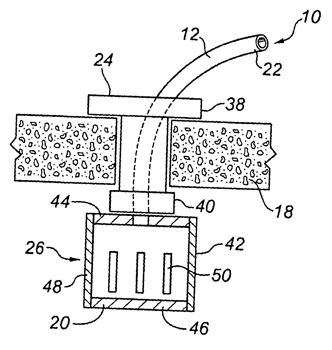
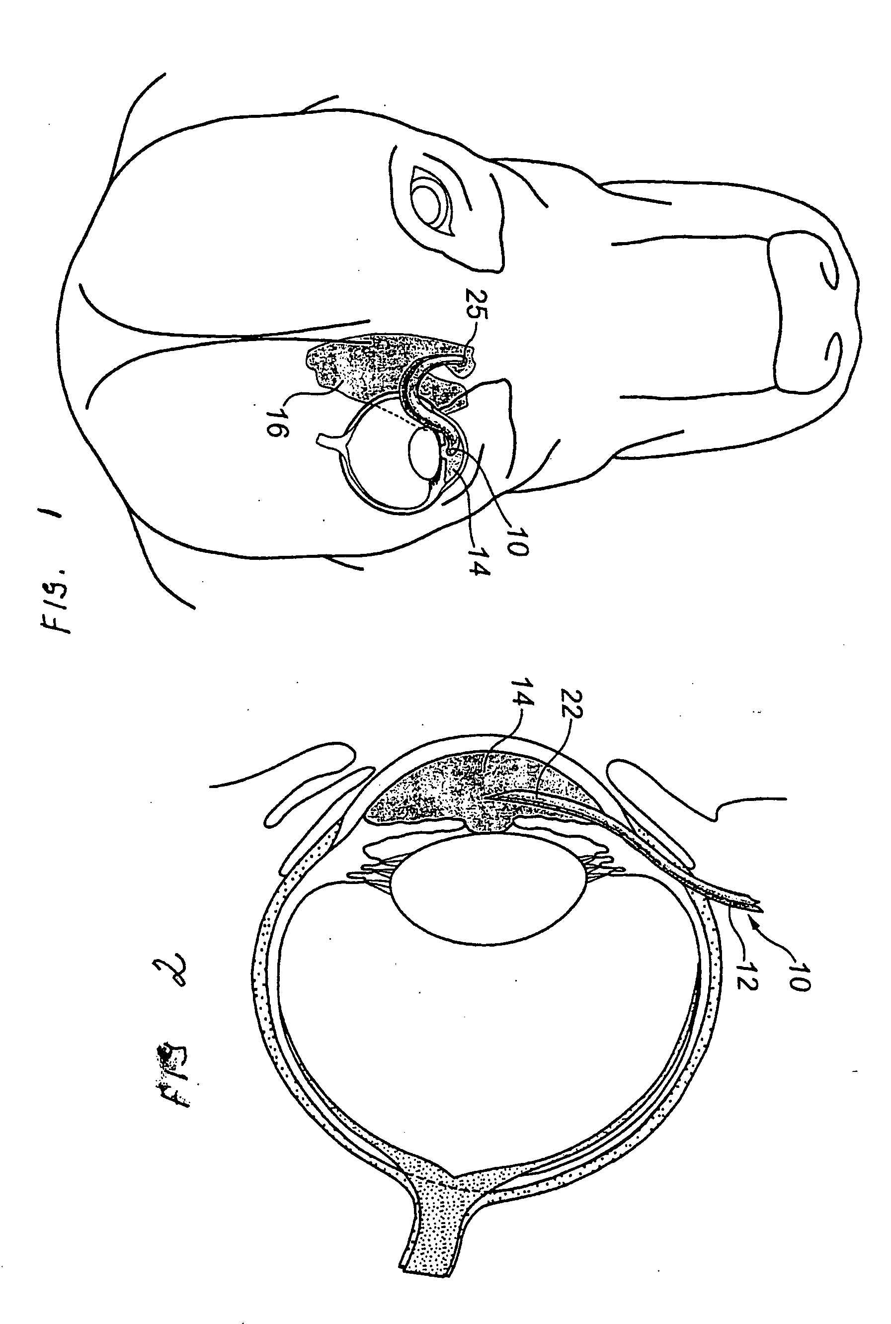
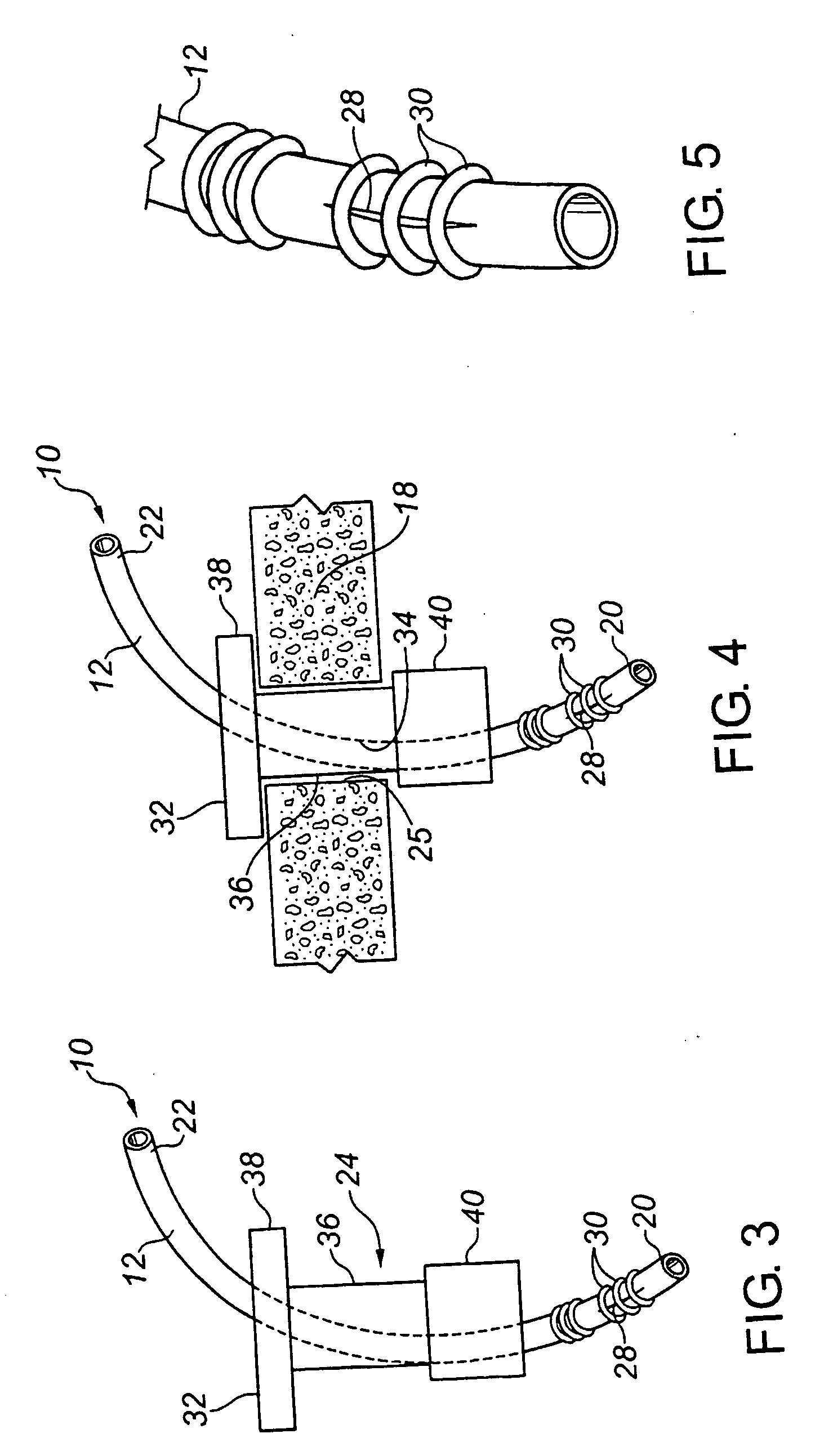
[0047] A shunt of the present invention was designed for and successfully implanted into four dogs having glaucoma. The details of the implanted shunt were as follows:

Tube and Slit Valve

[0048] Inner Diameter 0.02″ (0.5 mm) [0049] Outer diameter 0.037″ (0.94 mm) [0050] Length of Tube (rough cut to be 100 mm) then tailored to each eye in the operating room so that it extended approximately 1 / 3 away across the anterior chamber of the eye. [0051] Slit—1 mm length [0052] # Ligatures—4—to maintain predetermined fluid pressure, adjusted in the operating room [0053] Hole in the Frontal sinus bone (diameter)—(2 mm)

Plug (Sealing Device) [0054] Central Portion (that traversed the width of the bone)—(1.5 mm) [0055] Lower flange—2.5 mm [0056] Upper Flange—5 mm [0057] Length of Plug (Central Portion—traversing the bone)—3 mm

[0058] These patients did not experience frontal sinus, subcutaneous, conjunctival or intraocular infections; fibrosis, haemorrhage, or other untoward complications. Eac...


PUM


Abstract

This invention provides a shunt for implantation between the anterior chamber of the eye and the epithelial-lined space through the frontal sinus bone of a patient for the treatment of glaucoma. The shunt includes a tube having a length sufficient to span the distance between the anterior chamber of the eye and the epithelial-lined space of the patient, the tube having an open anterior chamber end and a closed epithelial-lined space end, and a seal device associated with the tube between the anterior chamber and epithelial-lined space ends, for sealing a hole in the frontal sinus bone, and for anchoring the tube against movement from the frontal sinus bone. The shunt also includes a fluid pressure openable valve in the tube, located at or near the epithelial-lined space sinus end, allowing for controlled flow of aqueous humor through the tube when implanted. The invention also extends to a method of treating glaucoma in a patient by surgically implanting the shunt between the anterior chamber of the eye and the frontal sinus.

Description

FIELD OF INVENTION [0001] The present invention relates to a method and device for controlling aqueous humor flow from the anterior chamber of the eye in the treatment of glaucoma. BACKGROUND OF THE INVENTION [0002] Glaucoma is a medical condition in which the intraocular pressure of the eye increases. The anterior chamber, the cavity located between the cornea and lens, is filled with a fluid called aqueous humor. The aqueous humor drains from the anterior chamber to the venous system through the canals of Schlemm and is replaced continuously by secretions from the ciliary body. Glaucoma occurs when aqueous humor does not drain properly from the anterior chamber, causing an increase in intraocular pressure, closure of surrounding blood vessels, and damage to the retina and optic nerve. This condition, left untreated, can be very painful and lead to blindness. It is estimated that 65 million people world-wide suffer from this condition (Glaucoma Research Foundation, 2002). Animals c...

Claims


Application Information

Patent Timeline
Patent Type & Authority Applications(United States)
IPC IPC(8): A61F9/00A61F9/007A61M5/00A61M27/00A61M39/22
CPCA61F9/0017A61F9/00781A61M27/002A61M39/22A61M2210/0612
Inventor GRAHN, BRUCE HAROLDSTOREY, ERIC SHADSTILLING, DENISE SHARON
Owner UNIVERSITY OF SASKATCHEWAN
Why Eureka
Try Eureka