Fetal RNA in amniotic fluid to determine gene expression in the developing fetus

a technology of amniotic fluid and fetal rna, which is applied in the direction of sugar derivatives, biochemistry apparatus and processes, organic chemistry, etc., can solve the problem that no other technology is available to determine the gene expression pattern of a living human fetus

Inactive Publication Date: 2006-01-05
NEW ENGLAND MEDICAL CENT HOSPITALS
View PDF1 Cites 10 Cited by
  • Summary
  • Abstract
  • Description
  • Claims
  • Application Information

AI Technical Summary

Benefits of technology

[0009] In general, the present invention involves isolating fetal RNA from a sample of amniotic fluid, and analyzing the fetal RNA obtained. In preferred embodiments, the analysis provides qualitative or quantitative information about fetal gene expression. In certain embodiments, fetal RNA is isolated at multiple time points during gestation. The present invention allows particular gene expression patterns, or elements of such patterns, to be correlated with developmental events, and further allows comparison of observed gene expression patterns or components with patterns or components for which such a correlation has been established.

Problems solved by technology

No other technology is available for determining gene expression pattern in a living human fetus.

Method used


Image

  • Fetal RNA in amniotic fluid to determine gene expression in the developing fetus
  • Fetal RNA in amniotic fluid to determine gene expression in the developing fetus
  • Fetal RNA in amniotic fluid to determine gene expression in the developing fetus

Examples


example 1

Preliminary Test—Fetal mRNA Extraction From Amniotic Fluid

[0225] Cell-free fetal mRNA has been successfully extracted and amplified from both fresh and frozen residual amniotic fluid samples. Amniotic fluid samples were initially collected for routine diagnostic purposes; the supernatant is usually discarded following karyotype analysis, while in therapeutic amniocentesis the entire sample is discarded. In the cytogenetics laboratory, samples were spun at 350×g for 10 minutes to remove cells for culture. Samples were centrifuged again at 13,000×g either upon receipt in the case of fresh samples, or immediately after thawing in the case of frozen samples. This ensured that the extracted RNA was truly extracellular.

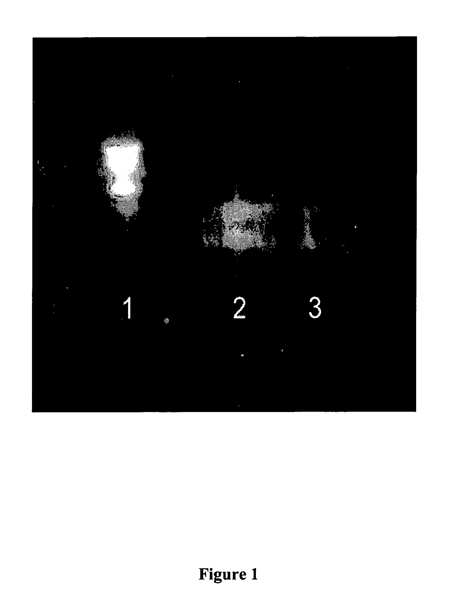
[0226] RNA was extracted using the Qiagen Viral RNA mini kit following the vacuum protocol as described above. Sample starting volumes were typically 420 μL. Synthetic poly-A RNA (15-25 μg) was added to the sample during extraction as a carrier. RNA was concentrated into ...

example 2

Large Volume Amniotic Fluid Samples—Processing and Storage

[0229] In some instances, large volumes (>1L) of amniotic fluid are drawn for therapeutic reasons (i.e., polyhydramnios). These samples, which are usually discarded, provide large starting quantities of fetal cell-free RNA. Nine of such high volume samples have been collected so far and are currently stored. Typically, the amniotic fluid was drawn into 1 L vacuum-sealed containers in a sterile manner. Upon receipt, the fluid was divided into 50 mL aliquots and centrifuged at 800×g for 15 minutes to remove any cellular material. The supernatant (45 mL) from the samples was pooled into 225 mL containers for storage at −80° C.

example 3

Large Volume Amniotic Fluid Samples—RNA Extraction and Preparation for Microarrays

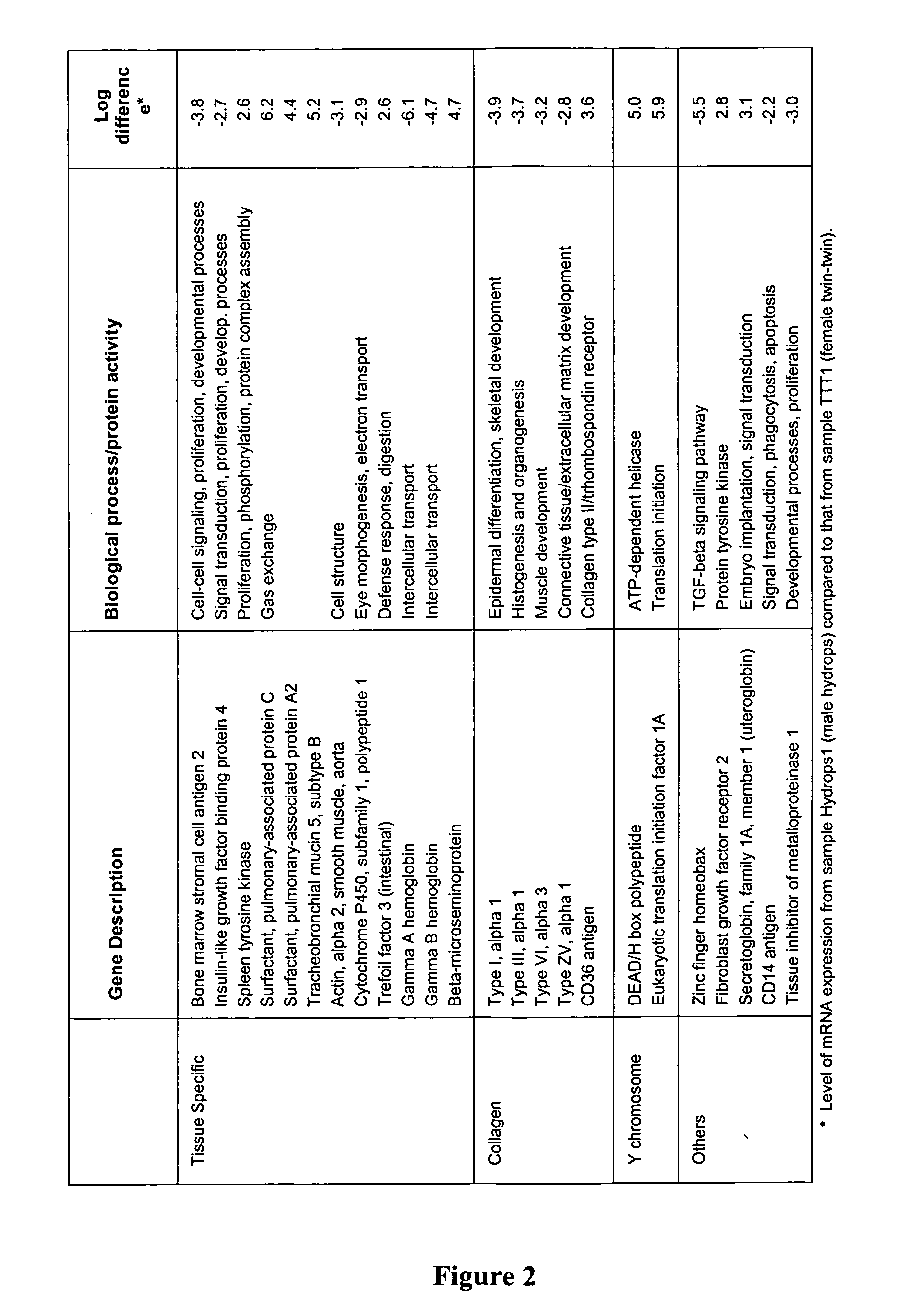
[0230] Two large volume amniotic fluid samples were obtained and processed as above. One was from the pregnant woman carrying twin female fetuses at 24 3 / 7 weeks of gestation (designated TTT1). The other sample was taken from the pregnant woman at 29 4 / 7 weeks of gestation whose male fetus had hydrops of unknown etiology (designated Hydrops1). In addition, 30 mL of each sample was taken (in 5 mL aliquots) and RNA was extracted in lieu of freezing the aliquot using a modification of the QIAamp Viral RNA Vacuum Protocol for Large Volumes (Qiagen, Inc., Valencia, Calif.) as described above. The two 30 mL samples yielded 7.2 ng and 12.7 ng, respectively. In order to obtain a larger quantity of RNA, 60 mL of previously frozen amniotic fluid supernatant from the above samples were used for an additional extraction. The samples were pooled, precipitated and resuspended in 10 μL of RNAse inhibitor free water,...


PUM

PropertyMeasurementUnit
volumeaaaaaaaaaa
volumesaaaaaaaaaa
volumesaaaaaaaaaa

Abstract

The present invention provides improved methods of prenatal diagnosis, monitoring, screening and/or testing. The invention is based, at least in part, on the discovery that amniotic fluid is a rich source of cell-free fetal RNA. Methods of isolation and analysis of fetal RNA are described, that can lead to information about fetal gene expression that is not available by other techniques. The inventive systems allow for a more comprehensive determination of a living human fetus' health, growth and development and for the prenatal diagnosis of a variety of diseases and conditions.

Description

PRIORITY INFORMATION [0001] This application claims priority to Provisional Patent Application No. 60 / 536,571, entitled “Fetal RNA in Amniotic Fluid to Determine Gene Expression in the Developing Fetus” and filed Jan. 15, 2004. The Provisional Patent Application is incorporated herein by reference in its entirety.GOVERNMENT INTERESTS [0002] The work described herein was funded by the National Institutes of Health (Grant No. NIH HD42053). The United States government may have certain rights in the invention.BACKGROUND OF THE INVENTION [0003] Genetic disorders and congenital abnormalities (also called birth defects) occur in about 3 to 5% of all live births (A. Robinson and M. G. Linden, “Clinical Genetic Handbook”, 1993, Blackwell Scientific Publications: Boston, Mass.). Combined, genetic disorders and congenital abnormalities have been estimated to account for up to 30% of pediatric hospital admissions (C. R. Scriver et al., Can. Med. Assoc. J., 1973, 108: 1111-1115; E. W. Ling et a...

Claims


Application Information

Patent Timeline
Patent Type & Authority Applications(United States)
IPC IPC(8): C12Q1/68C07H21/02
CPCC12Q1/6881C12Q2600/158
Inventor BIANCHI, DIANA W.LARRABEE, PAIGE B.JOHNSON, KIRBY L.
Owner NEW ENGLAND MEDICAL CENT HOSPITALS
Why Eureka
Try Eureka