
Optical apparatus and method of use for non-invasive tomographic scan of biological tissues

a tomographic scan and optical apparatus technology, applied in the field of optical imaging systems, can solve the problems of cws method not being able to distinguish light scattering and absorption properties, involving in these methods, and being usually noisy

Inactive Publication Date: 2006-01-19
O2 MEDTECH +1
View PDF14 Cites 193 Cited by
  • Summary
  • Abstract
  • Description
  • Claims
  • Application Information

AI Technical Summary

Benefits of technology

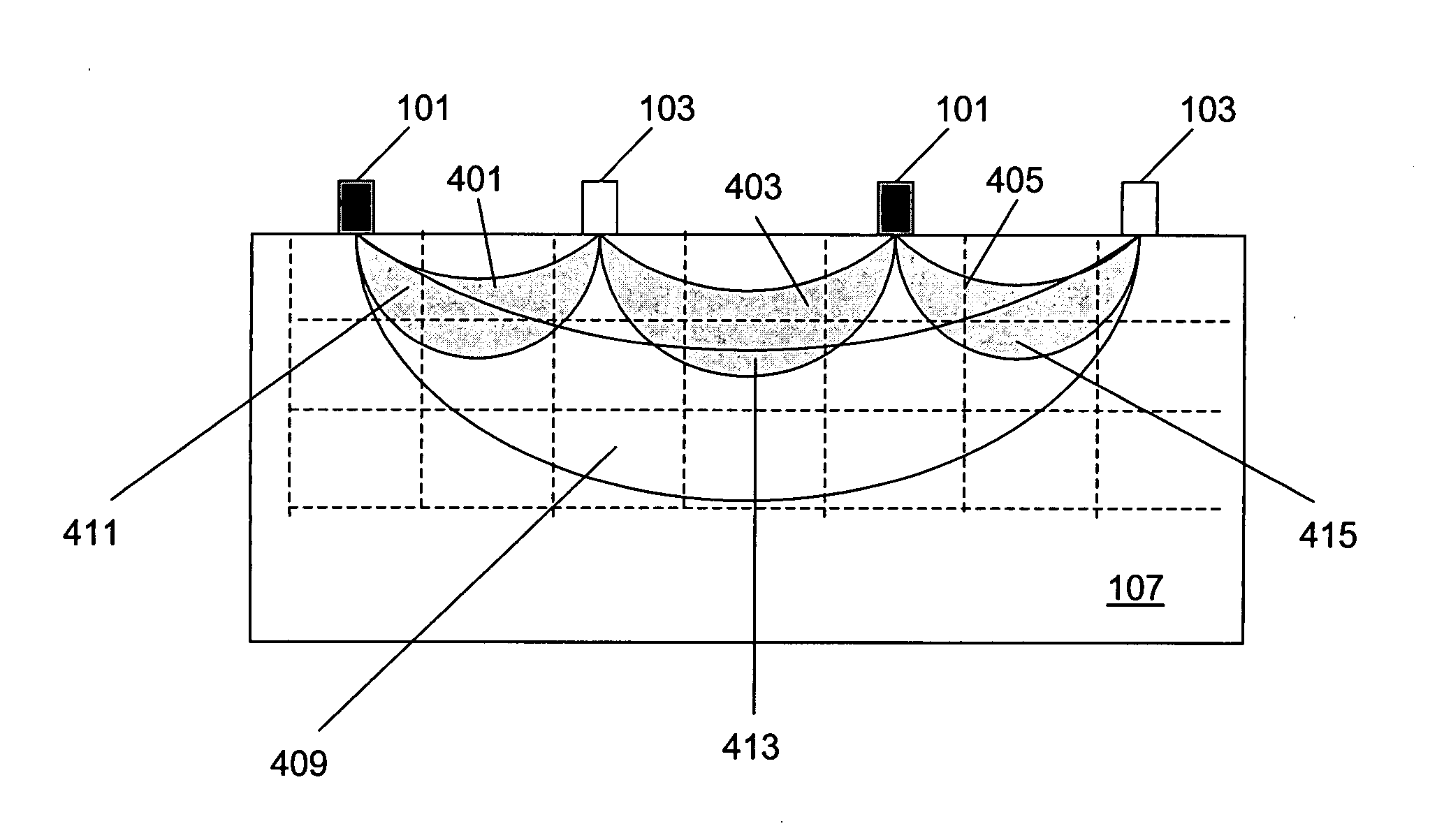
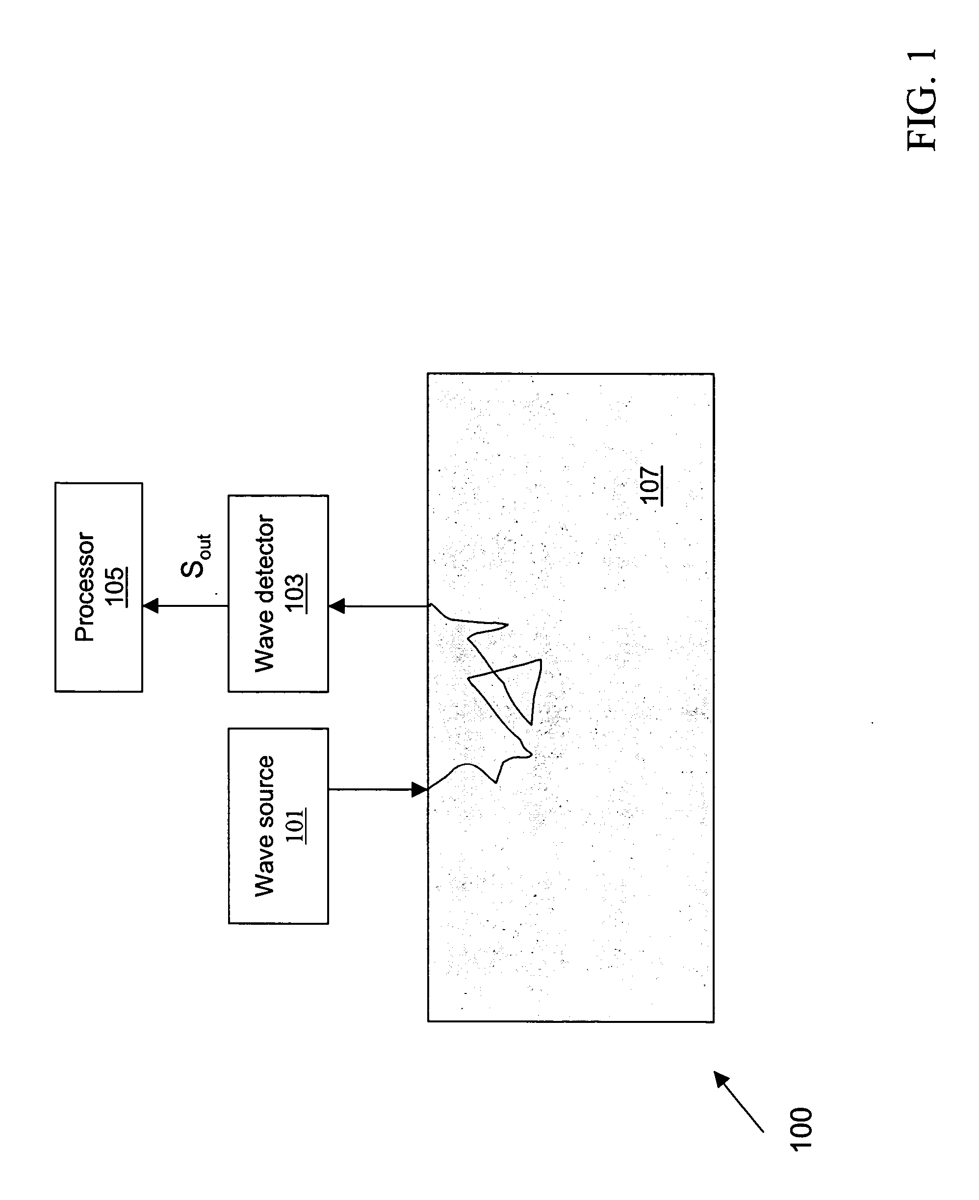
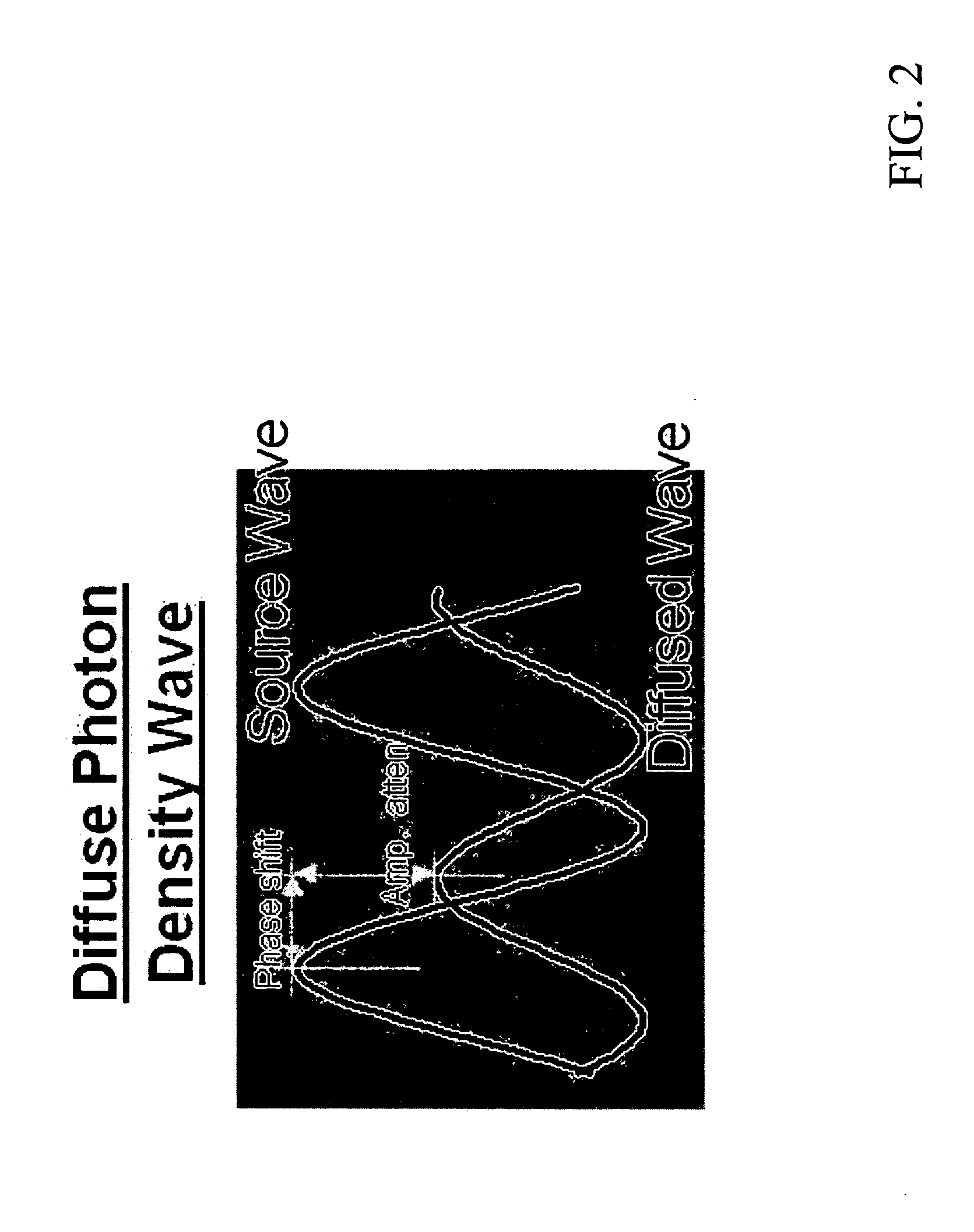
"The present invention is about optical imaging systems and methods for creating two- or three-dimensional images of highly scattering physiological media. The invention uses formulas and algorithms to determine absorption properties of these materials based on phase only measurements. The invention also includes methods for non-invasive tomographic scan of highly scattering media using back scattered photons, as well as simultaneous measurement of concentrations of biochemical substances and blood oxygen saturation inside tissue and artery. The technical effects of the invention include improved accuracy and accuracy in the measurement of absorption properties of highly scattering physiological media, as well as improved non-invasive tomographic scan and simultaneous measurement of biochemical substances and blood oxygen saturation."

Problems solved by technology

To the contrary, the CWS method cannot distinguish the light scattering and absorption properties and has generally been used to calculate relative values of or changes in the concentrations of the oxygenated or deoxygenated hemoglobins.
However, there are problems involving in these methods.
First, they are usually very noisy since the amplitude measurement is affected by many elements, such as source, detector drift, sensor attachment, etc.
Second, there is no analytical inverse solution for the diffusion equation which results in it being hard to conduct accurate calculation.
Finally, despite their capability of providing averaged quantitative hemoglobin concentrations and the oxygen saturation, the general problem of the TRS and PMS is that it is difficult to quantify concentrations of chromophores of a localized position inside tissues, such as the cortical tissue inside the head.
Nevertheless, there is no existing method which worked effectively, especially for the applications where only back reflected photons are detectable and transmitted photons are too weak to be detected, such as measurement on head or chest.
The major challenge has been due to the scattering effect.

Method used


Image

  • Optical apparatus and method of use for non-invasive tomographic scan of biological tissues
  • Optical apparatus and method of use for non-invasive tomographic scan of biological tissues
  • Optical apparatus and method of use for non-invasive tomographic scan of biological tissues

Examples


Embodiment Construction

[0023] The following detailed description is presented to enable any person skilled in the art to make and use the invention. For purposes of explanation, specific nomenclature is set forth to provide a thorough understanding of the present invention. However, it will be apparent to one skilled in the art that these specific details are not required to practice the invention. Descriptions of specific applications are provided only as representative examples. Various modifications to the preferred embodiments will be readily apparent to one skilled in the art, and the general principles defined herein may be applied to other embodiments and applications without departing from the scope of the invention. The present invention is not intended to be limited to the embodiments shown, but is to be accorded the widest possible scope consistent with the principles and features disclosed herein.

[0024] The primary object of present invention is to provide a non-invasive optical system, an op...


PUM


Abstract

The present invention relates to a non-invasive optical system equipped with optical tomographic scanning method and algorithm for quantifying scattering and absorption properties and chromophore concentrations of highly scattering medium such as biological tissues, for 3D mapping and imaging reconstruction of the spatial and temporal variations in such properties. The invention further relates to a method and an apparatus for simultaneous measurement of concentrations of biochemical substances and blood oxygen saturation inside a biological tissue and arterial blood.

Description

FIELD OF THE INVENTION [0001] The present invention relates to methods and optical imaging systems for images of spatial or temporal distribution of chromophores or properties thereof in a physiological medium. The optical imaging system is useful for the non-invasive measurement of various physiological properties in animal and human subjects and in particular, is useful in determining blood oxygen saturation of both tissue and arteries. BACKGROUND OF THE INVENTION [0002] Near-infrared spectroscopy has been used for non-invasive measurement of various physiological properties in animal and human subjects. The basic principle underlying the near-infrared spectroscopy is that a physiological medium, such as a tissue, includes a variety of light-absorbing chromophores and light-scattering substances which can interact with low energy near-infrared photons transmitted thereto and traveling therethrough. For example, deoxygenated and oxygenated hemoglobins in human blood are the most do...

Claims


Application Information

Patent Timeline
Patent Type & Authority Applications(United States)
IPC IPC(8): A61B5/00
CPCA61B5/14552A61B5/0073
Inventor CHENG, XUEFENG
Owner O2 MEDTECH
Learn More