
Dental retractor and method of use to produce anatomically accurate jaw models and dental prostheses

a retractor and anatomically accurate technology, applied in the field of dental retractors and methods of use to produce anatomically accurate jaw models and dental prostheses, can solve the problems of not being able to construct an anatomically correct model that contained gingival tissue, not being able to describe scanning process, etc., and achieve the effect of comfortable holding the tongue away

Inactive Publication Date: 2006-01-26
BIOMEDICAL MODELING
View PDF0 Cites 36 Cited by
  • Summary
  • Abstract
  • Description
  • Claims
  • Application Information

AI Technical Summary

Benefits of technology

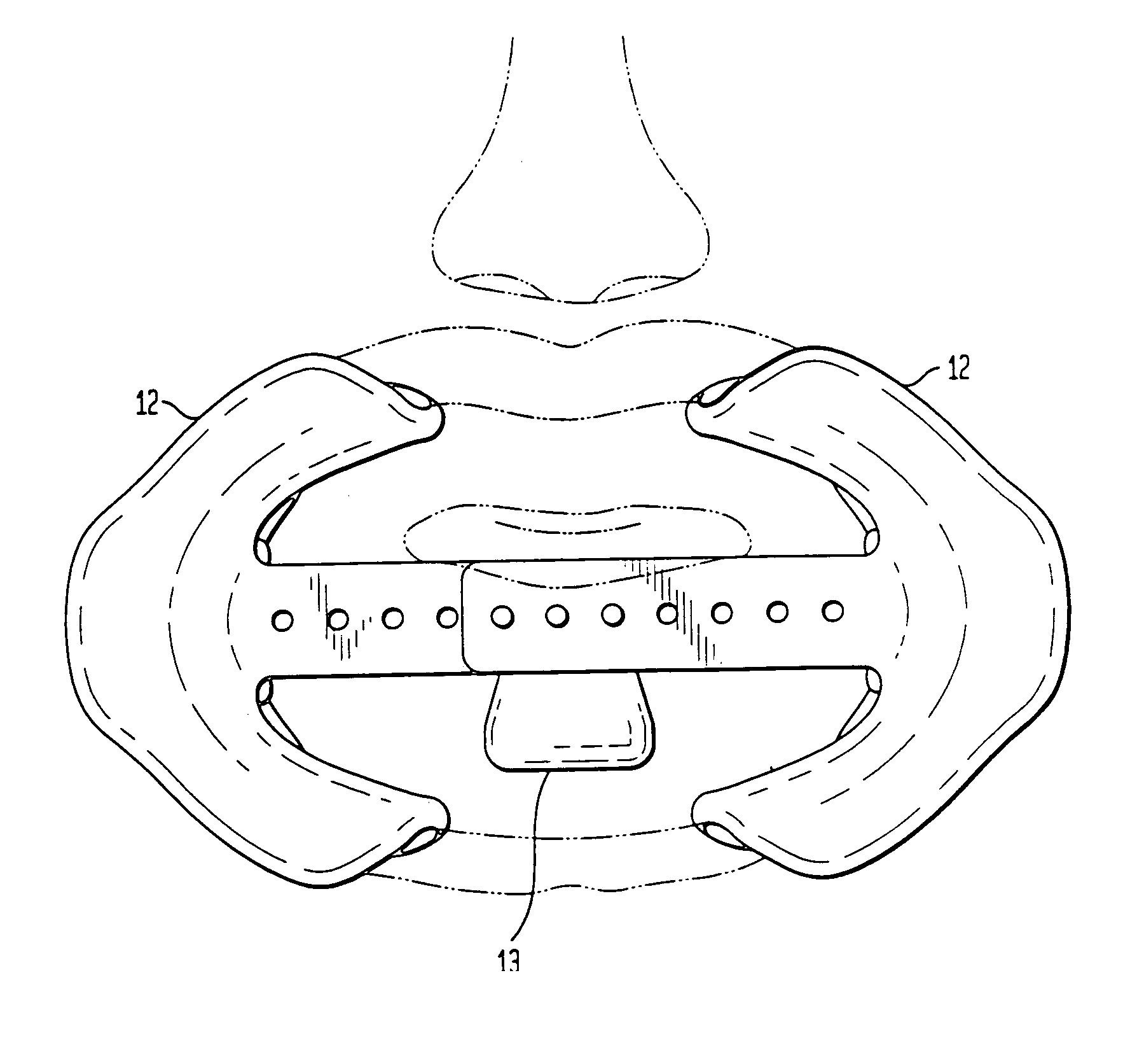
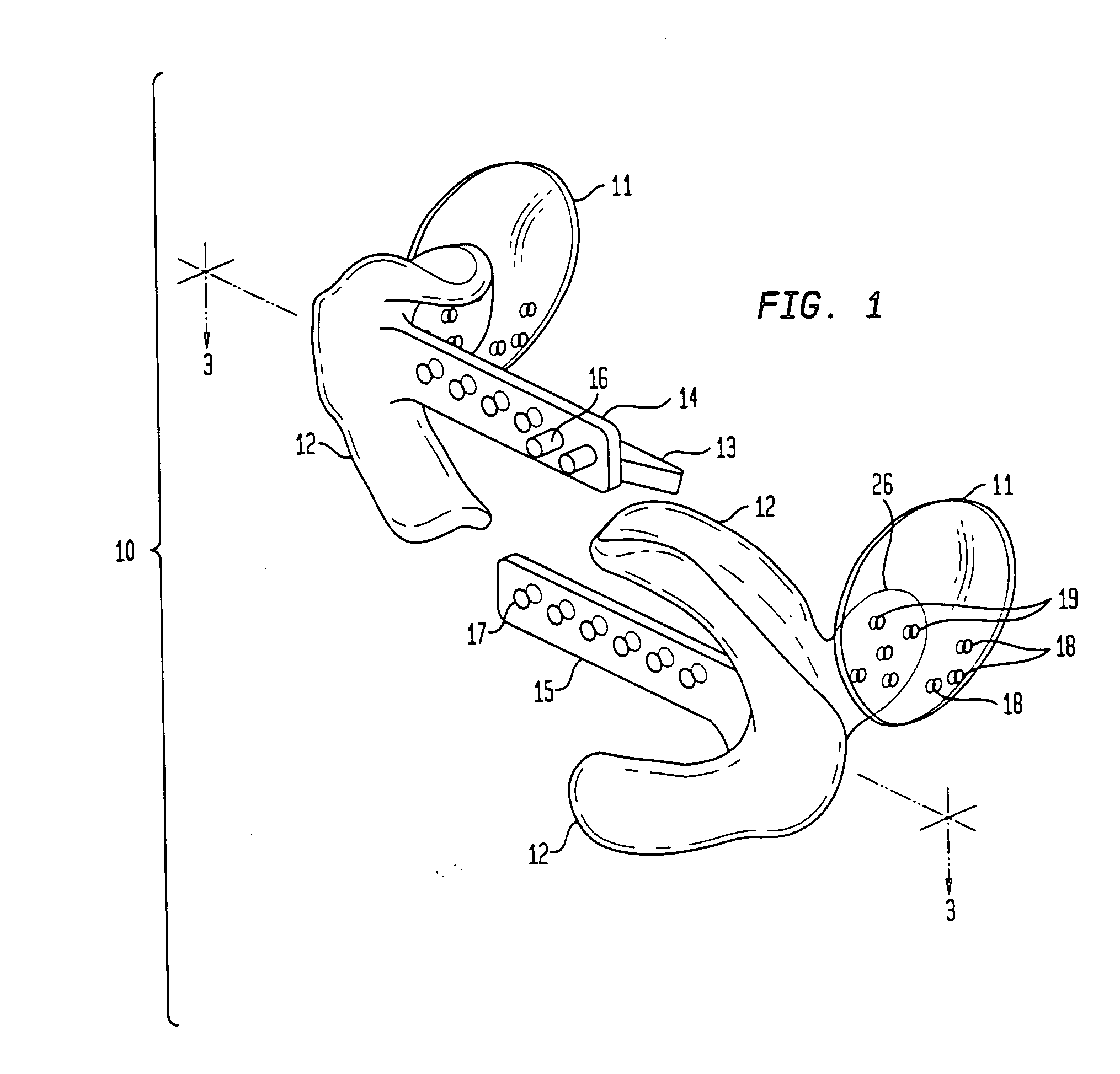
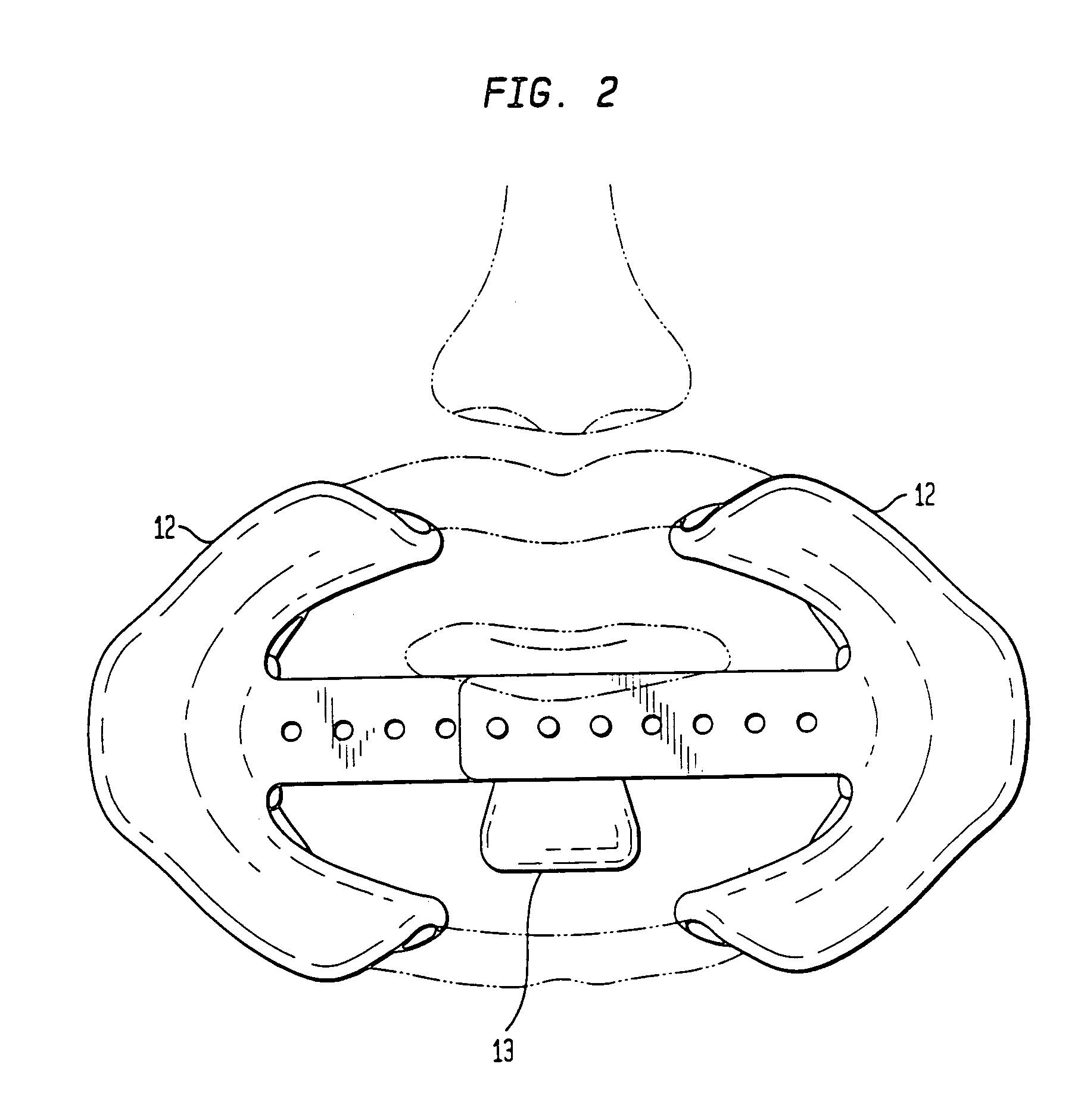
[0009] An improved retractor comprises one or more cheek positioning members and one or more lip positioning members has been developed. This retractor makes it possible to conduct X-ray CT or similar scans of the jaw and to construct anatomically accurate models of bone, teeth and gingival tissue. The cheek positioning member(s) applies pressure to the inside of the cheek pushing it away from the teeth and gingiva. The lip positioning member may push or cup the lips away from the gingiva. Alternatively, the lip positioning member may form a physical barrier or wall between the gingiva and the lips. Optionally and preferably, the improved retractor will have a tongue positioning member to comfortably hold the tongue away from gingival tissue. In still another optional embodiment, the retractor would also hold the tongue away from the palate. In alternative embodiments, the improved retractor may be constructed to permit unobstructed imagining of the entire jaw, upper jaw, the lower jaw, left side upper jaw, left side lower jaw or both and right side upper jaw, right side lower jaw or both. Preferably, the improved retractor will not contact the gingival tissue. More preferably, the improved retractor will be radiolucent. Most preferably, the improved retractor will provide a scan image having a different contrast than those of teeth, bone and gingival tissue.
[0011] The apparatus of the present invention solves the shortcomings in the art in a simple and straightforward device. What is provided is an improved, optionally radiolucent retractor useful when conducting CT scans of the jaw of an individual requiring a dental implant. The improved retractor allows CT scans to be conducted and models to be constructed that are anatomically accurate with respect to bone, teeth and gingival (soft) tissue. The retractor permits the patient's lips, cheeks and tongue to be held comfortably away from teeth and gingiva, thereby permitting an unobstructed X-ray CT image of jaw bone, teeth and gingiva to be taken and a model constructed therefrom accurately modeling bone, teeth and gingiva. Suitable model building techniques include:
[0014] FDM (Fused Deposition Modeling)—Process using molten plastics or wax extruded by a nozzle that traces the models cross sectional geometry layer by layer. FDM creates tough models that are ideal for functional usage.
[0015] Three-Dimensional Printing (3DP)—Ink-jet based process that prints the models cross sectional geometry on layers of powder spread on top of each other. This process enables models to be built quickly and affordably. Models may also be printed in color.
[0019] The advantages of advance planning are well known. They include: Improved functional and esthetic results, reduced chair time for the patient, lowered risk of implant and prosthetic failure, lowered risk of alveolar nerve damage, more accurate estimates of costs and time, lower cost of the entire procedure, reduced risk of unexpected difficulties during surgery & restorations, reduced risk of complications and reduced surgical time. The surgeon may rehearse the surgery and thereby refine procedures before operating on a patient. This is generally known as performing “model surgery”.

Problems solved by technology

The scanning process described in Pompa did not produce a model with soft gingival tissue.
The process of Pompa did not distinguish between gingival, lip, tongue and cheek tissue thereby making it impossible to construct an anatomically correct model that contained gingival tissue.

Method used


Image

  • Dental retractor and method of use to produce anatomically accurate jaw models and dental prostheses
  • Dental retractor and method of use to produce anatomically accurate jaw models and dental prostheses
  • Dental retractor and method of use to produce anatomically accurate jaw models and dental prostheses

Examples


Embodiment Construction

[0030] The improved, retractor of the present invention allows CT and similar imaging scans to be conducted of the jaw and models to be constructed that are anatomically accurate with respect to bone, teeth and gingival (soft) tissue. The retractor permits the patient's lips, cheeks and optionally the tongue to be held comfortably away from teeth and gingiva, thereby permitting an unobstructed image of jaw bone, teeth and gingiva to be taken and a stereolithographic or similar rapid prototyping model to be constructed there from accurately modeling bone, teeth and gingiva.

[0031] The improved retractor comprises one or more cheek positioning members and one or more lip positioning members. The cheek positioning member(s) apply pressure to the inside of the cheek pushing it away from the teeth and gingiva. The lip positioning member may push or cup and hold the lips away from the gingiva. Alternatively, the lip positioning member may form a physical barrier or wall between the gingiv...


PUM


Abstract

An improved retractor comprising one or more cheek positioning members and one or more lip positioning members has been developed. This retractor makes it possible to conduct X-ray CT or similar scans of the jaw and to construct anatomically accurate models of bone, teeth and gingival tissue. What is provided is an improved retractor useful when conducting CT scans of the jaw of an individual requiring a dental implant and models to be constructed that are anatomically accurate with respect to bone, teeth and gingival (soft) tissue. Drill guides, and prostheses are constructed with the jaw models.

Description

RELATED APPLICATION [0001] This Application claims priority of U.S. Provisional Ser. No. 60 / 589,460 filed Jul. 20, 2004, incorporated herein by reference.BACKGROUND OF THE INVENTION [0002] In modern dentistry, dental implants are the primary method of treatment to replace missing teeth. Much progress has been made since early attempts by Egyptians to replace pulled teeth with cleaned teeth. Prostheses and bridges have been developed to replace missing pieces. Materials suitable for implantation have been developed over the last twenty-five years and implants made from materials such as titanium and alloys of titanium have provided a significant improvement in implant therapy. The historical use of such modern implants has resulted in the confirmation that it is extremely important to determine the exact place where the hole must be drilled on the jaw bone, and the tilt thereof when designing and placing implants. Different radiographic tests have been used in order to study—prior to...

Claims


Application Information

Patent Timeline
Patent Type & Authority Applications(United States)
IPC IPC(8): A61C5/00
CPCA61C5/14A61B6/14A61C5/90A61B6/51
Inventor PRILUCK, EITANMAROTTA, LEONARDFRANCHELL, MICHAEL VINCENTPIGLIACELLI, STEVEN DEFABIOWEINBERG, CRISPIN BERNARDELSAID, KHALED
Owner BIOMEDICAL MODELING
Learn More