Rapid prion-detection assay

a prion detection and detection method technology, applied in the field of prion detection assays for animal and human diseases, can solve the problems of difficult detection of prions, inability to extract and analyze pathogenic prion proteins, and time-consuming and complex analysis methods

Inactive Publication Date: 2006-02-02
PRION DEVAL LAB
View PDF27 Cites 14 Cited by
  • Summary
  • Abstract
  • Description
  • Claims
  • Application Information

AI Technical Summary

Benefits of technology

[0011] The present invention is an assay for determining the presence and concentration of pathogenic prion protein in a biological sample obtained from a human or an animal. In each aspect of the invention, the pathogenic form of prion protein is readily extracted, ...

Problems solved by technology

Detection of prions is difficult because of the poor solubility of prions in many biological buffers and the tenacity of its aggregates in resisting dissolution.
As a result, the methodology used for analyzing prions is oftentimes time-intensive and complex.
The test strip and ...

Method used


Image

  • Rapid prion-detection assay
  • Rapid prion-detection assay
  • Rapid prion-detection assay

Examples


first embodiment

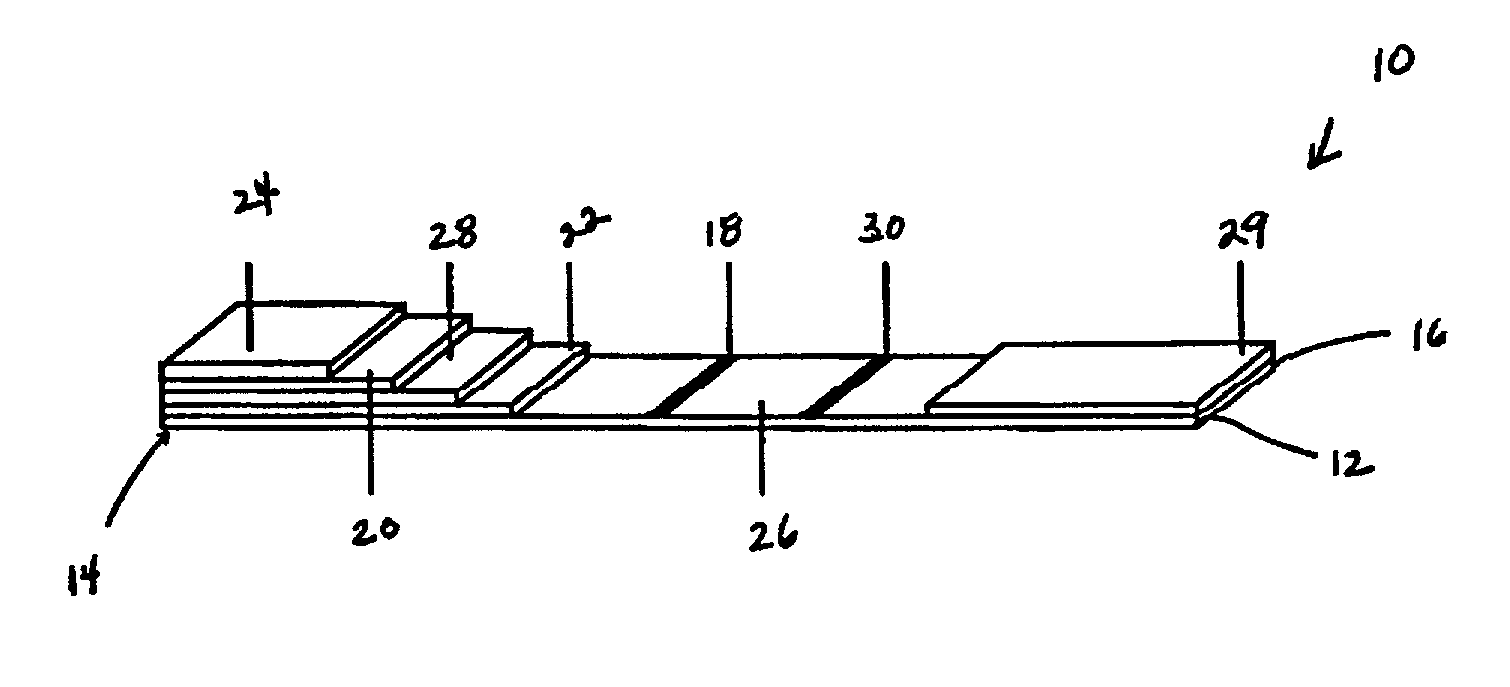
[0038] Shown in FIG. 1 is a test device 10 of a The test device 10 utilizes a pair of antibodies specific to PrPSC. These include (1) a labeled antibody that “detects” the PrPSC and (2) an immobilized antibody that “captures” the prion protein-antibody-label complex to form a “sandwich.” Briefly, in this invention, homogenized sample of a biological material is introduced to the test device. In the preferred embodiment, the sample first moves through a zone containing immobilized proteinase-K, which digests the nonpathogenic prion protein, leaving the PrPSC for analysis. The proteinase-K is immobilized to a solid support. The removal of the normal prion protein minimizes sample interference and results in a higher specificity for the analyte. As the treated sample moves through the test device, it encounters the first specific antibody conjugated to a label and affixed to a portion of the test device. In one embodiment, the label is a colored latex bead.

[0039] The fluid in the homo...

second embodiment

[0071] In a second embodiment, the assay is conducted using a test system or device where the support for the proteinase-K is external to the porous membrane. The support may be, e.g., magnetic beads, latex supports, filter tips, colloidal particles, conjugate supports, plastic surfaces, or glass surfaces.

[0072] The first embodiment of the assay includes homogenizing the sample with a suitable buffer, substantially as described above, and applying the homogenized sample to the test device, such as the composite described above and depicted in FIG. 1. The sample may be applied directly to the digestive pad or the filter pad, or it may be filtered onto either of such pads. Preferably, however, filtration is accomplished in situ directly by the device.

[0073] In the digestive pad, the homogenized sample is treated with the immobilized proteinase-K. As the homogenized sample and PrPSC flow through the device, the antibody conjugated to a label, such as a colored bead or other particulat...


PUM


Abstract

Assays are provided for rapid detection, with high specificity of the pathogenic form of prion protein responsible for neurodegenerative diseases affecting humans and animals, such as transmissible spongiform encephalopathy in bovine, sheep, and cats. Also provided are assays for testing animal feedstock, such as animal feed, for the presence or concentration of pathogenic prion protein. Results are available in from about 0.5 to about 20 minutes and preferably within from about 5 to about 10 minutes. The assays employ proteinase-K to remove normal prion protein from a biological sample, so that the sample may be analyzed by immunochromatography to determine the presence and concentration of pathogenic prion protein. Because the proteinase-K is immobilized on a solid support for in situ removal of interfering components, the present invention obviates the need for subsequent extraction of the desired analyte. All aspects of the present invention are suitable for quantifying the minimal detectable amount of pathogenic prion protein in a biological sample. Moreover, the simplicity of sample preparation makes the present invention suitable for use in the field.

Description

TECHNICAL FIELD [0001] This invention relates to rapid diagnostic assays for testing for disease in animals and humans, and more particularly to assays for detecting the pathogenic form of prion in biological fluids and tissues obtained from animals and humans suspected of having a prion-caused disease as well as foodstuffs made from such biological fluids and tissues. BACKGROUND OF INVENTION [0002] Humans and animals develop a variety of transmissible neurodegenerative disorders as a result of infection by prions—aberrant proteins that join bacteria, viruses, and viroids as infectious pathogens. Examples of prion diseases afflicting animals include scrapie in sheep and goats, and bovine spongiform encephalopathy (BSE) in cattle. Animals may contract a prion disease by consuming feed made from organs and other components from infected animals, such as cow udders and bone in the form of bone meal. Humans are subject to four prion diseases including kuru, Creutzfeldt-Jakob disease, Ge...

Claims


Application Information

Patent Timeline
IPC IPC(8): G01N33/53G01N33/531G01N33/543G01N33/545G01N33/68
CPCG01N33/6896Y10S435/97Y10S435/962G01N2800/2828
Inventor HAJIZADEH, KIAMARSMURTAZA, ZAKIR S.
Owner PRION DEVAL LAB
Why Eureka
Try Eureka