Area exposure dosimetry and area absorbed dosimetry
- Summary
- Abstract
- Description
- Claims
- Application Information
AI Technical Summary
Benefits of technology
Problems solved by technology
Method used
Image

Examples
first embodiment
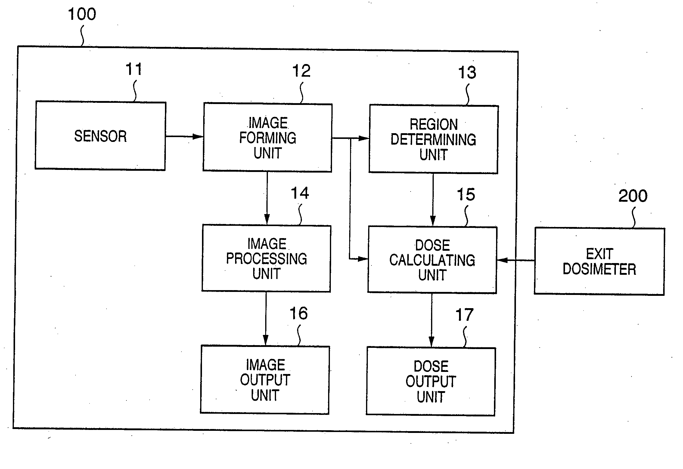
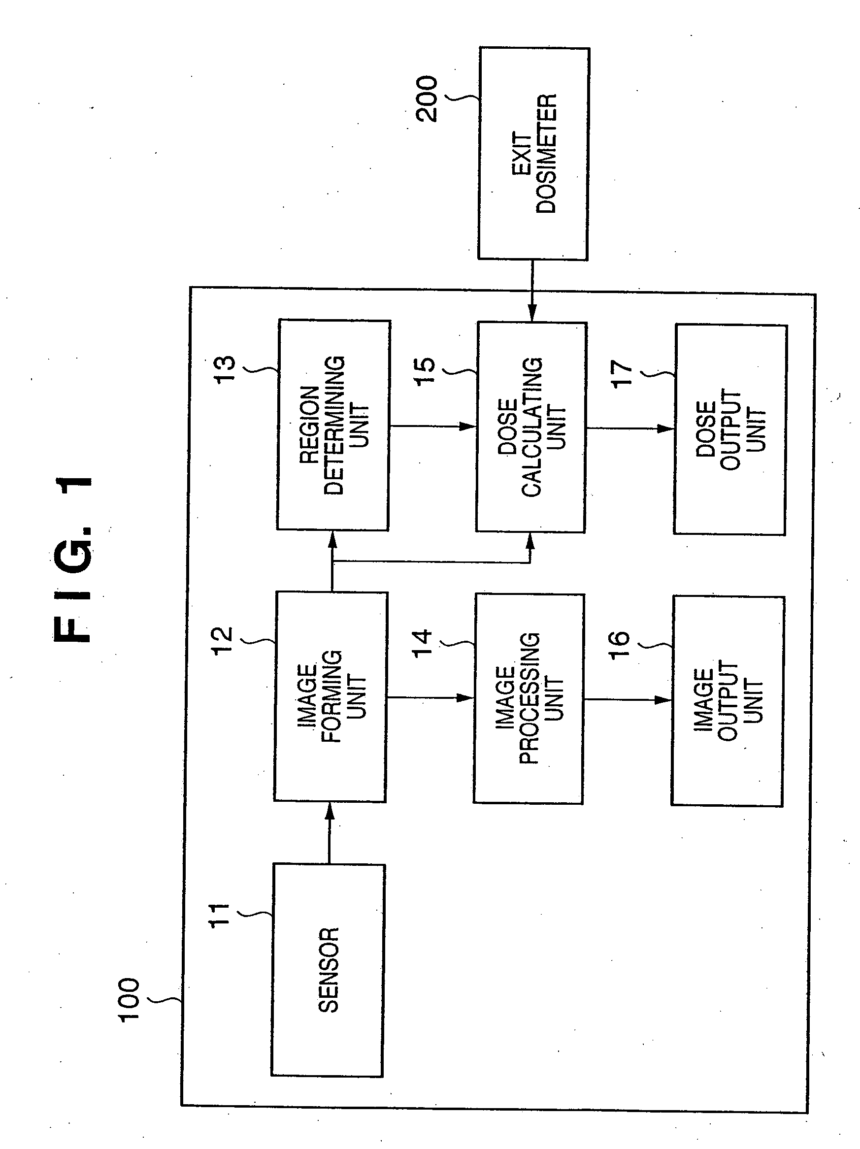
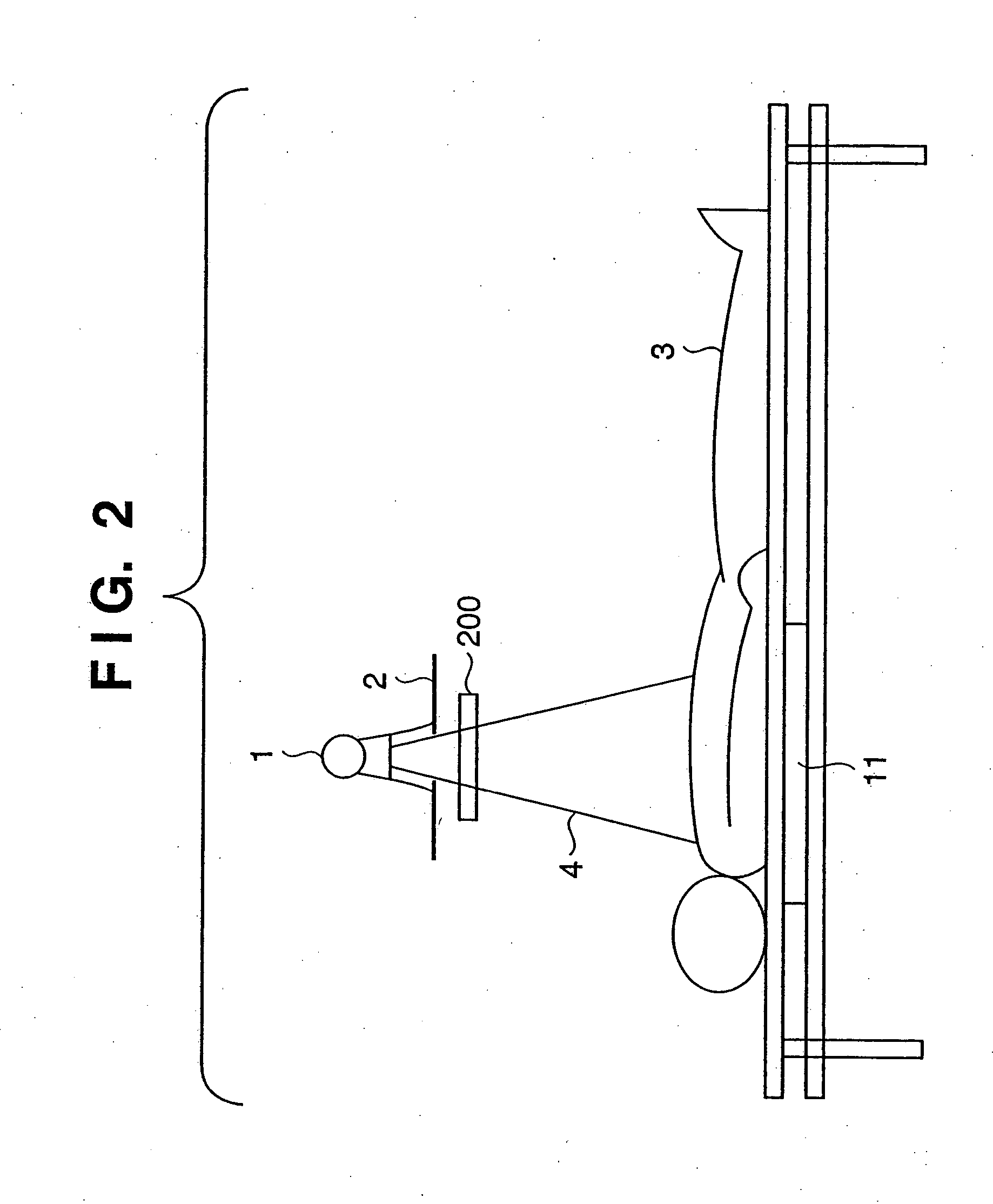
[0033]FIG. 1 is a block diagram showing the arrangement of an exposure dosimetry system according to the first embodiment of the present invention. The exposure dosimetry system of the first embodiment includes a digital radiography system 100 and an exit dosimeter (to be also referred to as an area dosimeter) 200 which is placed on the side of the sensor with respect to an X-ray stop and measures the area dose of X-rays emitted.
[0034] In the digital radiography system 100, reference numeral 11 denotes a sensor obtained by arranging, on a plane, elements for generating electric charges in accordance with the intensity of X-rays; 12, an image forming unit which forms an image by collecting electric charge quantities from the respective elements of the sensor 11; 13, a region determining unit which scans the image output from the image forming unit 12 and determines an X-ray irradiation region, non-object region, and object region; 14, an image processing unit which changes the image...
second embodiment
[0048] The second embodiment of the present invention will be described next.
[0049] In most medical examinations in Japan, only the front of the chest of a patient is radiographed. In this case, since the chin of the patient is rested and positioned on the upper side of the sensor unit, non-object regions always appear above the two shoulders.
[0050] An area exposure dose measuring method suitable for such a case, i.e., a case wherein a radiation digital image always includes non-object regions, will be described below. Assume that in the following case, a sensor sensitivity holding unit is calibrated in advance to allow calculation of an area exposure dose per unit area from the electric charge quantity generated per unit area by the sensor upon reception of X-rays.
[0051]FIG. 6 is a block diagram showing the arrangement of an exposure dosimetry system according to the second embodiment of the present invention. The arrangement of the exposure dosimetry system according to the sec...
third embodiment
[0056] The third embodiment of the present invention will be described next. A method of calculating the area absorbed dose of a patient in the third embodiment will be described with reference to FIG. 8. The system arrangement of this embodiment is the same as that of the second embodiment described with reference to FIGS. 6 and 2 (this embodiment does not include the exit dosimeter 200 or does not use it like the second embodiment), and hence a description thereof will be omitted.
[0057] Referring to FIG. 8, after a human body 3 is irradiated with X-rays 4, an image forming unit 12 acquires data from a sensor 11 on an element (pixel) basis to form a radiation digital image (step S401). A region determining unit 13 discriminates a non-object region on the basis of the information from the image forming unit 12 (step S402). The region determining unit 13 also calculates an area Sa of an irradiation region (step S403). A dose calculating unit 25 acquires an electric charge quantity e...
PUM
 Abstract
Description
Claims
Application Information
 - R&D Engineer
- R&D Manager
- IP Professional
- Industry Leading Data Capabilities
- Powerful AI technology
- Patent DNA Extraction
Browse by: Latest US Patents, China's latest patents, Technical Efficacy Thesaurus, Application Domain, Technology Topic.
© 2024 PatSnap. All rights reserved.Legal|Privacy policy|Modern Slavery Act Transparency Statement|Sitemap