Apparatus and method for enhancing quality of sectional plane image in 3 dimensional ultrasound data

a technology of ultrasound data and apparatus, applied in the field of ultrasound diagnostic systems, can solve the problems of uncomfortable and unnatural feelings, the resolution of the 3d data cannot be increased, and the sectional plane image is not suitable for diagnostic purposes, so as to achieve the effect of enhancing the quality of the sectional plane imag

Inactive Publication Date: 2006-05-11
MEDISON CO LTD
View PDF7 Cites 13 Cited by
  • Summary
  • Abstract
  • Description
  • Claims
  • Application Information

AI Technical Summary

Benefits of technology

[0011] In accordance with another aspect of the present invention, there is provided a method for enhancing quality of a sectional plane image in a 3-dimensional (3D) ultrasound image of a target object, comprising the steps of: a) storing indices of 3D data matched with Cartesian coordinates of a screen in a display device to display a ultrasound image of the target object and conical coordinates corresponding to the indices; b) selecting a reference sectional plane image in 3D data of a target object and calculating a display region to display the selected reference sectional plane image; c) scan-converting conical coordinates of a plurality of sectional plane images with offsets adjusted to a predetermined value into Cartesian coordinates by referencing the indices; d) rendering the scan-converted sectional plane images; and e) compounding a predetermined number of sectional plane images in which the reference sectional plane image is positioned at a center thereof.

Problems solved by technology

The resolution of the 3D data cannot be increased due to the limitation of a 3D data acquisition process.
Also, since the sectional plane image is a virtual image acquired through a reconstruction of the 3D data, the sectional plane image is not suitable for diagnostic purposes.
However, a problem may arise in that the ultrasound image with the speckle noise, which is removed according to the conventional post-processing, produces uncomfortable and unnatural feelings.

Method used


Image

  • Apparatus and method for enhancing quality of sectional plane image in 3 dimensional ultrasound data
  • Apparatus and method for enhancing quality of sectional plane image in 3 dimensional ultrasound data
  • Apparatus and method for enhancing quality of sectional plane image in 3 dimensional ultrasound data

Examples


Embodiment Construction

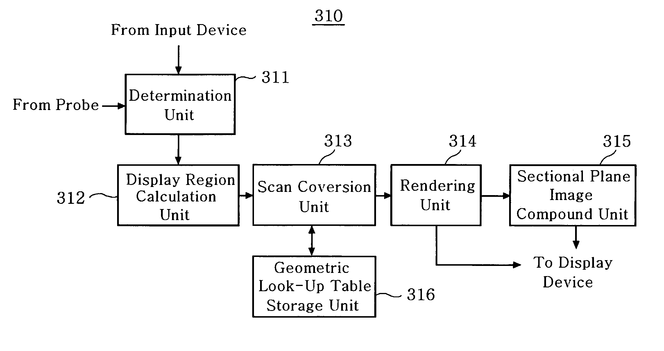
[0022]FIG. 4A is a diagram illustrating an ultrasound diagnostic system, which is constructed in accordance with the preferred embodiment of the present invention.

[0023] As illustrated in FIG. 4A, an ultrasound diagnostic system 300 includes a probe 301, a display device 303 and a body 302. The probe 301 is used to acquire 3D ultrasound data from a target object to be displayed. A mechanical scanning process (scan by moving a mechanical arm or rotating a stepping motor) or a hand-free process (scan by a user's hand) may be applied to the probe 301. The display device 303 (e.g., a monitor) is used to display the ultrasound data acquired from the probe 301. It should be noted herein that as long as the display device can display a 3D ultrasound image in accordance with the present invention, any type of display device may be used. The body 302 includes an ultrasound image processing device 310 for enhancing the quality of the sectional plane image in the 3D ultrasound image.

[0024]FI...


PUM


Abstract

The present invention relates to an apparatus for enhancing the quality of a sectional plane image in a 3-dimensional ultrasound image. The apparatus for enhancing a sectional plane image in a 3-dimensional ultrasound image, includes: a display region calculation unit for selecting a reference sectional plane image in 3D ultrasound data of a target object received from a probe and calculating a display region to display the selected reference sectional plane image; a geometric look-up table storage unit for storing indices matched with 3-dimensional Cartesian coordinates of a screen displaying the target object and conical coordinates corresponding to the indices; a scan conversion unit for scan-converting conical coordinates of a plurality of sectional plane images with offsets between the sectional plane images adjusted to a predetermined value into Cartesian coordinates by referencing the geometric look-up table storage unit; a rendering unit for rendering the scan-converted sectional plane images including the reference sectional plane image; and a sectional plane image compound unit for compounding a predetermined number of sectional plane images in which the reference sectional plane image is positioned at a center thereof.

Description

FIELD OF THE INVENTION [0001] The present invention generally relates to an ultrasound diagnostic system, and more particularly to an apparatus and a method for enhancing the quality of a sectional plane image in 3D ultrasound data of a target object in an ultrasound diagnostic system. BACKGROUND OF THE INVENTION [0002] Generally, a 3-dimensional (3D) ultrasound diagnostic system is medical equipment for providing clinical information such as spatial information and anatomical information, which cannot be provided from a conventional 2-dimensional image. The 3D ultrasound diagnostic device acquires the 3D ultrasound data of a target object by using a probe. The 3D ultrasound diagnostic device then displays a 3D image of the target object on a screen of a display device by converting conical coordinates of the acquired data to Cartesian coordinates suitable for display (scan conversion). [0003] A region displaying ultrasound images of the target object on the screen of a display devi...

Claims


Application Information

Patent Timeline
Patent Type & Authority Applications(United States)
IPC IPC(8): A61B8/00G06T1/00G06T3/00G06T5/20
CPCG06T2207/30004G06T5/20G06T5/002G06T2207/10136G06T15/00A61B8/13G06T5/00
Inventor LEE, JAE KEUN
Owner MEDISON CO LTD
Why Eureka
Try Eureka