
Method and device to obtain percutaneous tissue samples

a percutaneous tissue and biopsy method technology, applied in the field of percutaneous biopsy devices, can solve the problems of serious cognitive defects of patients, unstable design, and inability to accurately target the small lesions of the core biopsy devi

Inactive Publication Date: 2006-06-08
DAUM WOLFGANG
View PDF4 Cites 164 Cited by
  • Summary
  • Abstract
  • Description
  • Claims
  • Application Information

AI Technical Summary

Benefits of technology

[0011] The suggested biopsy mechanism will cut the specimen in front of the tip of the guiding needle

Problems solved by technology

Bending: Due to the beveled tip of the obdurator and the thin notch strap, which makes the design unstable, the obdurator bends during its forward movement through the tissue towards the opposite side of the tip bevel.
This bending makes the core biopsy devices imprecise in targeting smaller lesions.
The same problem occurs, when targeted breast tumor lesions are close to the lung pleura.
In brain tissue every needle penetration in healthy tissue may cause serious cognitive defects of the patient.
Even if more MRI compatible material like titanium alloys are used for the material, this solid part causes a rather large artifact at the tip of the obdurator, especially when the needle is used in higher magnetic flux MRI tomographers, like 1.5 or 3.0 Tesla.
Torn Sample: While core biopsy devices work well in fatty tissue, they often have difficulties in more dense tissue.
Here the cutting needle often rather tears the tissue resulting in an insufficient tissue sample for the pathological analysis.
Vacuum aspiration biopsy devices lack the disadvantage that they tear out the tissue and give imprecise specimen cuts.
Further this type of biopsy can only be accomplished with softer and easier to tear tissue.

Method used


Image

  • Method and device to obtain percutaneous tissue samples
  • Method and device to obtain percutaneous tissue samples
  • Method and device to obtain percutaneous tissue samples

Examples


Embodiment Construction

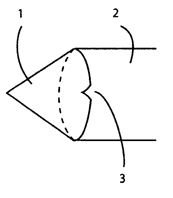
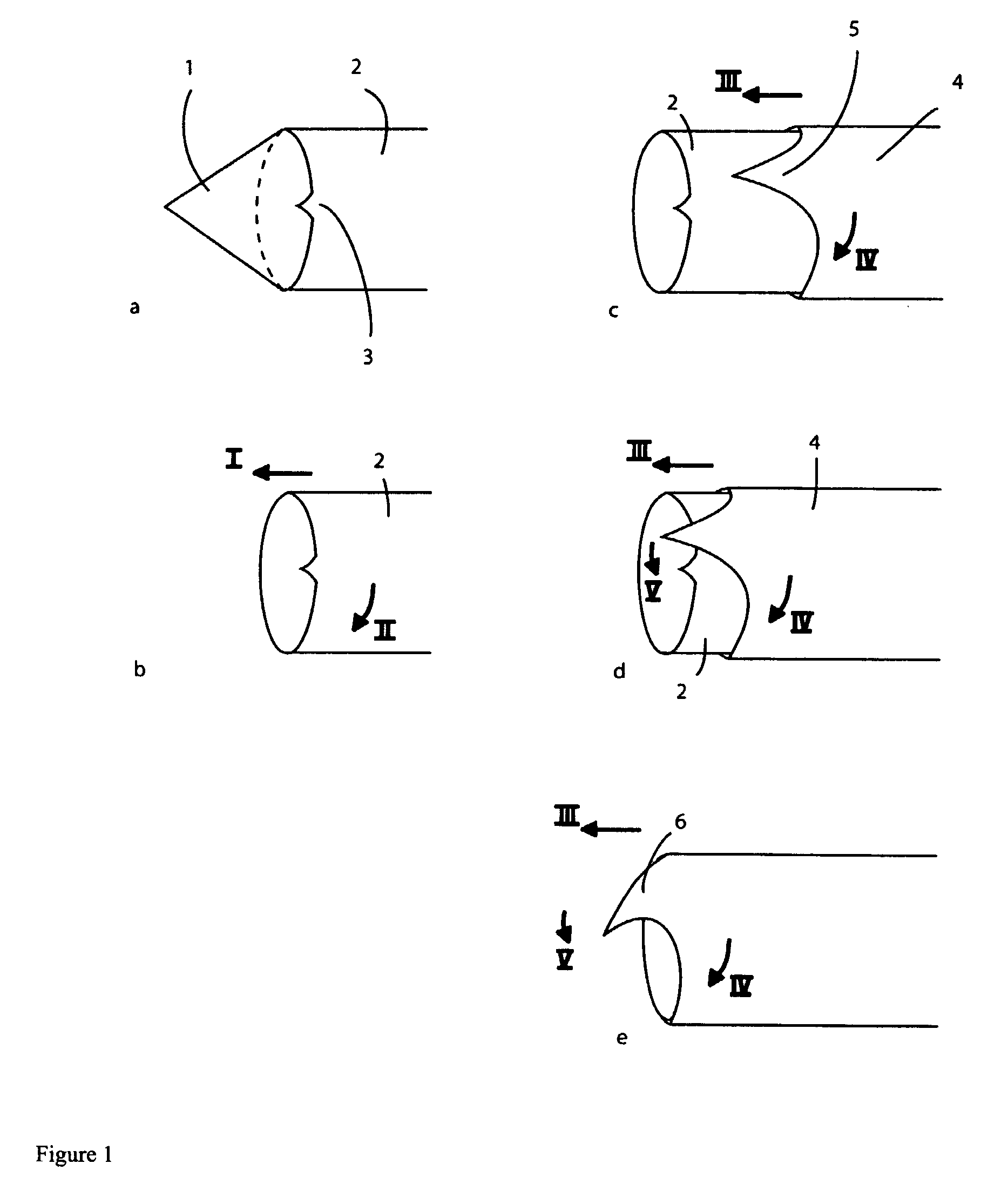
[0017] The new cutting mechanism cuts at the tip of a needle a dome like specimen. FIG. 1 illustrates the basic new biopsy concept operating in five time snap shot stages. Only the distally located parts of the instrument are shown; the proximal parts (handle) are not shown.

[0018]FIG. 1a shows the access tube 2 in which an inner stylet 1 with a trocar like tip is positioned. This needle set is percutaneously pushed through the patients tissue until the tip of the stylet 1 is positioned at the location of planned biopsy.

[0019] In FIG. 1b the inner stylet is withdrawn backwards and removed from the access tube 2. The access tube 2 now is rotated (arrow II) and pushed forward (arrow I) in distal direction. During this procedure tissue is cut by the cutting blade of the access tube 2 and a pillar like specimen collects within the access tube 2. The diameter of the specimen equals the inner diameter of the access tube 2, typically 1 mm to 4 mm. This collecting of specimen can be suppor...


PUM


Abstract

A new method and design for a percutaneous biopsy system that cuts only the tissue lesion specimen and that does not penetrate through or beyond the targeted tissue into intact tissue. The proposed mechanism operates only in the targeted lesion space and leaves healthy or unsuspicious tissue intact. The proposed biopsy mechanism will cut the specimen in front of the tip of the guiding needle. The device may be image guided by ultrasound, any x-ray based modality or magnetic resonance (MRI).

Description

FIELD OF THE INVENTION [0001] The present invention relates to a percutaneous biopsy device that cuts a suspicious desired tissue specimen, leaving undesired tissue unharmed. This application claims priority to U.S. Ser. No. 60 / 634,386, which incorporated herewith by reference. BACKGROUND OF THE INVENTION [0002] Medical biopsy is a technique to obtain tissue samples for pathologic diagnostics. Open surgical biopsies are still the standard techniques in many medical fields. There are basically two principle percutaneous biopsy techniques for the interventional or minimally invasive biopsy market, which are fine needle aspiration and core biopsy. Approximately two million biopsies are performed in the United States each year. [0003] The core biopsy technique—also known as Temno technique—is the oldest and most common biopsy technique on the market. Core biopsy devices are available as manually operated spring loaded or as fully automatic systems. All devices use a coaxial needle set c...

Claims


Application Information

Patent Timeline
IPC IPC(8): A61B10/00
CPCA61B10/02A61B10/0266A61B10/04A61B2010/0208A61B2010/045
Inventor DAUM, WOLFGANG
Owner DAUM WOLFGANG
Learn More