Medical wire device

a wire device and wire technology, applied in the field of medical wire devices, can solve problems such as adverse effects, and achieve the effects of high radiopaque properties, high flexibility, and high strength

Inactive Publication Date: 2006-07-20
KANEKA MEDIX
View PDF7 Cites 96 Cited by
  • Summary
  • Abstract
  • Description
  • Claims
  • Application Information

AI Technical Summary

Benefits of technology

[0027] According to the medical wire devices of the present invention, the filter making up the filter unit is made up of a metal having radiopaque property, whereby a surgeon is able to easily confirm under radiography that the filter is in a state surely developed in a lumen and brought into close contact with an inner wall surface without any clearance when it is developed.
[0028] According to the medical wire devices of the present invention, the filter making up the filter unit is made up of a metal having high strength, whereby the metallic metal material making up the filter can be made diametrally small or shin. As a result, the filter itself can be made thin and made up as that having high flexibility. Accordingly, the filter can be developed or folded following the deformation of the supporting frame, such as expansion or diametrical contraction, without impeding the deformation of the supporting frame, so that the filter can be surely developed to an expected form in a lumen.
[0029] In addition, the metallic wire material is made diametrally small or thin, whereby the opening diameter of the filter can be made small while retaining the expected opening rate, so that the embolismic debris can be surely captured under a state that perfusion on a downstream side from a constriction lesion is scarcely impeded. Accordingly, a function as the filter unit can be improved.
[0030] In particular, the filter is made up of tungsten or an alloy composed mainly of tungsten, whereby such effects as described above can be surely achieved because tungsten or the alloy composed mainly of tungsten has high radiopaque property upon radiography, high strength and excellent working ability by itself compared with other metals.
[0031] In addition, the supporting frame making up the filter unit is made up of the metal having radiopaque property, whereby sufficient effects can be achieved from the viewpoint of practical use.
[0032] Further, the supporting frame making up the filter unit is composed of that having a thin wall not thicker than the specified thickness, whereby the function inherent in the filter unit that the free embolismic debris in the lumen are captured can be surely achieved as described above, and, at the same time, the filter unit itself can be made up as that having high flexibility, thereby being capable to deform the filter unit following the form of a lumen into which it should be inserted. As a result, high housing ability into the lumen and high operating ability within the lumen are achieved, and thus the filter unit can be made up as that adequately applicable to, for example, blood vessels having a small inner diameter and a complicated form, or the like.

Problems solved by technology

As a result, there is a possibility that the free embolismic debris may be deposited on another site in the lumen to form a thrombus at the site or may reach a vital organ to adversely affect it.

Method used


Image

  • Medical wire device
  • Medical wire device
  • Medical wire device

Examples


production example 1

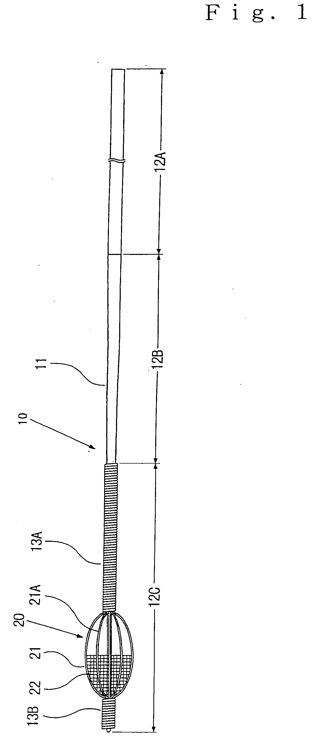
[0096] Following the construction shown in FIG. 1, a medical wire device (10) according to the present invention was produced. This medical wire device is referred to as “Wire Device 1”. A specific construction is as follows.

[0097] A core wire (11) is composed of stainless steel and has an overall length of 2,700 mm, a proximal-side region (12a) has a length of 2,350 mm and a diameter of 0.35 mm, a tapered region (12B) has a length of 200 mm, and a distal-side region has a length of 150 mm and a diameter of 0.25 mm

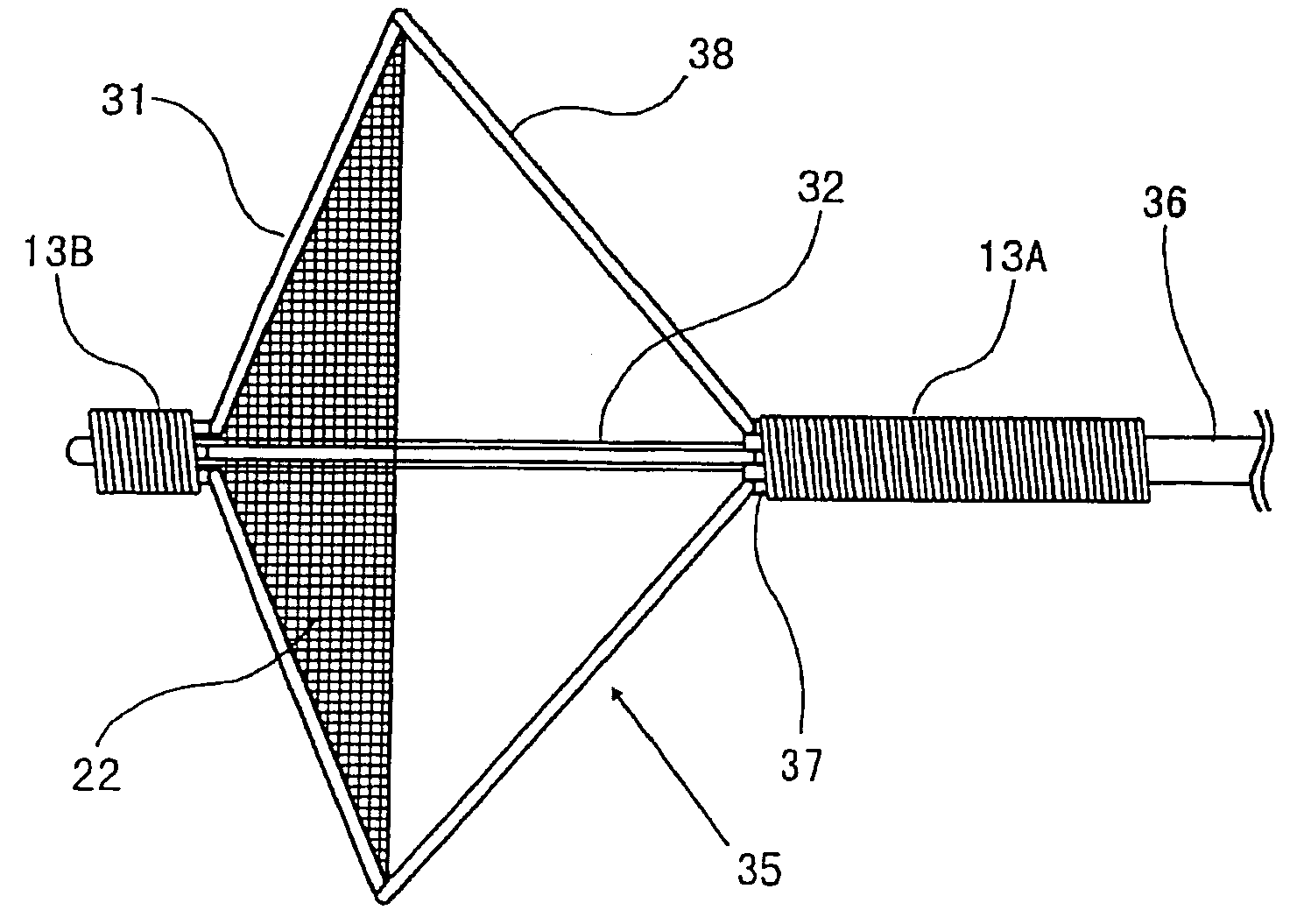
(1) Supporting Frame:

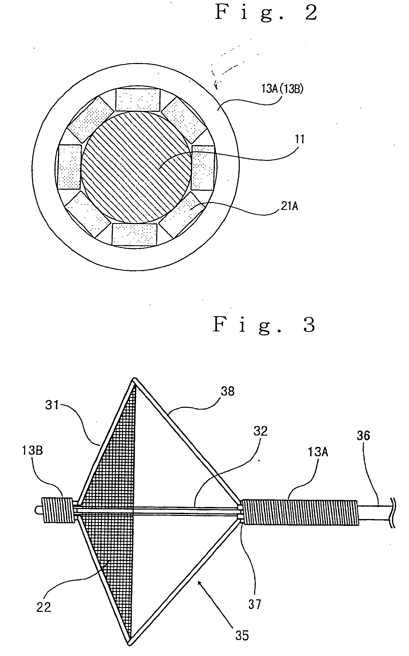
[0098] A supporting frame (21) has a spheroidal frame structure that 8 frame components (21A), which are belt-like board materials 0.05 mm×0.1 mm in sizes composed of tungsten, are provided so as to extend in a longitudinal direction along the core wire in a state that a distal-side end of each of the frame components has been fixed to the core wire by silver soldering, and the proximal-side end thereof has been made freely movable to the core wire...

production example 2

[0102] A medical wire device of the same construction as in Production Example 1 except that a platinum-tungsten alloy (Pt / W=92 / 8) was used in place of tungsten as the metallic wire materials making up the supporting frame and filter in Production Example 1 was produced. This medical wire device is referred to as “Wire Device 2”. The tensile break strength of the metallic wire material making up the filter in Wire Device 2 is 1,500 N / mm2.

production example 3

[0103] A medical wire device of the same construction as in Production Example 1 except that a tube for expanding a frame (developing means), which covered the core wire and extended in a longitudinal direction, was provided at a proximal-side end of the-supporting frame making up the filter unit in Production Example 1 so as to contract the supporting frame by pulling this developing means on the proximal side and expand the supporting frame by pressing the developing means on the distal side, and a wire in a straight form having a diameter of 0.20 mm was used as the core wire, was produced. The tube for expanding the frame is composed of a Ti—Ni alloy and has an outer diameter of 0.36 mm, an inner diameter of 0.21 mm and a length of 2,250 mm. This medical wire device is referred to as “Wire Device 3”.


PUM


Abstract

Disclosed herein is a novel medical wire device which is equipped with a filter unit, capable of surely capturing free embolismic debris upon angioplasty of a constriction lesion in a lumen, capable of easily confirming a developed state of a filter under radiography by a surgeon, and thus improved in function of the filter unit. The medical wire device of the present invention comprises a filter unit which is to be located, upon angioplasty of a constriction lesion in a lumen, on a distal side from the constriction lesion for capturing free embolismic debris separated from an inner wall of the constriction lesion, and a wire provided with the filter unit at its distal-side end. A filter is made up of a metal having radiopaque property.

Description

TECHNICAL FIELD [0001] The present invention relates to a medical wire device used in, for example, angioplasty for a constriction lesion in a blood vessel and other lumens in a vital body. BACKGROUND ART [0002] As a treatment method for dilating or reopening a constriction lesion in a blood vessel or any other lumen, which has been formed by deposition of embolismic debris such as plaque or other substances on a vessel wall, there is currently known angioplasty. In such angioplasty, for example, a method of forming a lumen by introducing an expansible balloon into a constriction lesion in a contracted state and expanding the balloon, a method of forming a lumen by introducing a stent in a contracted state in a diameter into a constriction lesion and expanding it, or the like is utilized. [0003] In such angioplasty, in some cases, embolismic debris located at a constriction lesion in a lumen may unexpectedly separate from an inner wall of the lumen and be caused to flow in a periphe...

Claims


Application Information

Patent Timeline
Patent Type & Authority Applications(United States)
IPC IPC(8): A61M29/00A61B17/00A61B17/12A61F2/01
CPCA61B17/12022A61B17/12172A61F2/013A61F2002/018A61F2230/0006A61F2230/0067A61F2230/0076A61F2230/008A61F2230/0093A61F2002/016A61F2250/0098
Inventor SAKAI, SHINICHIOGAWA, ATSUSHI
Owner KANEKA MEDIX
Why Eureka
Try Eureka