
Biopsy forceps

a forcep and biopsy technology, applied in the field of medical devices, can solve the problems of high cost, time-consuming, difficult, etc., and achieve the effect of facilitating the removal of tissue samples

Inactive Publication Date: 2006-08-10
WILSONCOOK MEDICAL
View PDF99 Cites 35 Cited by
  • Summary
  • Abstract
  • Description
  • Claims
  • Application Information

AI Technical Summary

Benefits of technology

[0006] Accordingly, it is an object of the present invention to provide a medical device having features that resolve or improve upon one or more of the above-described drawbacks.
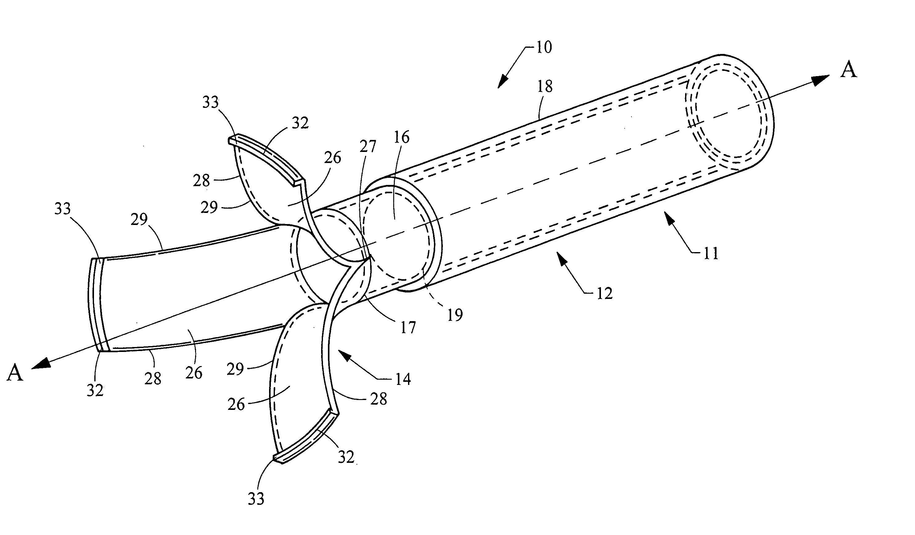
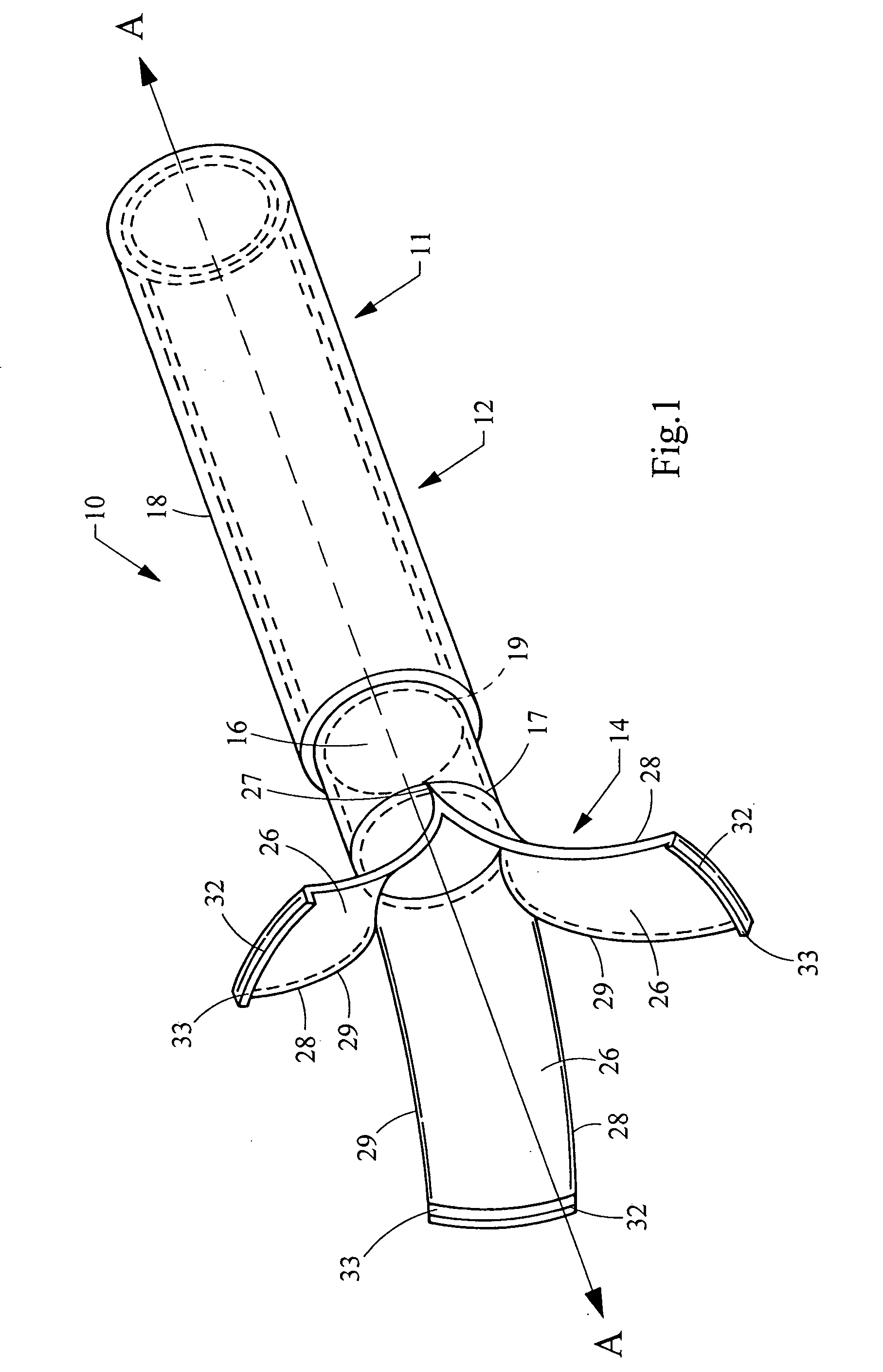
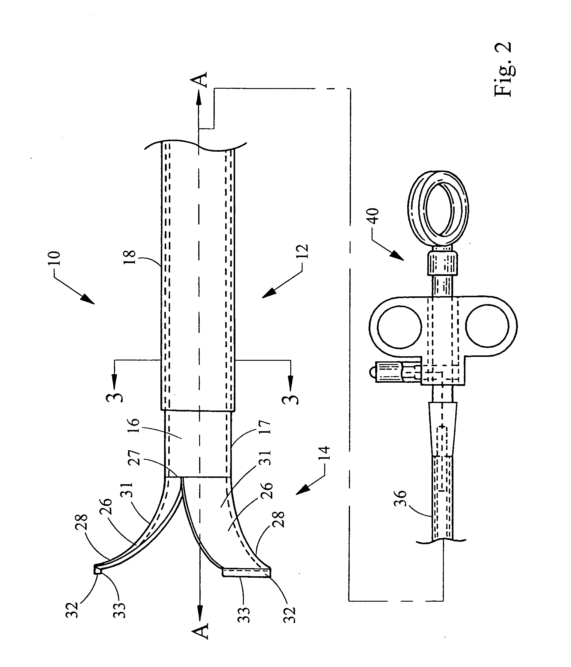
[0007] According to one aspect of the present invention, the foregoing object is obtained by providing a biopsy forceps having a sheath and an inner shaft slidably disposed within the sheath. The shaft has a longitudinal axis defined therethrough and a plurality of grasping members that are movable between an open configuration and a closed configuration. At least one of the plurality of grasping members is biased outwardly from the longitudinal axis when in the open configuration. At least one of the plurality of grasping members is unrestricted by the sheath when in the open configuration and is constrained by the sheath when in the closed configuration. The plurality of grasping members can be formed so that when they are in a closed configuration, they form a receptacle for retaining one or more biopsy samples. One or more of the grasping members may further be provided with a cutting edge to more easily remove a tissue sample.
[0008] According to another aspect of the present invention the shaft may be connectable to an electrocautery device. As a result, the shaft may be energized by the electrocautery device to electrosurgically cut the tissue.

Problems solved by technology

The manufacture of biopsy forceps is therefore expensive, difficult, and time consuming.

Method used


Image

  • Biopsy forceps
  • Biopsy forceps
  • Biopsy forceps

Examples


Embodiment Construction

[0023] The invention is described with reference to the drawings in which like elements are referred to by like numerals. The relationship and functioning of the various elements of this invention are better understood by the following detailed description. However, the embodiments of this invention as described below are by way of example only, and the invention is not limited to the embodiments illustrated in the drawings. It should also be understood that the drawings are not to scale and in certain instances details that are not necessary for an understanding of the present invention have been omitted, such as conventional details of fabrication and assembly. Moreover, it should be noted that the invention described herein includes methodologies that have a wide variety of applications.

[0024] Referring to the drawings, FIGS. 1-3 depict an illustrative embodiment of the present invention. Generally, a medical device 10 is provided to take tissue samples for medical analysis. As ...


PUM


Abstract

A biopsy forceps and method of using the biopsy forceps. The biopsy forceps includes a plurality of grasping members extending from an inner shaft. The plurality of grasping members are biased toward an open configuration. Sliding a sheath over the grasping members constrains the grasping members to a closed configuration. A method of performing a tissue biopsy is also disclosed.

Description

[0001] This application claims the benefit of U.S. Provisional Application No. 60 / 646,104, filed Jan. 20, 2005.FIELD OF THE INVENTION [0002] This invention generally relates to medical devices, and particularly to forceps used for obtaining biopsy samples. BACKGROUND [0003] Physicians in many specialties commonly obtain biopsy samples from patients to determine the presence of tissue abnormalities, such as cancerous cells. Sometimes biopsies are taken without the need for an invasive procedure. For example, physicians can take skin biopsies to test for melanoma. In many cases, however, a physician must access a biopsy location inside a patient's abdominal cavity, thoracic cavity, or gastrointestinal system. For such procedures, physicians often use an endoscope to avoid more traumatic open surgery. Modern endoscopes are long, flexible instruments having a viewing system and a working channel through which a biopsy forceps can be passed. [0004] Common endoscopic biopsy forceps are fo...

Claims


Application Information

Patent Timeline
IPC IPC(8): A61B17/28
CPCA61B10/06A61B17/320016A61B2017/2937A61B2017/2944
Inventor SURTI, VIHAR C.
Owner WILSONCOOK MEDICAL
Learn More