Detection of activation of endothelial cells as surrogate marker for angiogenesis

a technology of endothelial cells and surrogate markers, which is applied in the field of detection of endothelial cells as surrogate markers for angiogenesis, can solve the problems of no quantitative method for determining the total angiogenic output of a patient's tumor burden, bleeding and blindness, tumor regression and dormancy, etc., and achieves the effect of sensitive and convenient measurement of angiogenesis

Inactive Publication Date: 2006-09-07
MONOGRAM BIOSCIENCES
View PDF61 Cites 29 Cited by
  • Summary
  • Abstract
  • Description
  • Claims
  • Application Information

AI Technical Summary

Benefits of technology

[0013] The present invention provides an innovative approach to assessing angiogenesis through sensitive, direct detection of activation of cells that contribute to or promote angiogenesis, e.g., endothelial cells, stroma cells or tumor cells, at a molecular level. In general, activation of such cells is detected by measuring the levels of cellular components and their protein complexes participating in a specific angiogenesis signaling pathway in these cells. Compared with the currently available techniques for assessing angiogenesis, the methods provided in the present invention can sensitively an

Problems solved by technology

In diabetes, new capillaries formed in the retina invade the vitreous humor, causing bleeding and blindness.
Indeed, in some protocols, the application of such inhibitors leads to tumor regression and dormancy even after cessation of treatment (O'Reilly et al.
At present, there is no quantitative method for determining the total angiogenic output of a patient's tumor burden, so surrogate markers, such as angiogenic proteins present in serum, plasma, and urine.
There are various disadvantages associated with above approaches to asses

Method used


Image

  • Detection of activation of endothelial cells as surrogate marker for angiogenesis
  • Detection of activation of endothelial cells as surrogate marker for angiogenesis
  • Detection of activation of endothelial cells as surrogate marker for angiogenesis

Examples


example 1

Immunomagnetic Isolation of Human Endothelial Cells

[0281] Substantially pure CEC and CECP can be isolated by using the immunomagnetic isolation / enrichment technique described in Kinzler et al. (2000) Science 289:1197-1202. Briefly, the epithelial and hematopoietic cell fractions in the peripheral blood samples are sequentially removed via negative selection via antibody-linked magnetic beads (BerEP4 beads- Epithelial, CD45 beads- Pan leukocyte, CD64 beads-Macrophages, and CD14 beads- Monocytes). The remaining cells are stained with P1H12 antibodies and are isolated via positive selection with magnetic beads.

[0282] In this example, CEC and CECP were isolated by using a protocol modified from Voest et al. (2004) Annals of Oncology 15:139-145 as follows: [0283] 1) Magnetic beads (Dynal M450 IgGi, Dynal AS, Oslo, Norway) were conjugated to a monoclonal antibody for CD146, MCAM, an endothelial cell surface junction protein. [0284] 2) Whole blood was diluted 1:3 with 0.9% NaCl and incub...

example 2

Lysis of CEC or CECP

[0309] The isolated CEC or CECP were lysed according to the following protocol:

Protocol Step

[0310] 1) Seed 2 15 cm dishes with 6×10⁶ cells in 20 mL EGM [0311] 2) Prepare fresh lysis buffer [0312] 3) Starting now, always work on ice. Aspirate the medium. [0313] 4) Add 600 ul of fresh lysis buffer. [0314] 5) Swirl the plates to distribute the buffer evenly. [0315] 6) Scrap the cells. [0316] 7) Collect the crude lysate in 1.5-ml tube. [0317] 8) Wait for 30 min. [0318] 9) Spin down at 4 C at 14000 rpm for 10 min [0319] 10) Collect the supernatant in 1.5-ml new tube. [0320] 11) Store at −80 C for later use.

[0321] The lysis buffer was prepared by mixing the following reagents:

Lysis10 mlbufferReagentsStock con.Final concVol1)10% triton X-1001011.002)1M Tris pH 7.510.050.503)1M NaF10.050.504)5M NaCl50.10.205)2M B-Glycerolphosphate10.050.506)0.1M Na3VO40.10.0010.107)1 mg / ml pepstatin10.010.108)Complete mini protease1 tablet9)0.5M EDTA0.50.0050.10Total (ml)3.00Water...

example 3

Analysis of Cell Lysate for Protein Complexes of Angiogenic Receptors and Phosphorylation of Downstream Effector Proteins

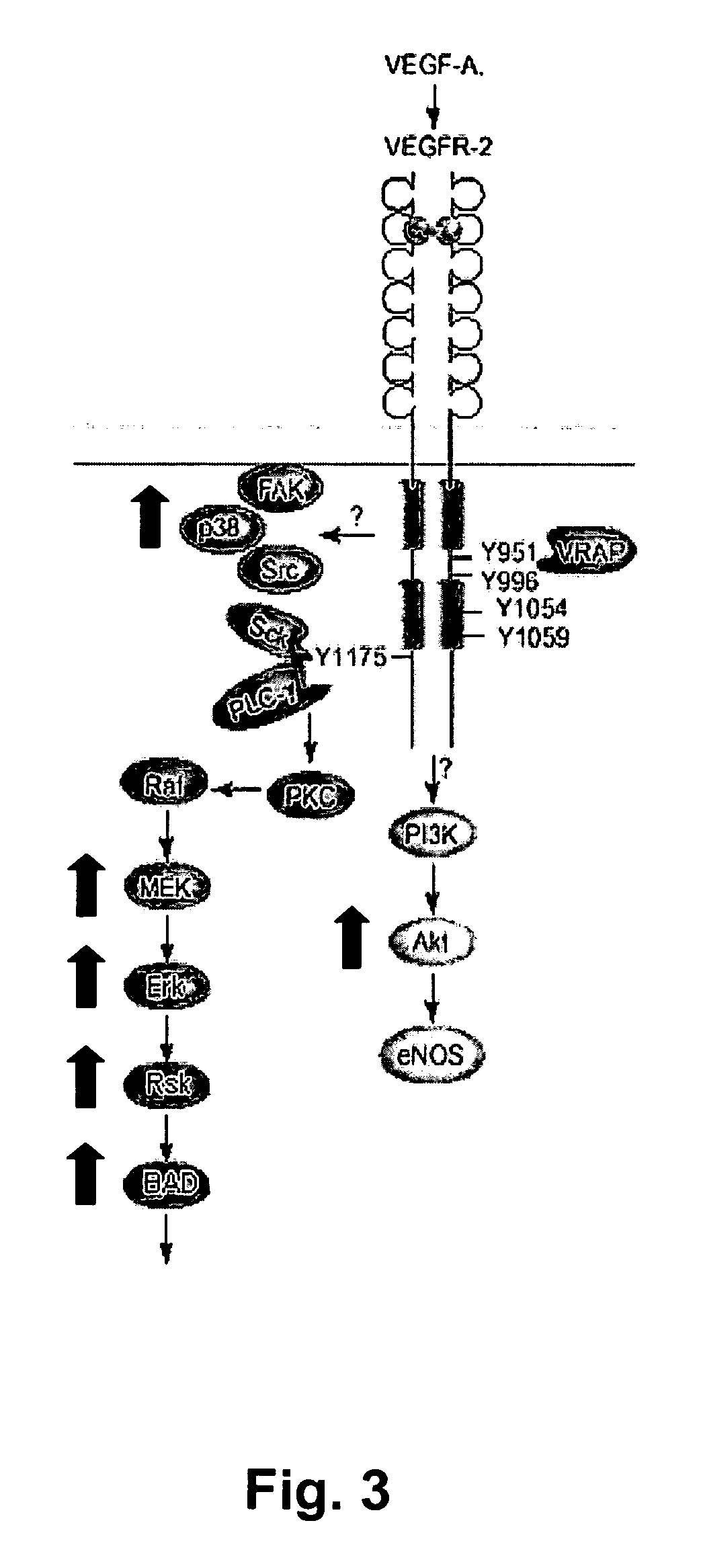
[0322] Protein complexes formed by angiogenic receptors (e.g., VEGFR, Tie), such as the homodimers and heterodimers formed by VEGFR, were measured in cell lysates from CEC, CECP, and cell lines such as human umbilical vein endothelial cells (HUVEC), and human vulval carcinoma cell line (A431). Measurements were made using three binding compounds and a cleaving probe by using the following protocol. [0323] 1) Add Lysis Buffer and Lysate into 96-well PCR plate to 30 uL final volume. [0324] 2) Add 5 uL antibody mix to each well. [0325] 3) Incubate reactions at RT for 2 hours on plate shaker. [0326] 4) Add 5 uL steptavidin-scissor mix to each well in dark room. Mix is 32.5×. Added 15 uL to 472.5 uL Dilution Buffer (1% BSA in PBS) [0327] 5) Incubate reactions at RT for 45 minutes on plate shaker in dark room. [0328] 6) Pre-wet 96-well 2 micron membrane filter plates w...


PUM

PropertyMeasurementUnit
Electrical resistanceaaaaaaaaaa
Surfaceaaaaaaaaaa
Separationaaaaaaaaaa

Abstract

Methods, compositions and kits are provided for assessing angiogenesis through sensitive, direct detection of activation of endothelial cells at molecular levels. In general, activation of endothelial cells is detected by measuring the levels of cellular components and their protein complexes participating in a specific angiogenesis signaling pathway in endothelial cells. The methods can be used for assessing status of diseases associated with undesirable angiogenesis, such as the likelihood of developing the disease, presence or absence of the disease, prognosis of the disease and the likelihood of response or resistance to a particular anti-angiogenic therapy. The methods can also be used to guide the design of effective therapeutic regimens targeting a specific angiogenic signaling pathway, as well as in conjunction with therapeutic intervention of diseases or conditions associated with undesirable angiogenesis.

Description

[0001] This application claims the benefit of U.S. Provisional Application No. 60 / 625,694, filed Nov. 4, 2004, the contents of which is incorporated by reference in its entirety.FIELD OF THE INVENTION [0002] The present invention relates generally to methods for accessing angiogenesis through detection of the status of endothelial cell activation and more particularly to methods of determining angiogenic signal transduction pathway activation at a molecular level, and for using such status information to select patients responsive to pathway-specific drugs, and to rationally design effective therapy with drugs that specifically modulate angiogenesis signal transduction pathways. Description of Related Art [0003] Angiogenesis is the fundamental process by which new blood vessels are formed. The process involves the migration of vascular endothelial cells into tissue, followed by the condensation of such endothelial cells into vessels. Angiogenesis may occur naturally or be induced by...

Claims


Application Information

Patent Timeline
IPC IPC(8): G01N33/574
CPCG01N33/5041G01N33/5064G01N33/5091G01N33/574G01N2333/515G01N2333/71G01N2800/52Y10S436/824
Inventor MOORE, SEAN C.SINGH, SHARATSALIMI-MOOSAVI, HOSSEINCAO, LICHINGSPERINDE, JEFF
Owner MONOGRAM BIOSCIENCES
Why Eureka
Try Eureka