Method and apparatus for automated analysis of biological specimen

a biological specimen and automatic classification technology, applied in biochemistry apparatus and processes, image data processing, material testing goods, etc., can solve the problems of computational difficulty in processing large-scale agglomeration of cells, independent analysis of objects, etc., and achieve the effect of improving the recognition accuracy

Inactive Publication Date: 2006-09-14
MEDISINE
View PDF25 Cites 27 Cited by
  • Summary
  • Abstract
  • Description
  • Claims
  • Application Information

AI Technical Summary

Benefits of technology

[0023] According to a first aspect, the invention uses aggregation features of objects in an image to assist in object classification. Typically, an aggregation feature is obtained on the basis of neighbourhood objects and their relationship to the given object, thus characterising the n

Problems solved by technology

A significant limitation of the above prior art systems is that the objects are analysed independently of each other

Method used


Image

  • Method and apparatus for automated analysis of biological specimen
  • Method and apparatus for automated analysis of biological specimen
  • Method and apparatus for automated analysis of biological specimen

Examples


hardware implementation example

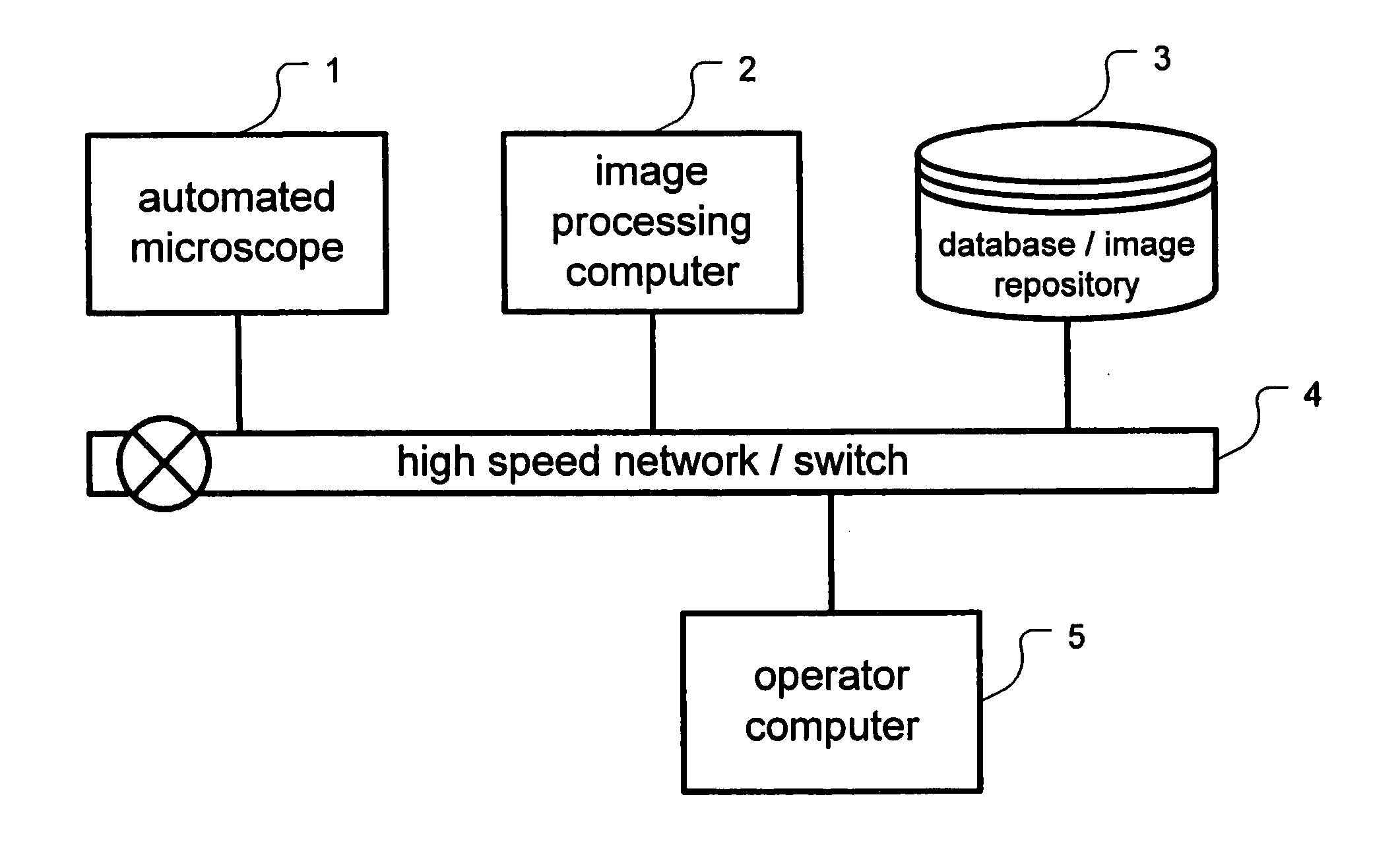
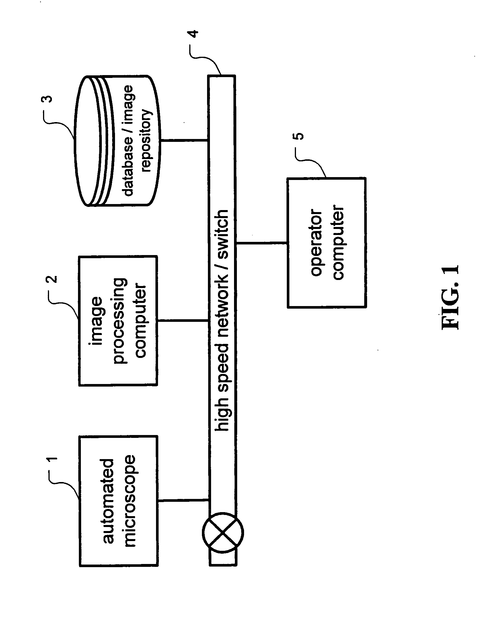
[0120] In a preferred embodiment of the embodiment of the invention shown in FIG. 1, the scanning microscope 1 is an Aperio ScanScope T2 supplied by Aperio Technologies, Vista, Calif., USA, and the computer 2 is a dual processor server running a Microsoft Windows operating system. The scanning microscope and the computer are interconnected using a Gigabit Ethernet. The parallelisation of image recognition is implemented by means of Windows multithreading. The image repository 3 is a Redundant Array of Independent Disks (RAID) connected to the computer 2.

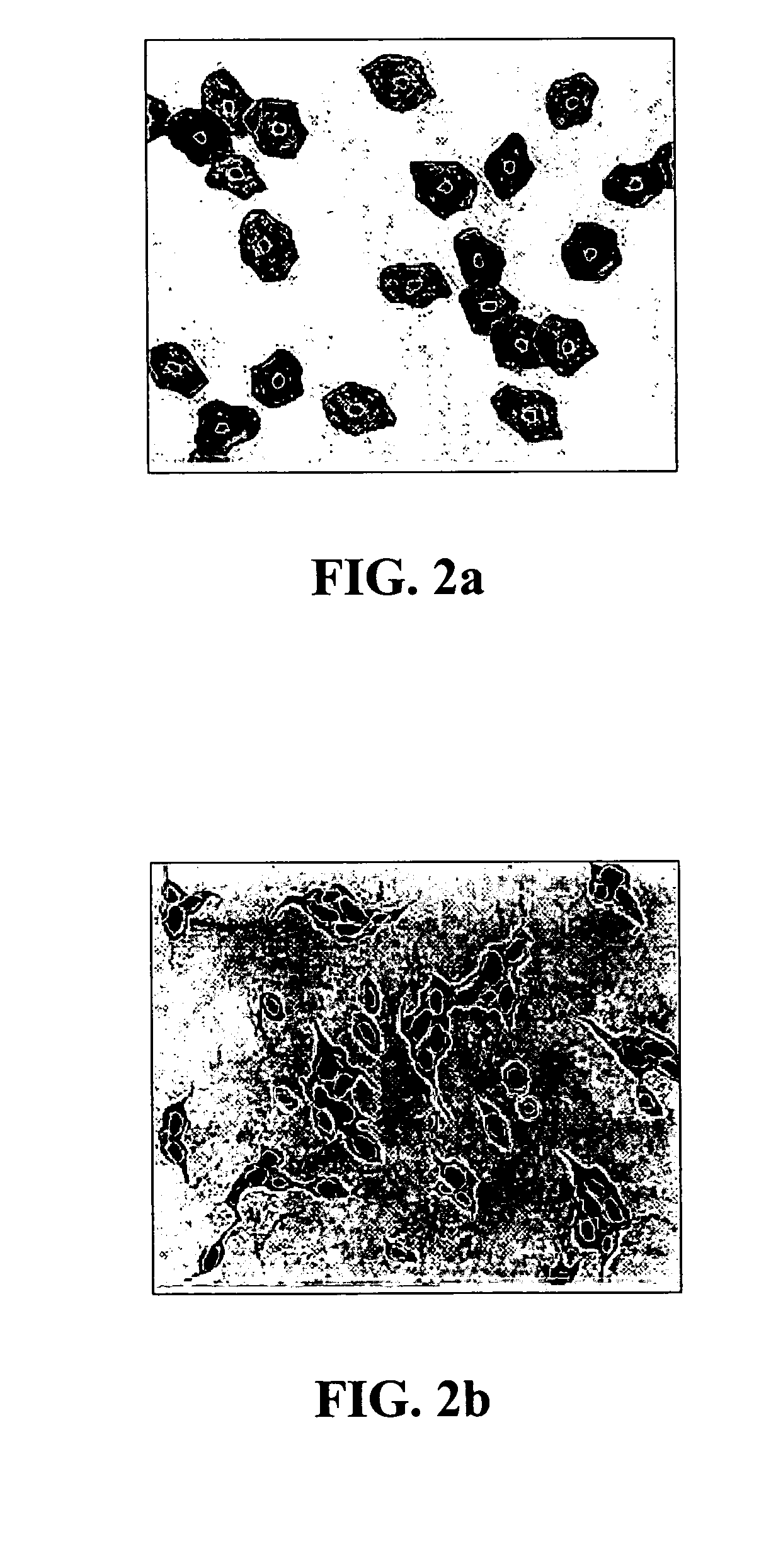
[0121] Referring now to FIG. 12 there is illustrated, in schematic form, an apparatus 20 for putting the image processor computer 2 of FIG. 1 into effect. An image 22 of biological objects such as cells is passed to a segmentor 24 which seeks to identify separate objects, for example as discussed above and as illustrated by segmented image 26. The segmented image, or data relating to the segments is passed to analyser 28 which ident...


PUM


Abstract

Apparatus and methods for automatic classification of cells in biological samples is disclosed. Aggregation parameters of identified objects are calculated using relative spatial positions of the objects, and the aggregation parameters are used in the classification. Centres of cells may be located using an edge detector and parameterisation across at least one more dimension than the spatial dimensions of the image.

Description

FIELD OF THE INVENTION [0001] The present invention relates to the automatic classification of cells in biological specimens, and more particularly, but not exclusively, to a method for quantitative microscopical analysis in cytology and pathology applications, and an associated computer system therefor. BACKGROUND OF THE INVENTION [0002] The recognition of cancer or other diseases or conditions manifested through cellular abnormalities is currently a highly skilled task. For example, the cervical smear test is performed to detect cervical cancer at its earliest stages when it can be treated and the chances of a complete cure are high. [0003] Currently, the analysis and interpretation of cervical smears is dependent upon trained cytotechnologists (also known as screeners) who examine the thousands of cells in a smear under the microscope for evidence of cancer. The cancer cells can be distinguished from the normal cells in the smear by their shape and size and also by the structure ...

Claims


Application Information

Patent Timeline
IPC IPC(8): C12Q1/00G06F19/00G06K9/00G06T5/00G06T7/00
CPCG06K9/00127G06T7/0012G06T7/0083G06T2207/10056G06T2207/20061G06T2207/30024G06T7/12G06V20/69
Inventor PTITSYN, NIKOLAI
Owner MEDISINE
Why Eureka
Try Eureka