Ultrasonic doppler system for determining movement of artery walls

a doppler system and ultrasonic imaging technology, applied in ultrasonic/sonic/infrasonic image/data processing, instruments, ultrasonic/sonic/infrasonic diagnostics, etc., can solve the problem that the method disclosed in the cited document for calculating artery dilation cannot be directly used, and the system is not suitable for the study of deep and thick

Inactive Publication Date: 2006-09-21
KONINKLIJKE PHILIPS ELECTRONICS NV
View PDF5 Cites 15 Cited by
  • Summary
  • Abstract
  • Description
  • Claims
  • Application Information

AI Technical Summary

Benefits of technology

[0007] It is also an object of the invention to propose an ultrasound apparatus having such an ultrasonic imaging system. It is a further object of the invention to propose an image processing method for displaying an image sequence of deep arteries. In particular, it is an object of the invention to propose an image processing method for evaluation of parameters related to the tension and strain of the ...

Problems solved by technology

A problem is that the cited document relates to an image processing method based on image acquisition with ultrasound scanning lines that are perpendicular to the artery axis.
This kind of system is not appropriate for the study of a deep and thick artery such as the aorta and particul...

Method used


Image

  • Ultrasonic doppler system for determining movement of artery walls
  • Ultrasonic doppler system for determining movement of artery walls
  • Ultrasonic doppler system for determining movement of artery walls

Examples


Embodiment Construction

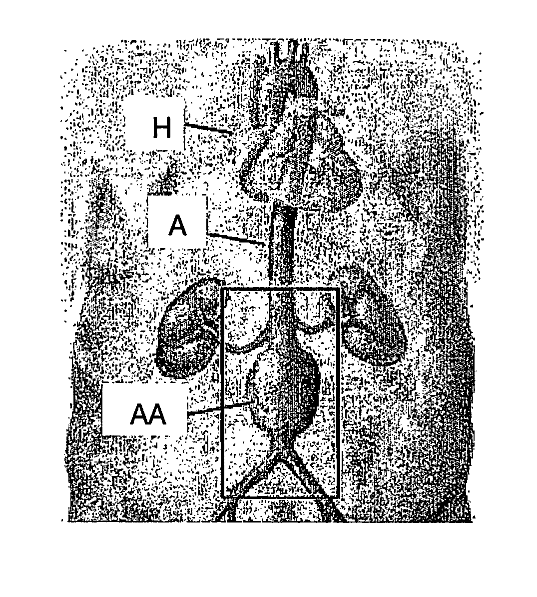
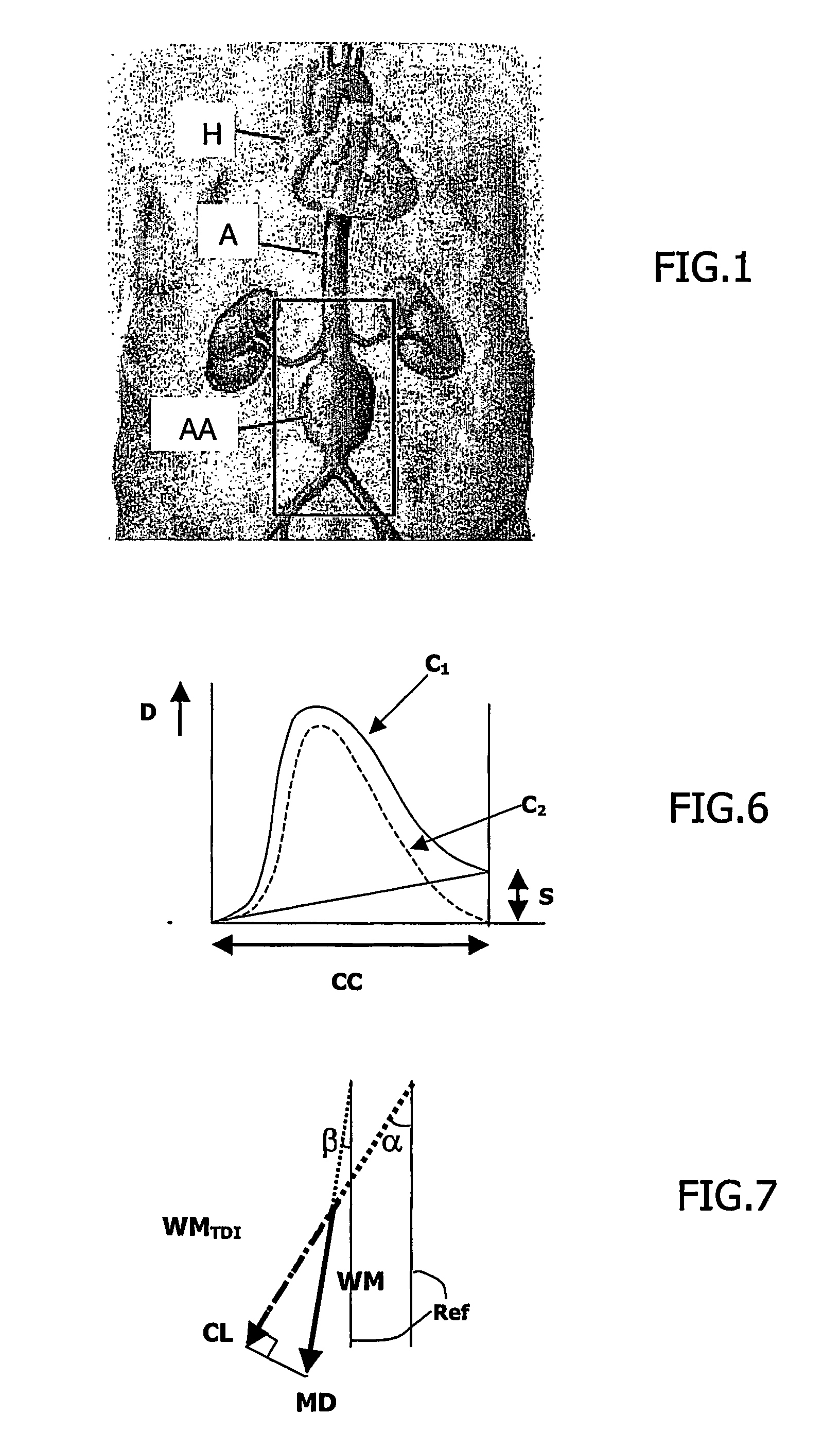
[0019] Referring to FIG. 1A, Abdominal Aortic Aneurysm AAA is defined by a doubling of the normal diameter of the infra-renal aorta A. The heart is denoted by H. The AAA abnormality is present in 5% of men aged over 65 years. Rupture of the aneurysm, the most common complication of AAA, is responsible for about 2% of deaths in men in this age group and is the tenth leading cause of death in men in Europe. Since most AAAs are asymptomatic until rupture occurs, up to 50% of all AAAs repairs are performed as an emergency operation. As the operative mortality for ruptured AAA is around 50%, and only a small fraction of patients with ruptured AAAs survive to reach hospital, the overall community mortality for ruptured AAAs is estimated at over 90%. For this reason, there is an increasing interest in the clinical and cost effectiveness of mass screening programs for AAAs. Acquired abdominal aortic aneurysms classically are characterized anatomically by an unparallelism of the aorta edges,...


PUM


Abstract

The invention relates to an ultrasonic viewing system, for displaying images of an artery using a curved array of transducer elements, comprising means for acquiring (51) of an ultrasonic image sequence and a Doppler color sequence of a segment of artery explored along its longitudinal axis and having walls moving in relation with the cardiac cycle; and comprising processing means for: estimating the velocity and motion amplitude (53, 54, 55) of the artery walls along Doppler color ultrasound scanning lines; estimating the motion amplitude (58) of the artery walls along lines perpendicular to the artery global axis; and further comprising: display means for displaying (60) curves of this last artery wall amplitude on a dedicated display on which the user may have interaction. The invention further relates to an image processing method having steps to be carried out using this system.

Description

FIELD OF THE INVENTION [0001] The invention relates to an ultrasonic imaging system, and / or an ultrasonic examination apparatus, for acquiring a sequence of medical images of an artery segment using a curved array of transducer elements. The invention further relates to an image processing method for processing the images produced by said system and particularly for displaying an ultrasonic image sequence of an artery segment with indications of arterial parameters in function of the cardiac cycle. The invention is used in the field of ultrasonic imaging, to provide a cardio-vascular non-invasive medical tool for examining patients suspected to present anomalies of arteries and notably anomalies of the aorta such as aortic aneurysms. BACKGROUND OF THE INVENTION [0002] An ultrasonic image processing method for calculating dilation curves related to an artery segment is already known from the U.S. Pat. No. 05,579,771 (Bonnefous, Dec. 3, 1996). This document describes a method for char...

Claims


Application Information

Patent Timeline
IPC IPC(8): G06K9/00A61B8/08
CPCA61B8/08A61B8/488A61B8/02
Inventor GERMOND-ROUET, LAURENCEBONNEFOUS, ODILE
Owner KONINKLIJKE PHILIPS ELECTRONICS NV
Why Eureka
Try Eureka