

Method of constructing nuclear-transplanted egg parthenogenetic embryo and parthenogenetic mammal
a technology of nuclear transplantation and embryo, which is applied in the field of nuclear transplantation embryo and parthenogenetic mammal construction, can solve the problems of its growth after the fact, and achieve the effect of ensuring the survival of the egg
- Summary
- Abstract
- Description
- Claims
- Application Information
AI Technical Summary
Benefits of technology
Problems solved by technology
Method used
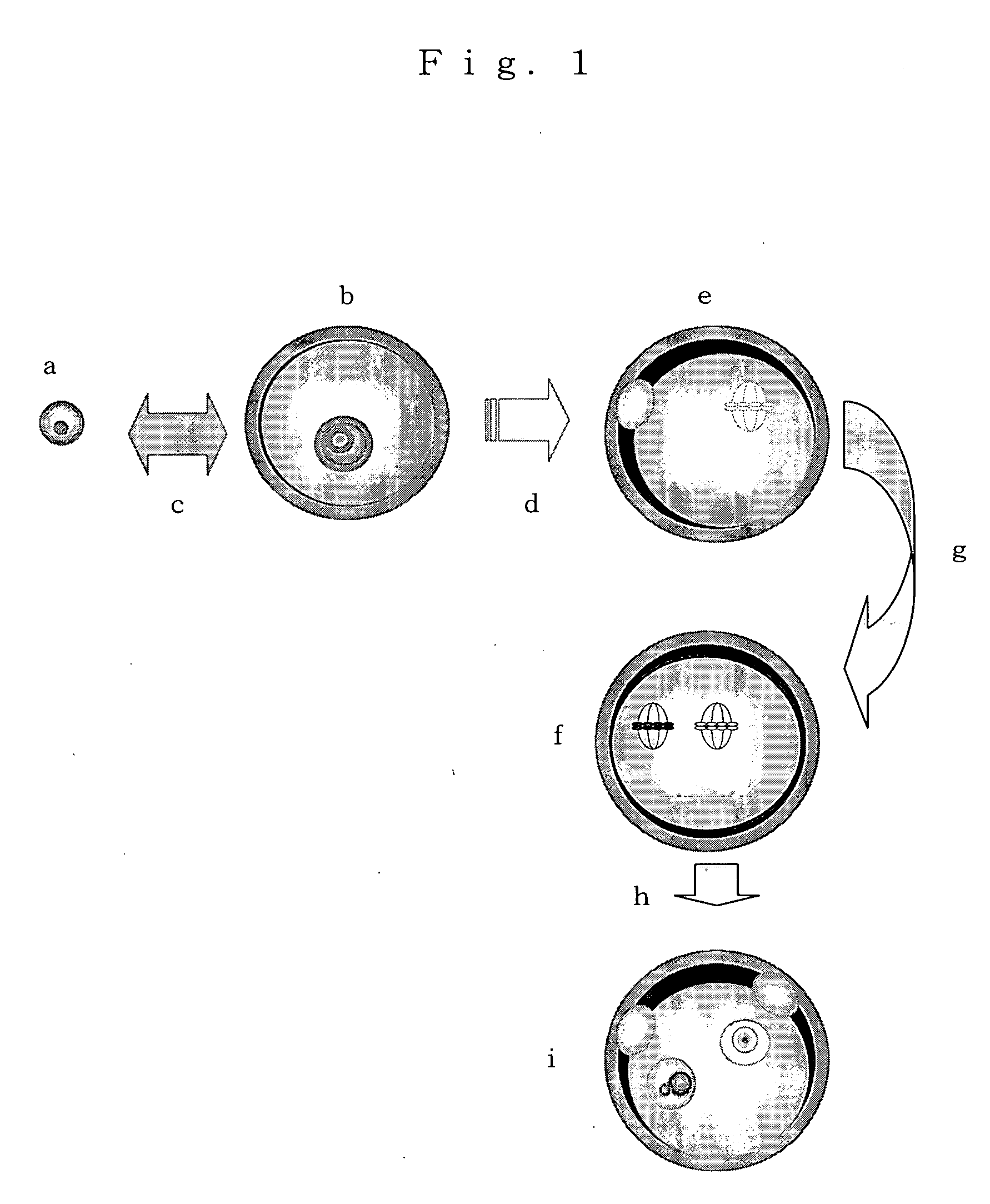
Image

Examples
example 1
(Collection of Ng Ova)
[0056] Ovaries were collected from one-day-old neonates of mice (Leighton et al, Nature 375: 34-39, 1995) from which 13 Kb of H19 genes and upstream regions thereof had been deleted according to a target gene recombination method (Methods in Enzymology 225: 803-890, 1993). The collected ovaries were transferred into a 0.02% EDTA solution and cultured at 37° C. for 10 minutes. Then, the ovaries were cut apart with an injection needle and dissociated non-grown stage ova were collected and used as ng ova as a donor.
(Collection of GV Stage Egg)
[0057] Matured mice (8 to 12 weeks old, B6D2F1, Charles River / Claire) were administered with pregnant mare ciliary gonadotropic hormone at a dose of 5 to 7.5 IU, and then grown ovarian follicles in the ovaries were cut apart with a 27-gage injection needle to collect GV stage eggs covered each with cumulus cells. The cumulus cells were removed by pipetting, and then the GV stage eggs were cultured in an M2 culture medium...
example 2
(Construction of Parthenogenetic Embryo)
[0062] The thus-obtained 212 second nucleus-implanted eggs were cultured in an M16 culture medium containing 10 M of strontium chloride at 37° C. for 3 hours to artificially induce activation of the ova. As a result, 189 ova were activated. 158 Ova in which two each of the second polocytes and pronuclei were generated were in vitro cultured in an M16 culture medium at 37° C. for 3 days. As a result, 131 blastocysts were produced.
example 3
(Construction of Parthenogenetic Mouse)
[0063] The thus-obtained 131 blastocysts were implanted in an uterus of a female mouse at day 2.5 of pseudopregnancy (female mouse that was mated with a vasoligated male according to a conventional method and a day when its copulatory plug was confirmed was a day 0.5), to give birth to 2 normal female neonates. Analysis of genes of these showed that they had H19 gene (derived from the fg ovum) and Neor gene (derived from ng ovum), so that it was confirmed that the above neonates were neonates from the second nucleus-implanted-eggs.
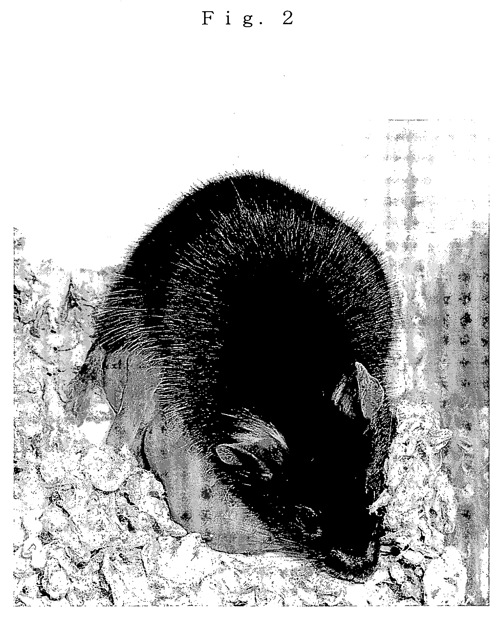
[0064] One of these was sacrificed by euthanasia for gene analysis after its anabiosis was confirmed. The other one was named “Kaguya”, and it normally grown to adulthood. “Kaguya” delivered neonates by mating with a male, so that it was confirmed that “Kaguya” had normal reproduction ability. FIG. 2 shows a photograph taken when “Kaguya” safely delivered the neonates.
PUM
| Property | Measurement | Unit |
|---|---|---|
| diameter | aaaaa | aaaaa |
| diameter | aaaaa | aaaaa |
| diameter | aaaaa | aaaaa |
Abstract
Description
Claims
Application Information