Real time vascular imaging during solid organ transplant

a solid organ transplant and real-time vascular imaging technology, applied in the field of medical imaging, can solve the problems of ineffective, expensive and inconvenient, and traditional intra-operative imaging techniques

Inactive Publication Date: 2006-10-26
NOVADAG TECH INC
View PDF26 Cites 131 Cited by
  • Summary
  • Abstract
  • Description
  • Claims
  • Application Information

AI Technical Summary

Benefits of technology

[0006] In certain embodiments the invention provides a portable system useful for imaging at least one vessel which is surgically joined to a transplanted organ in a recipient subject comprising: a) a fluorescent dye; b) an energy source capable of emitting sufficient energy such that the fluorescent dye fluoresces; and c) an imaging head.
[0007] In certain embodiments the invention provides a portable system useful for imaging at least one vessel which is surgically joined to a transplanted organ in a recip

Problems solved by technology

Traditional intra-operative imaging techniques, are

Method used


Image

  • Real time vascular imaging during solid organ transplant
  • Real time vascular imaging during solid organ transplant

Examples


Embodiment Construction

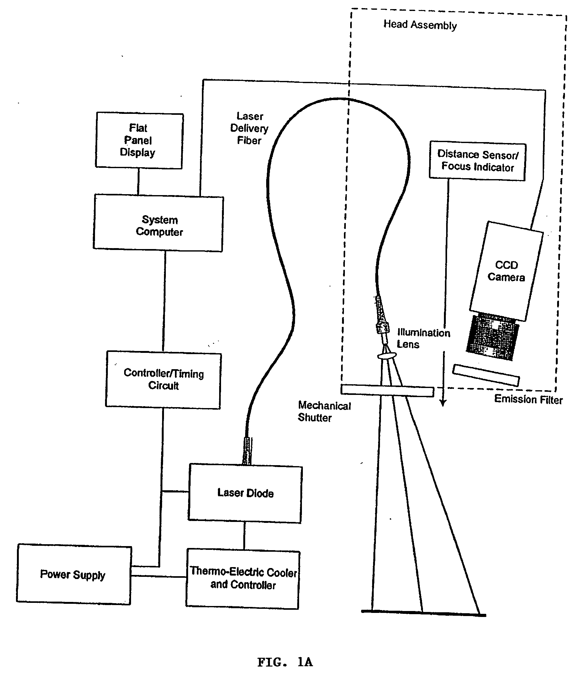
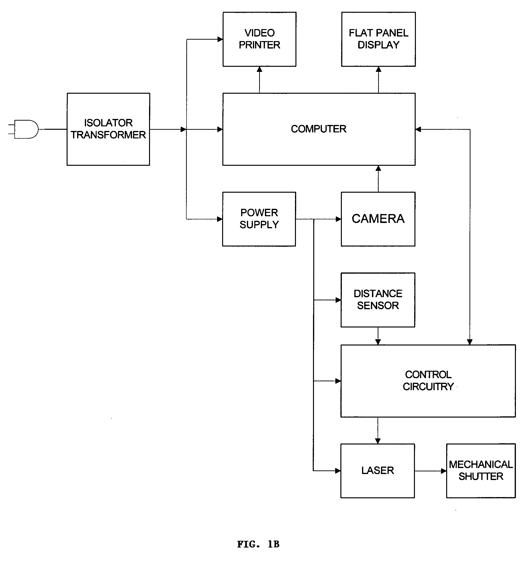
[0011] Organ transplants of various types are performed routinely. Solid organ transplants of all types require the joining of vessels, e.g., blood vessels in order to attach the donor organ. Some organ transplants, such as a pancreas transplant also require the joining of a duct to a lumen, e.g. the pancreatic duct to the digestive tract. Occlusion, due to, thrombosis, plaque, or free floating endothelial material, for example, is a risk associated with transplant surgery. The invention described herein, in certain embodiments, provides a method and a system which a surgeon can use intra-operatively to determine if an anastomosis created during transplant surgery is patent. The invention also provides a method and system to determine, pre-surgery, if a donor organ, or a vessel attached to a donor organ, is patent.

[0012] Subject as used herein, refers to any animal. The animal may be a mammal. Examples of suitable mammals include, but are not limited to, humans, non-human primates,...


PUM


Abstract

The invention provides methods and systems for imaging vessels in a subject. In certain embodiments the vessels may be associated with a solid organ transplant.

Description

FIELD OF THE INVENTION [0001] The invention relates generally to the field of medical imaging. Certain embodiments of the invention provide methods for imaging of vasculature in a subject. Certain other embodiments provide systems which are useful for imaging vasculature in a subject. BACKGROUND OF THE INVENTION [0002] Imaging of biological tissues and organs assists doctors in both diagnosis and treatment. A variety of medical techniques which are suitable for imaging biological tissues and organs are known. These include traditional x-rays, ultra-sound, as well as magnetic resonance imaging (MRI), and computerized tomography (CT). A variety of dyes used in medical imaging have also been described including radio opaque dyes, fluorescent dyes, as well as, colormetric dyes (see e.g., U.S. Pat. Nos. 5,699,798; 5,279,298; 6,351,663; and U.S. patent application Ser. No. 10 / 365,028). Imaging techniques and systems using fluorescent dyes have been described for the heart and eye (see, U....

Claims


Application Information

Patent Timeline
IPC IPC(8): A61K49/00
CPCA61K49/0034A61K49/0019A61K49/0045A61K49/0041A61K49/0043A61K49/0032
Inventor MANGAT, GURPREETBRZOZOWSKI, LUKASZCROFT, RICHARD L.
Owner NOVADAG TECH INC
Why Eureka
Try Eureka