Apparatus and method for rendering volume data

a volume data and apparatus technology, applied in the field of ultrasonic diagnostic systems, can solve the problems of difficult enhancement of rendering speed and decrease of rendering speed, and achieve the effect of enhancing rendering speed

Active Publication Date: 2006-11-09
MEDISON CO LTD
View PDF8 Cites 15 Cited by
  • Summary
  • Abstract
  • Description
  • Claims
  • Application Information

AI Technical Summary

Benefits of technology

[0011] Therefore, it is an object of the present invention to provide an apparatus and a method for enhancing the rendering speed and ensuring that a minute object, which is to be sampled in volume ray casting, is included by adjusting the sampling interval in volume data.

Problems solved by technology

However, since the volume data are sampled even for a space, which is unnecessary to form the 3D ultrasound image, in a constant interval in the conventional volume ray casting, there is a problem in that it is difficult to enhance the rendering speed.
Thus, there is a problem in that the rendering speed becomes decreased.
Also, since the volume data are sampled at a constant sampling interval in the conventional volume ray casting, there is a problem in that objects, which are smaller than the sampling interval, can be excluded during the sampling process.

Method used


Image

  • Apparatus and method for rendering volume data
  • Apparatus and method for rendering volume data
  • Apparatus and method for rendering volume data

Examples


Embodiment Construction

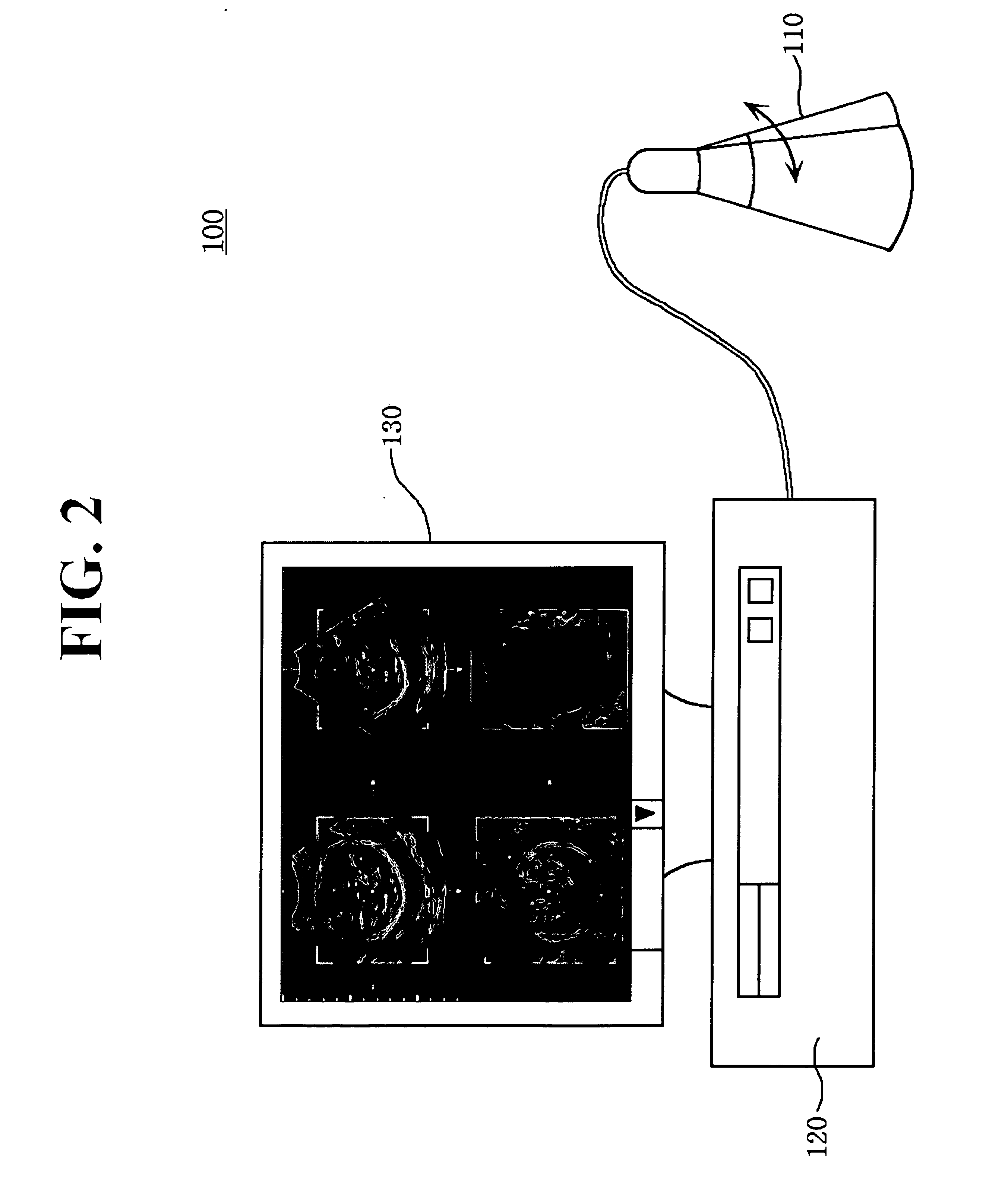
[0022]FIG. 2 is a block diagram showing an ultrasound diagnostic system, which is constructed in accordance with the present invention.

[0023] As shown in FIG. 2, a 3D ultrasound diagnostic system 100 includes a probe 110, a body 120 and a display unit 130.

[0024] The probe 110 is used to acquire 3D ultrasound data from a target object to be displayed. A mechanical scanning process (scan by moving a mechanical arm or rotating a stepping motor) or a hand-free process (scan by a user's hand) may be applied to the probe 110.

[0025] The display device 130 (e.g., a monitor) is used to display the ultrasound data acquired from the probe 110. It should be noted herein that as long as the display device can display a 3D ultrasound image in accordance with the present invention, any type of display device may be used.

[0026] As shown in FIG. 3, the body 120 comprises the following: an interface unit 121; a determination unit 122; a display region calculation unit 123; a scan conversion unit ...


PUM


Abstract

The present invention relates to an apparatus and a method for rendering volume data in an ultrasound diagnostic system. A method for rendering volume data including an object space and an empty space acquired from ultrasound data, comprises the following steps: a) casting a virtual ray from at least one pixel comprising a viewing plane into the volume data; b) sampling voxels of the volume data along the virtual ray in a first sampling interval; c) checking whether a currently sampled voxel corresponds to the object space or the empty space by using opacity based on a voxel value of a sampled voxel; d) at step c), if it is determined that the currently sampled voxel corresponds to the object space, then returning to the previously sampled voxel and sampling voxels in a second sampling interval shorter than the first sampling interval; e) checking whether accumulated opacity calculated based on the sampled voxels along the virtual ray is greater than a critical value; f) at step e), if it is determined that the accumulated opacity is greater than the critical value, then completing the sampling process for the current virtual ray and calculating a rendering value by using the voxel values and the opacity of the sampled voxels; and g) repeating the steps a) to f) until the sampling process for the pixels comprising the viewing plane is completed.

Description

FIELD OF THE INVENTION [0001] The present invention generally relates to ultrasound diagnostic systems, and more particularly to an apparatus and a method for rendering volume data in the ultrasound diagnostic system. BACKGROUND OF THE INVENTION [0002] Generally, an ultrasound diagnostic system transmits an ultrasound signal from the surface of a human body toward a desired portion within a target object. This is so that an ultrasound image of soft tissues or blood flow can be obtained through non- invasive means by using information obtained through ultrasound echo signals. [0003] Compared to other medical imaging systems (e.g., X-ray diagnostic system, X- ray CT scanner, MRI and nuclear medicine diagnostic system), the ultrasound diagnostic system is advantageous since it is small in size and fairly inexpensive. Further, the ultrasound diagnostic system is capable of providing real-time display and is highly safe without any dangerous side effects such as exposure to X-rays, etc. ...

Claims


Application Information

Patent Timeline
Patent Type & Authority Applications(United States)
IPC IPC(8): G06T17/00
CPCG06T15/06G06T2210/41G06T15/08A61B8/00
Inventor KWON, EUI CHULSHIN, BYEONG SEOK
Owner MEDISON CO LTD
Why Eureka
Try Eureka