Flat panel detector based slot scanning configuration

a detector and slot scanning technology, applied in the field of diagnostic radiology, can solve the problems of reducing contrast, low speed scanning techniques are not practical in diagnostic radiology, and the general effect of unsatisfactory techniques for obtaining high image quality

Active Publication Date: 2006-11-09
GENERAL ELECTRIC CO
View PDF7 Cites 6 Cited by
  • Summary
  • Abstract
  • Description
  • Claims
  • Application Information

AI Technical Summary

Benefits of technology

[0016] Still another embodiment comprises forming an image of an object using an X-ray device. This embodiment comprises producing an X-ray beam using a conventional X-ray source and creating a plurality of beam segments from the X-ray beam. The method further comprises scanning the object using at least one of the beam portion and producing an X-ray image on a flat panel detector. This method may comprises at least one of using a removable pre-patient collimator to form the plurality of beam portions and using a post-patient collimator passing at least one of the beam portion. Further, the method may comprise minimizing acquisition time.

Problems solved by technology

One of the most significant challenges in using standard flat panel imaging is that, after performing X-ray imaging of an object (a patient for example), the X-ray image is composed of more scattered X-rays than direct transmitted X-rays, thus reducing contrast.
However, such known techniques have generally proved to be unsatisfactory in obtaining high image quality while maintaining rapid scanning rates and low exposure times. While it is possible to obtain images of good quality with very slow scanning speeds, as with a single scanning beam for example, such low speed scanning techniques are not practical in diagnostic radiology in view of the fact that the patients' body parts and organs move while the patients are being X-rayed.
Thus, if relatively long exposure times are required to obtain an X-ray image, the images may be blurred due to the movement of the organs and body parts being X-rayed and are useless for diagnostic purposes.
One limitation associated with previously used post-patient collimators is that they do not totally reduce or eliminate scattered radiation and may block X-rays that have passed through the patient, forming useful doses.
These scattered beam portions carry no significant information, and thus tend to blur or reduce the contrast in any resulting X-ray image.
However, due to the thickness, dense concentration of material in the abdomen, and the large radiation field necessary to image the abdominal area, a large amount of X-ray scattering is created, thus making it difficult to obtain clear radiographic images of the abdomen, as mentioned previously.
The materials used to detect the X-rays in the such previous designs (single crystal Si and Xe gas for example) do not work effectively for high kV imaging used for certain applications (radiography of R&F for example).
Finally, the mechanical scanning used to form the image makes high frame rate imaging such as fluoro imaging difficult if not impossible.

Method used


Image

  • Flat panel detector based slot scanning configuration
  • Flat panel detector based slot scanning configuration
  • Flat panel detector based slot scanning configuration

Examples


Embodiment Construction

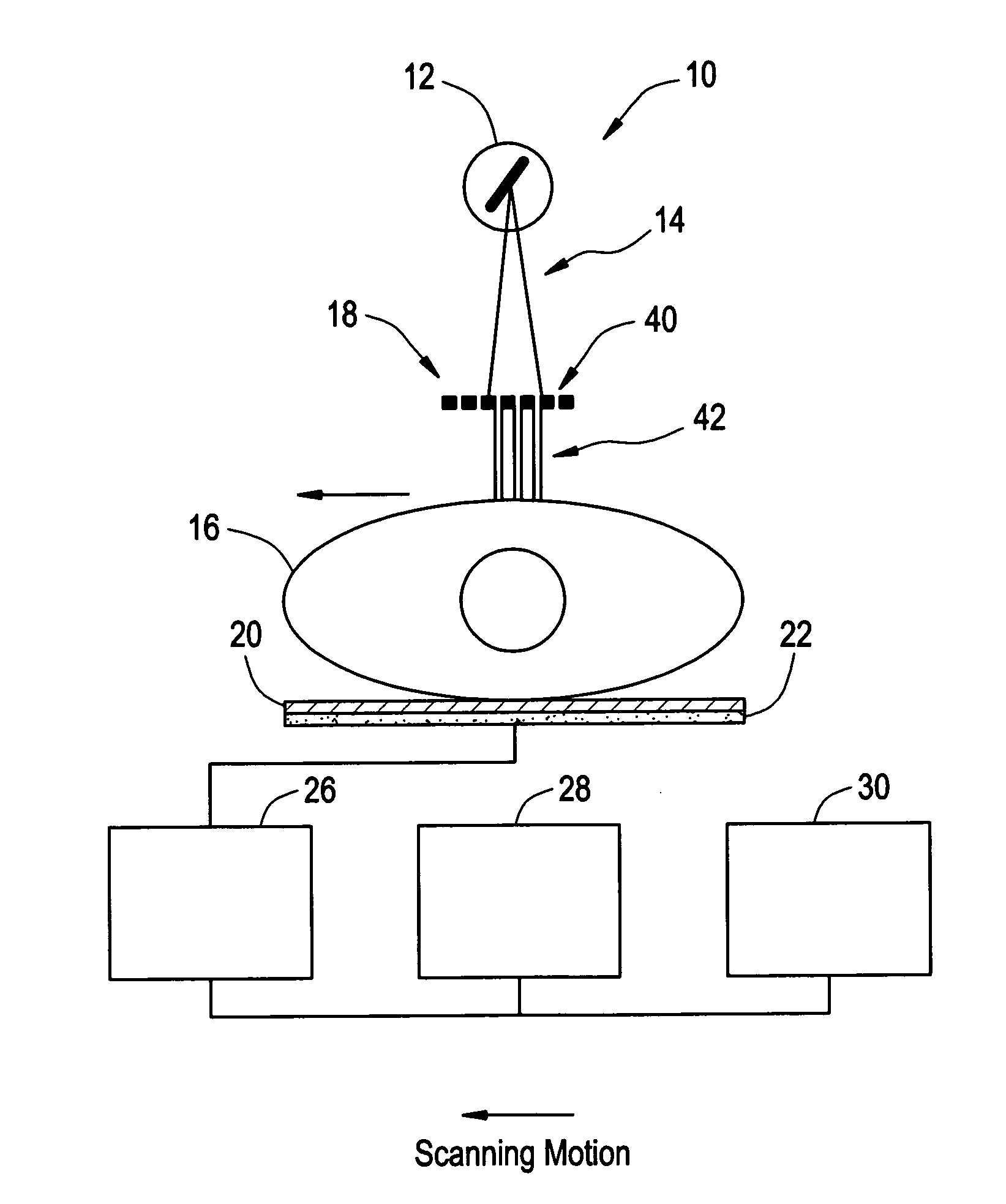
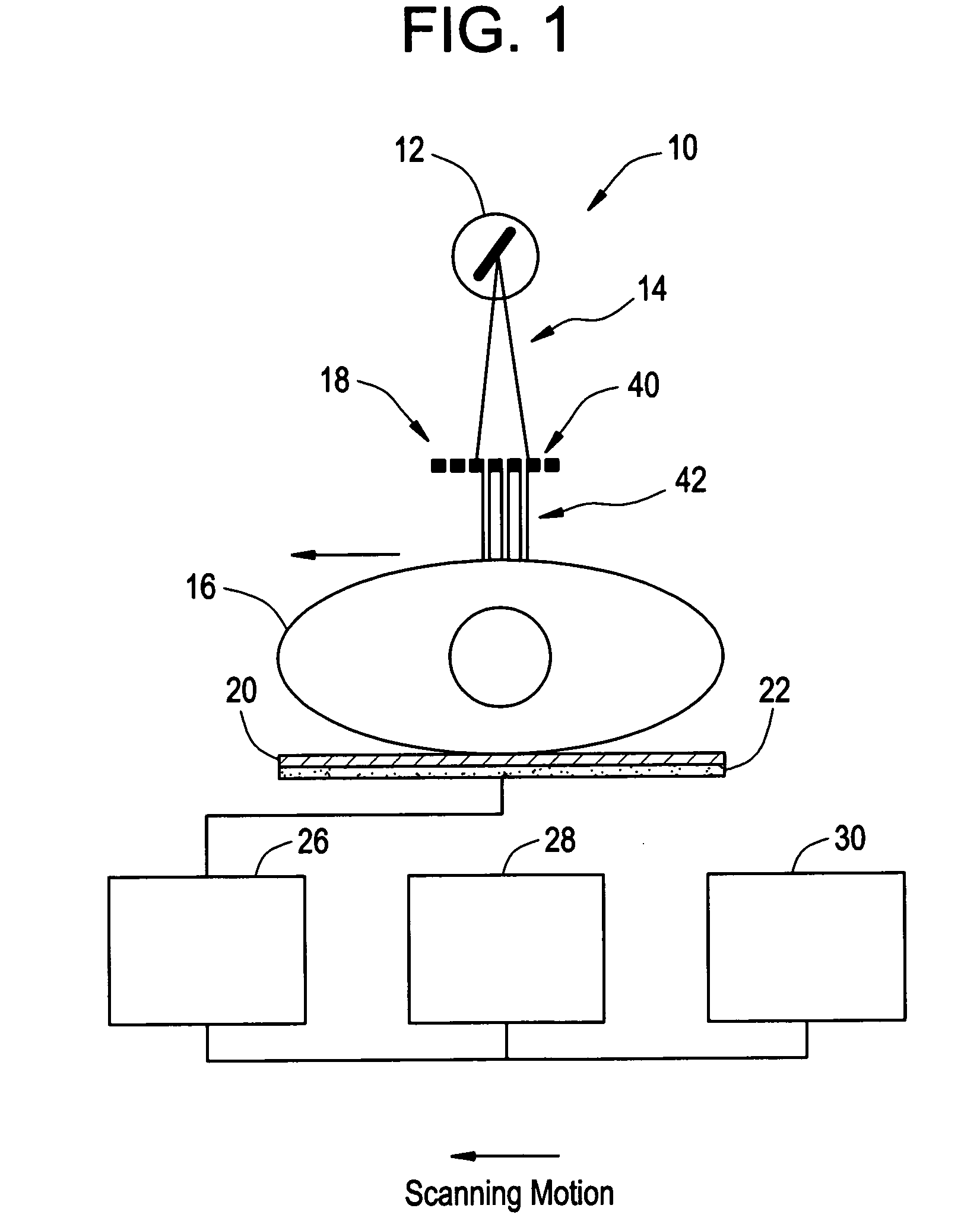
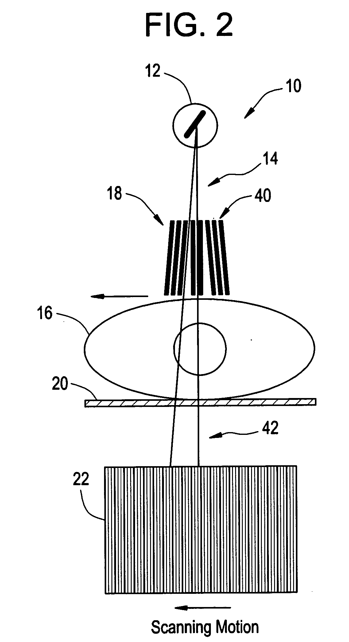
[0028] Clear, high quality X-ray images are necessary to obtain the degree of detail needed to permit accurate diagnosis of disease and detection of tumors and other improper growths in the crowded abdominal area, for example. However, due to the thickness and dense concentration of material in the abdomen, and the large radiation field necessary to image that area, a large amount of X-rays are scattered, thus making it difficult to obtain clear images, as provided previously. Accordingly, it is highly desirable and important to the advancement of diagnostic radiography (abdominal diagnostic radiography for example) that clear X-ray images (i.e., having enhanced contrast) be obtained.

[0029] At least one embodiment relates in general to configuring a diagnostic X-ray device, system or apparatus and performing diagnostic radiology using such a configured X-ray device, system or apparatus. More particularly, embodiments are directed to an X-ray device, system or apparatus having at le...


PUM

PropertyMeasurementUnit
focal spot sizeaaaaaaaaaa
widthaaaaaaaaaa

Abstract

The present invention relates to a diagnostic X-ray device, system or apparatus for performing diagnostic radiology and a method of configuring such a diagnostic X-ray device, system or apparatus. More specifically, the present invention relates to a diagnostic system for forming at least one image of an object having enhanced contrast. The system comprises a beam source adapted to produce an imaging beam and a masking member adapted to form at least one beam portion from the imaging beam and adapted to image the object. The system further comprises a flat panel detector positioned in a path of at least one beam portion penetrating the object and adapted to form at least one image of the object.

Description

RELATED APPLICATIONS [0001] This application is a continuation of U.S. application Ser. No. 10 / 990,267, filed Nov. 16, 2004, which is hereby incorporated by reference in its entirety.BACKGROUND OF THE INVENTION [0002] This application is directed in general to diagnostic radiology using an X-ray device, system or apparatus. This application is directed in particular to an X-ray device, system or apparatus using a flat panel detector in a slot scanning configuration. [0003] It is known that scattered X-rays have contrast reducing effects. One of the most significant challenges in using standard flat panel imaging is that, after performing X-ray imaging of an object (a patient for example), the X-ray image is composed of more scattered X-rays than direct transmitted X-rays, thus reducing contrast. This is especially true for thick objects (i.e., heavy patients). [0004] Efforts have been made to improve contrast in diagnostic image radiology systems, including using air gaps, improved ...

Claims


Application Information

Patent Timeline
Patent Type & Authority Applications(United States)
IPC IPC(8): G01N23/04
CPCA61B6/027A61B6/06A61B6/4233A61B6/4291G01N23/04G01N23/20G01T1/2018A61B6/588G01T1/2006
Inventor UNGER, CHRISTOPHER D.HOFFMAN, DAVID M.BHATT, VIVEK
Owner GENERAL ELECTRIC CO
Why Eureka
Try Eureka