Mammography method and apparatus for generating digital tomosynthetic 3D image

a digital tomosynthetic and image technology, applied in the field of mammography, can solve the problems of difficult identification of women and inability to achieve a suitable degree of exposure control, and achieve the effect of quick reading and evaluation

Active Publication Date: 2006-11-30
SIEMENS HEALTHCARE GMBH
View PDF8 Cites 38 Cited by
  • Summary
  • Abstract
  • Description
  • Claims
  • Application Information

AI Technical Summary

Benefits of technology

[0011] In a preferred embodiment of the method, the preliminary image is recorded with a spatial resolution that equals the spatial resolution of the subsequent individual images. This makes it possible to use the preliminary image, which has been made for the purpose of determining the radiographic parameters, in the tomosynthetic reconstruction process, while taking into account the radiographic parameters set for this preliminary image, doing so either directly or by merging the image With

Problems solved by technology

In such a projection the subsequent tissue layers in the direction of the X-ray are superimposed, and thus strongly absorbing benign structures may cover a malignant tumor and make the identification th

Method used


Image

  • Mammography method and apparatus for generating digital tomosynthetic 3D image
  • Mammography method and apparatus for generating digital tomosynthetic 3D image
  • Mammography method and apparatus for generating digital tomosynthetic 3D image

Examples


Embodiment Construction

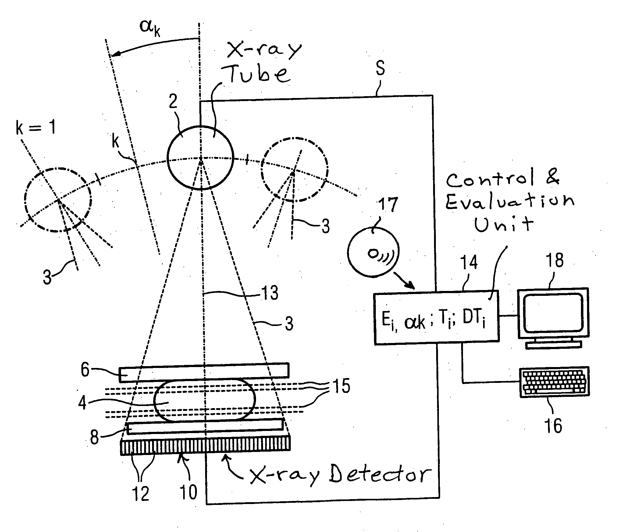
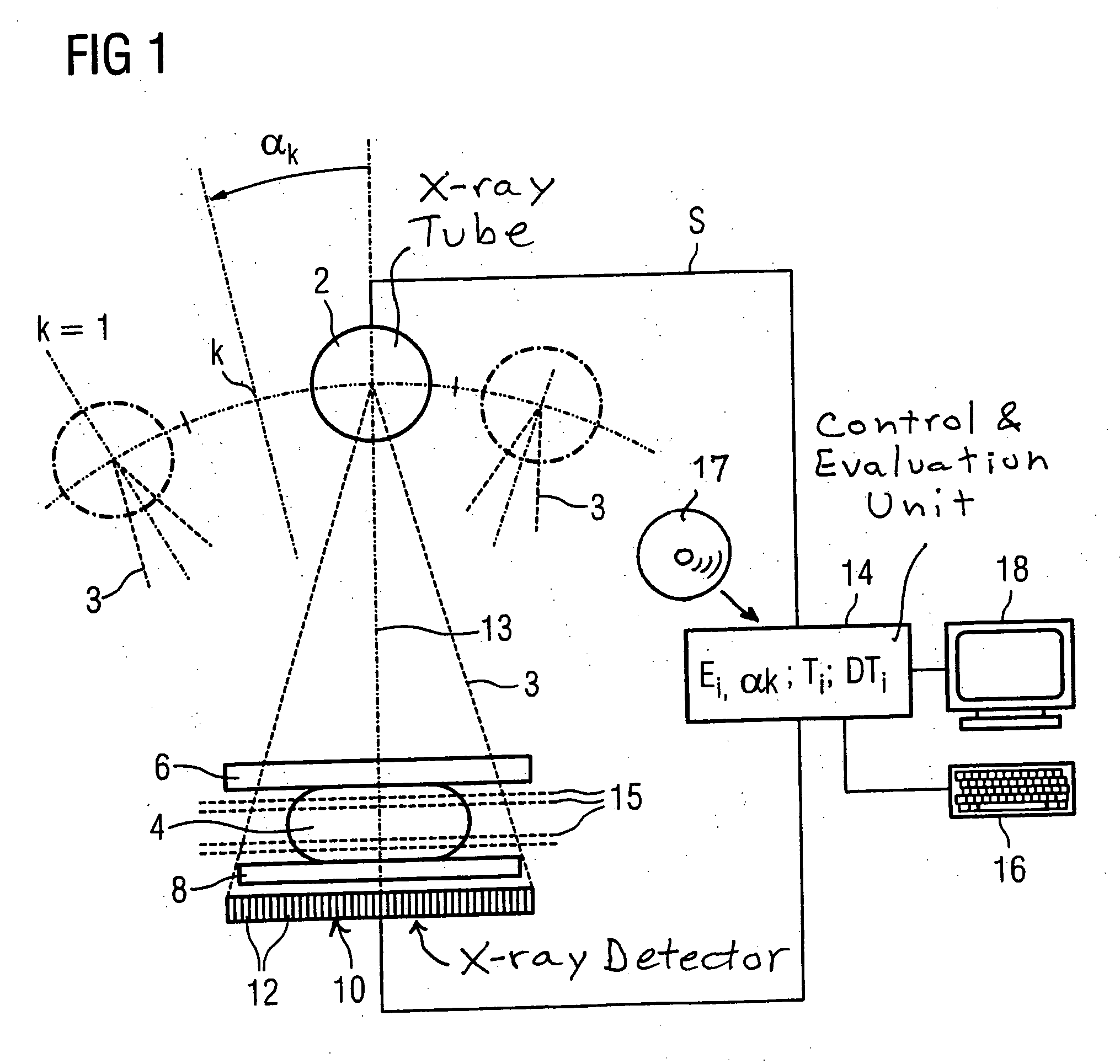
[0017] As shown in FIG. 1, a mammography device has an X-ray tube 2 for the generation of an X-ray beam 3 that passes through an examined object. The examined object 4 in this case is a female breast, which is held between a compression plate 6 and a patient examination table 8. As the X-ray tube is moved through a number of projection angles, X-ray beams pass through the examined object 4, the compression plate 6, and the patient examination table 8, are received by a large-surface digital X-ray detector 10, which is composed of a number of individual detectors 12 arranged in a matrix-like array. The X-ray tube 2 can be rotated from a starting position k=0 through various angle positions k up to a maximum projection angle of αmax (maximum displacement), so that individual images Ek of the examined object 4 can be made at different projection angles αk relative to the normal 13 of the patient examination table 8 of the X-ray detector 10. In the starting position k=0 (projection angl...


PUM


Abstract

In a method and a device for generating a digital tomosynthetic 3D X-ray image of an examined object, a number of individual images are made of the examined object at different projection angles relative to the normal of the patient examination table by moving the X-ray tube. In a starting position, the X-ray tube is operated with a dose that is lower than that used for subsequent individual images. At least one preliminary image is recorded at the starting position, which is then evaluated to determine the radiographic parameters required for the recording of the subsequent individual images.

Description

BACKGROUND OF THE INVENTION [0001] 1. Field of the Invention [0002] The present invention relates to a method and a device used to generate a digital tomosynthetic 3D X-ray image of an examined object, particular in mammography. [0003] 2. Description of the Prior Art [0004] When an X-ray image of an examined object is being prepared, for example, for mammography, it must ensure that the radiographic parameters, particularly the exposure time, are set correctly so as to guarantee an image quality suitable for diagnostic evaluation. In order to limit the exposure of the area of the examined object that is being irradiated to the minimum radiation required for diagnostic purposes, it is desirable to be able to correctly set the radiographic parameters with only one attempt. For this purpose, a so-called automatic exposure control (AEC) is conventionally used. In analog mammography, as seen in the direction of propagation of the X-rays, a number of solid-state detectors are arranged beh...

Claims


Application Information

Patent Timeline
IPC IPC(8): A61B6/04
CPCA61B6/025G01N23/06A61B6/502A61B6/0414
Inventor MERTELMEIER, THOMAS
Owner SIEMENS HEALTHCARE GMBH
Why Eureka
Try Eureka