Phase based digital imaging

a digital imaging and phase technology, applied in the field of phase based digital imaging, can solve the problems of large safety limitations, resolution, cost, lack or limited specificity of key chemicals or structures necessary for functional body monitoring, and difficulty in large-scale interpretation of screening mammograms, so as to achieve enhanced diagnostic image

Inactive Publication Date: 2006-12-28
UNIV OF MASSACHUSETTS
View PDF4 Cites 35 Cited by
  • Summary
  • Abstract
  • Description
  • Claims
  • Application Information

AI Technical Summary

Benefits of technology

[0007] The present invention relates generally to the field of medical imaging in which digital images can be acquired and used in the diagnosis and / or treatment of medical conditions. A preferred embodiment of the invention uses image processing techniques to separate the phase information from the acquired digital image to provide an enhanced diagnostic image. A preferred embodiment of the invention is particularly useful for the identification and imaging of those features of the animal or human body which cause high spatial frequency features in the acquired image. Hard tissue structures such as bone or calcified or crystalline masses, lesions or cysts can cause such a high spatial frequency response making them suitable for phase component imaging.

Problems solved by technology

Although an impressive array of body-imaging techniques, such as x-ray imaging, x-ray computed tomography, magnetic resonance imaging, thermal infrared imaging (TIR), ultrasound, and radioisotope imaging are currently available to yield useful information, there are important limitations of safety, resolution, cost, and lack or limited specificity to key chemicals or structures necessary for functional body monitoring.
At times, even for qualified personnel, it is difficult to interpret screening mammograms in large numbers.

Method used


Image

  • Phase based digital imaging
  • Phase based digital imaging
  • Phase based digital imaging

Examples


Embodiment Construction

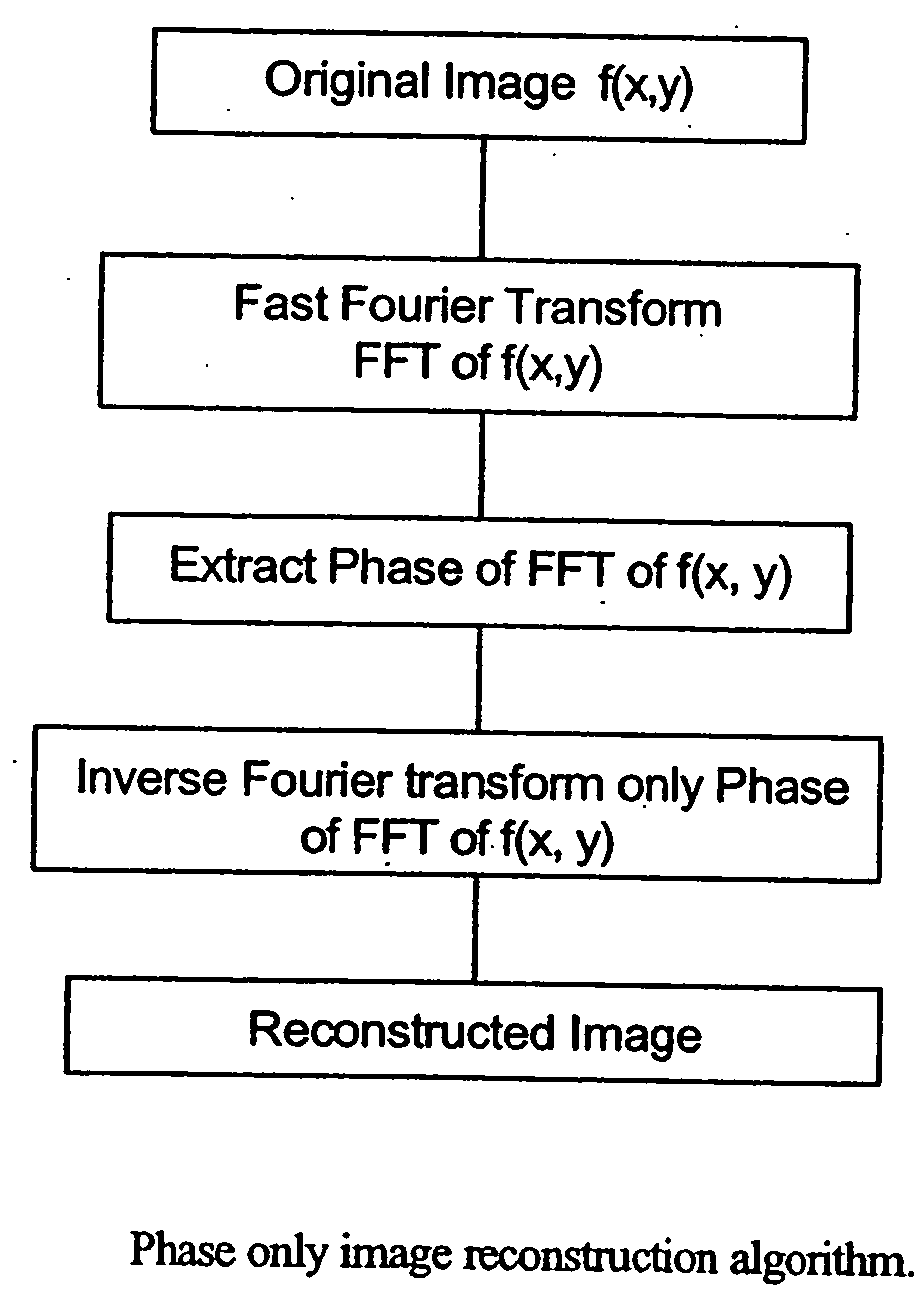
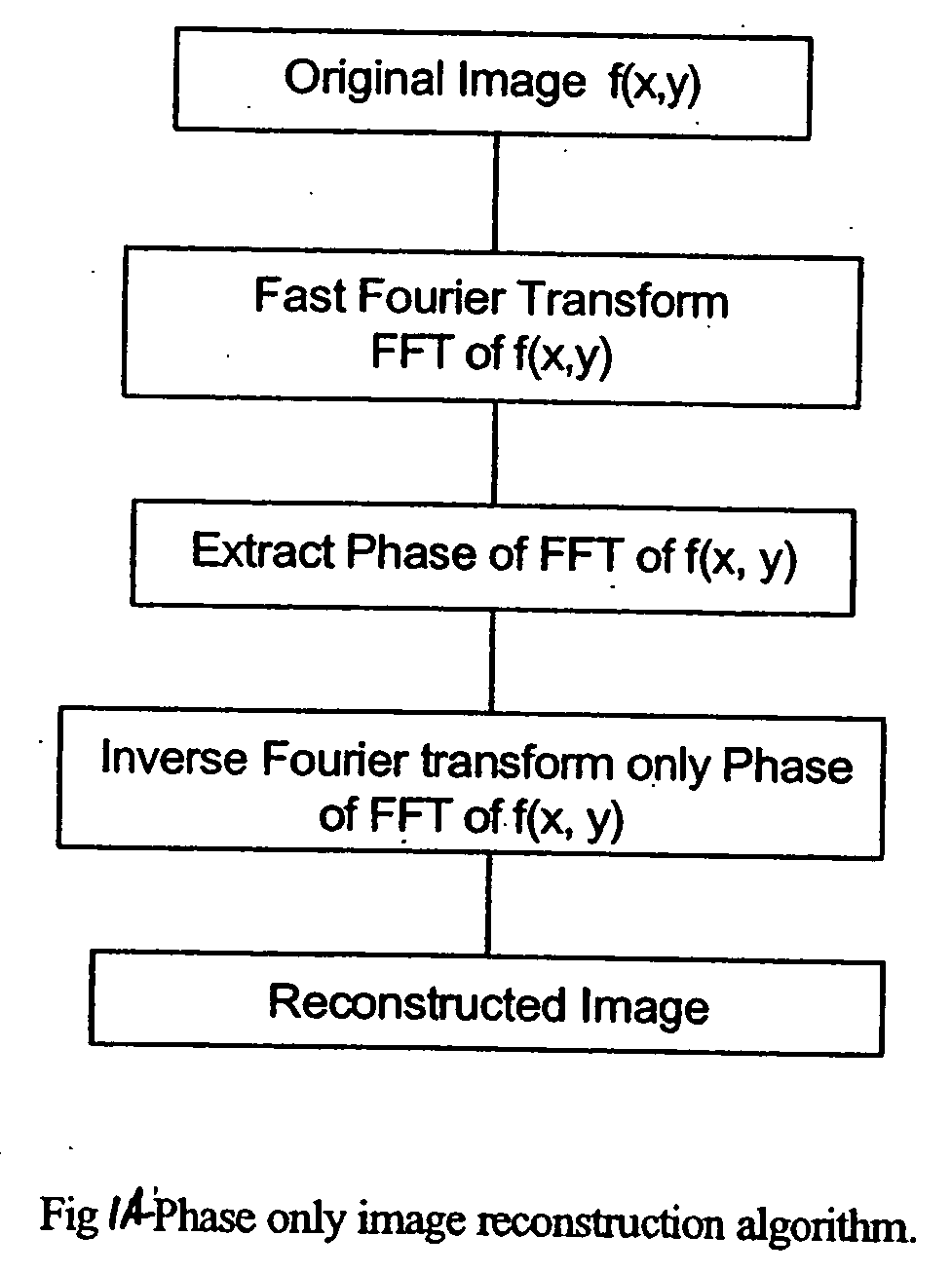
[0043] A preferred embodiment of the present invention is the phase characteristics of Fourier transform of medical images for computer aided diagnosis (CAD). We propose phase-only image reconstruction, original image reconstruction from phase-only information, phase-only correlations, spectral phase subtraction techniques for comprehensive CAD.

[0044] The method for phase-only image reconstruction is shown in FIG. 1A. Original digital image (digital mammogram, digital chest x-ray or in general any digital radiograph) is Fast Fourier Transformed using a programmed FFT sequence stored on a computer. The phase angle of the FFT spectrum is calculated. The phase of low spatial frequencies in the Fourier spectrum is zero or close to zero, while the phase of high spatial frequencies is in the neighborhood of ±π. From this phase angle, a phase-only function with unit amplitude transmittance is generated. The phase-only function is inverse Fourier transformed using another FFT operation to ...


PUM


Abstract

The present invention relates to systems and methods for medical imaging. Digital images are processed to provide phase images of a region of interest to aid in the diagnosis and treatment of various conditions. A preferred embodiment of the invention provides improved mammography screening for cancerous or precancerous conditions.

Description

CROSS REFERENCE TO RELATED APPLICATIONS [0001] The present application claims priority to U.S. Provisional Patent Application No. 60 / 648,637 filed Jan. 28, 2005. The entire contents of the above application is incorporated herein by reference.STATEMENT REGARDING FEDERALLY SPONSORED RESEARCH AND DEVELOPMENT [0002] This invention was supported, in whole or in part, by grant 1R21CA89673-01A1 from The National Institutes for Health. The Government has certain rights in the invention.BACKGROUND OF THE INVENTION [0003] There are numerous features in the human body in which medical imaging techniques can be used effectively to assist in the diagnosis and treatment of medical conditions. Various diseases or conditions involve calcified materials, structures or deposits within the human body that are indicative of the medical condition of the patient. These can include features of the human skeleton, such as the spine, calcified deposits within the arterial system such as obstructions to blo...

Claims


Application Information

Patent Timeline
Patent Type & Authority Applications(United States)
IPC IPC(8): G06K9/00G06K9/36
CPCG06T7/0012A61B6/583G06T2207/30068
Inventor KOTHAPALLI, SRI-RAJASEKHARYELLESWARAPU, CHANDRA S.WU, PENGFEIRAO, D.V.G.L.N.
Owner UNIV OF MASSACHUSETTS
Why Eureka
Try Eureka