Abnormality detection in medical images

a medical image and abnormality technology, applied in the field of digital image processing, can solve the problems of complex two-dimensional (2-d) views of superimposed anatomy, x-ray imaging can also have difficulty in resolving soft tissue features, and mri does not image bone well

Inactive Publication Date: 2007-02-15
CARESTREAM HEALTH INC
View PDF41 Cites 147 Cited by
  • Summary
  • Abstract
  • Description
  • Claims
  • Application Information

AI Technical Summary

Benefits of technology

[0023] An object of the present invention is to provide a system and method for computer assisted detection and / or diagnosis of disease or abnormalities utilizing imagery from multiple modalities.

Problems solved by technology

For example, X-ray imaging has high spatial and intensity resolutions, shows bony anatomy with high detail, and is relatively inexpensive to use; however, it also presents the viewer with complex two-dimensional (2-D) views of superimposed anatomy.
X-ray imaging can also have difficulty resolving soft tissue features.
MRI has the advantage of displaying three-dimensional (3-D) images of soft tissues with high contrast and high spatial resolution, and it does not involve ionizing radiation (as does X-ray and CT); however, MRI does not image bone well.
A disadvantage of ultrasound is that it cannot easily image through gas or bones, making it difficult to obtain images of some organs.
Nuclear medicine provides images depicting metabolic information that can be early indicators of pathological conditions; however, it can be difficult to accurately pinpoint the location of anomalies in the body due to the lack of structural information in the images.
However, PET systems are very costly, because of the need for a cyclotron to produce positron-emitting nuclides. fMRI is not frequently used in clinical applications, with the exception of surgical planning, which aims to determine the areas of the brain that respond to specific cognitive tasks, in order to avoid those areas during surgery.
However, the capsule and / or camera cannot be controlled or fixed on certain areas of interest, as can be done with a scope.
However, even though medical imaging modalities provide a wide variety of visual information, many diseases are difficult to detect or diagnose.
Although these methods and systems related to aligning and fusing images from multiple modalities, they do not address how to utilize the multimodal imagery to provide an enhanced method for detecting and / or diagnosing disease.
Existing CAD methods and systems apply to individual medical images or to multiple medical images from a single modality, and as such, they are not optimal when applied to a collection of images from multiple modalities.
Registration and fusion techniques, while providing visually meaningful information to the medical practitioner, do not provide the same level of assistance as CAD systems.

Method used


Image

  • Abnormality detection in medical images
  • Abnormality detection in medical images
  • Abnormality detection in medical images

Examples


Embodiment Construction

[0042] The following is a detailed description of the preferred embodiments of the invention, reference being made to the drawings in which the same reference numerals identify the same elements of structure in each of the several figures.

[0043] In the following description, various aspects of the present invention will be described. For purposes of explanation, specific configurations and details are set forth in order to provide a thorough understanding of the present invention. However, it will also be apparent to one skilled in the art that the present invention may be practiced without the specific details presented herein. Furthermore, well-known features may be omitted or simplified in order not to obscure the present invention.

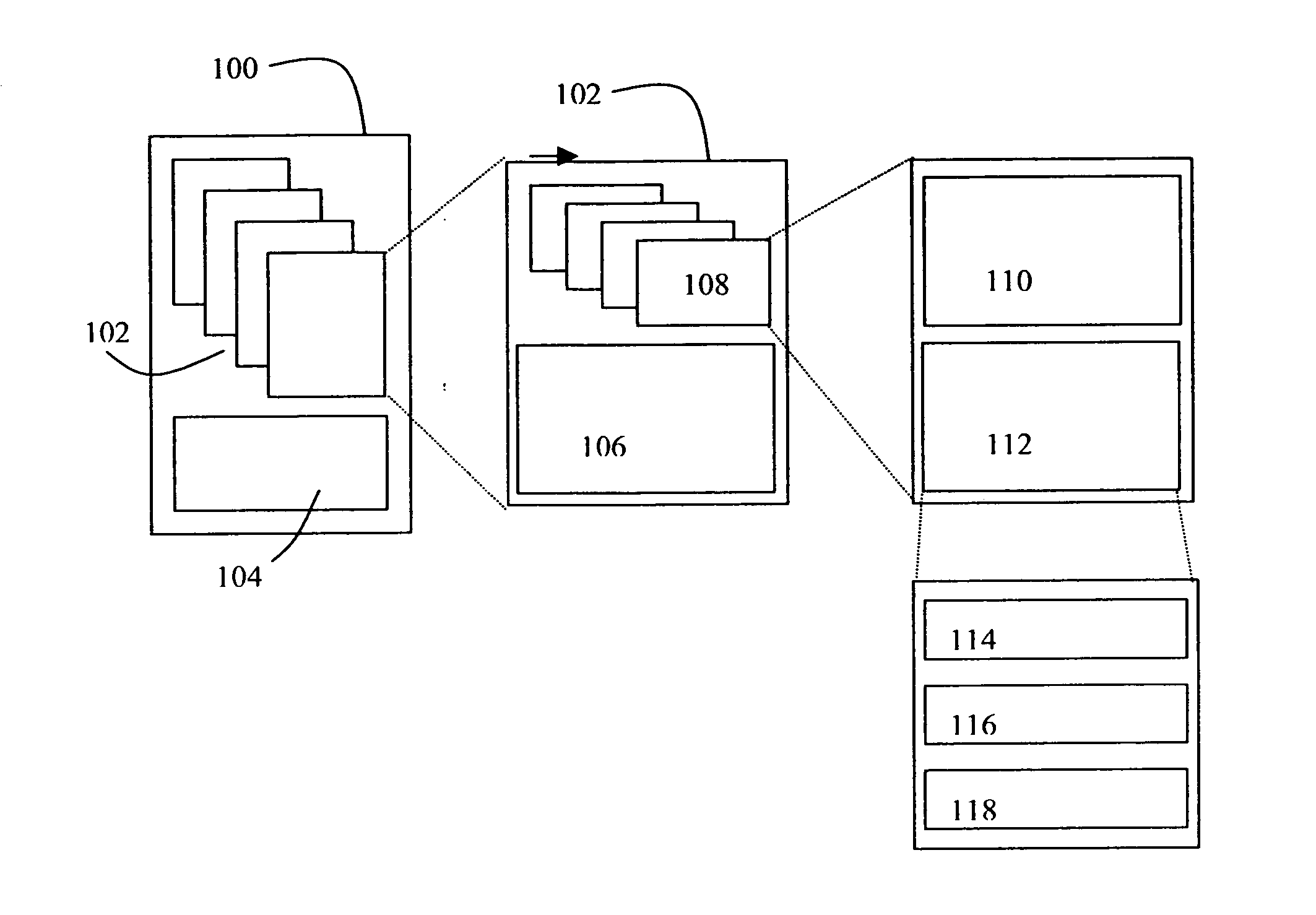
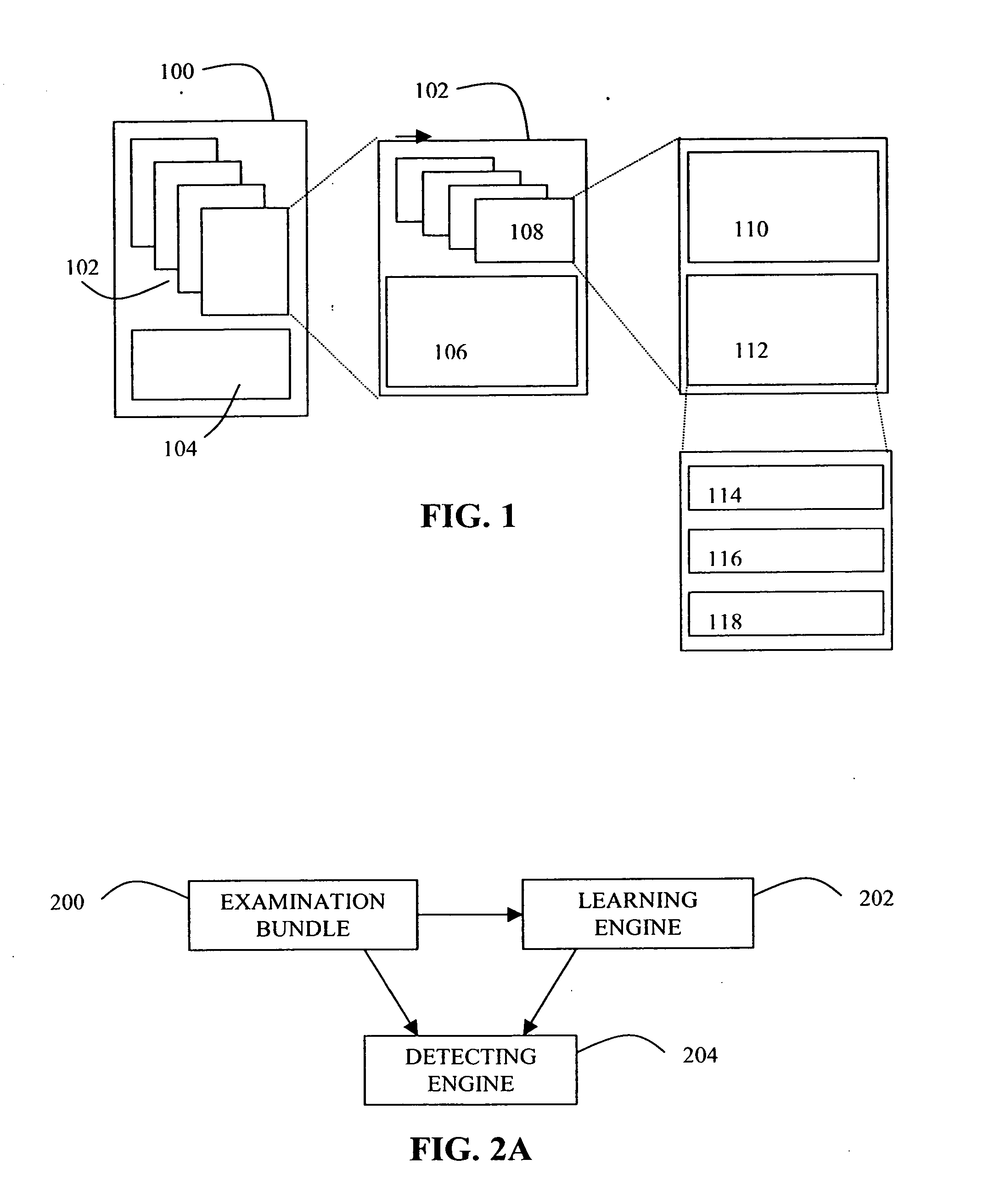
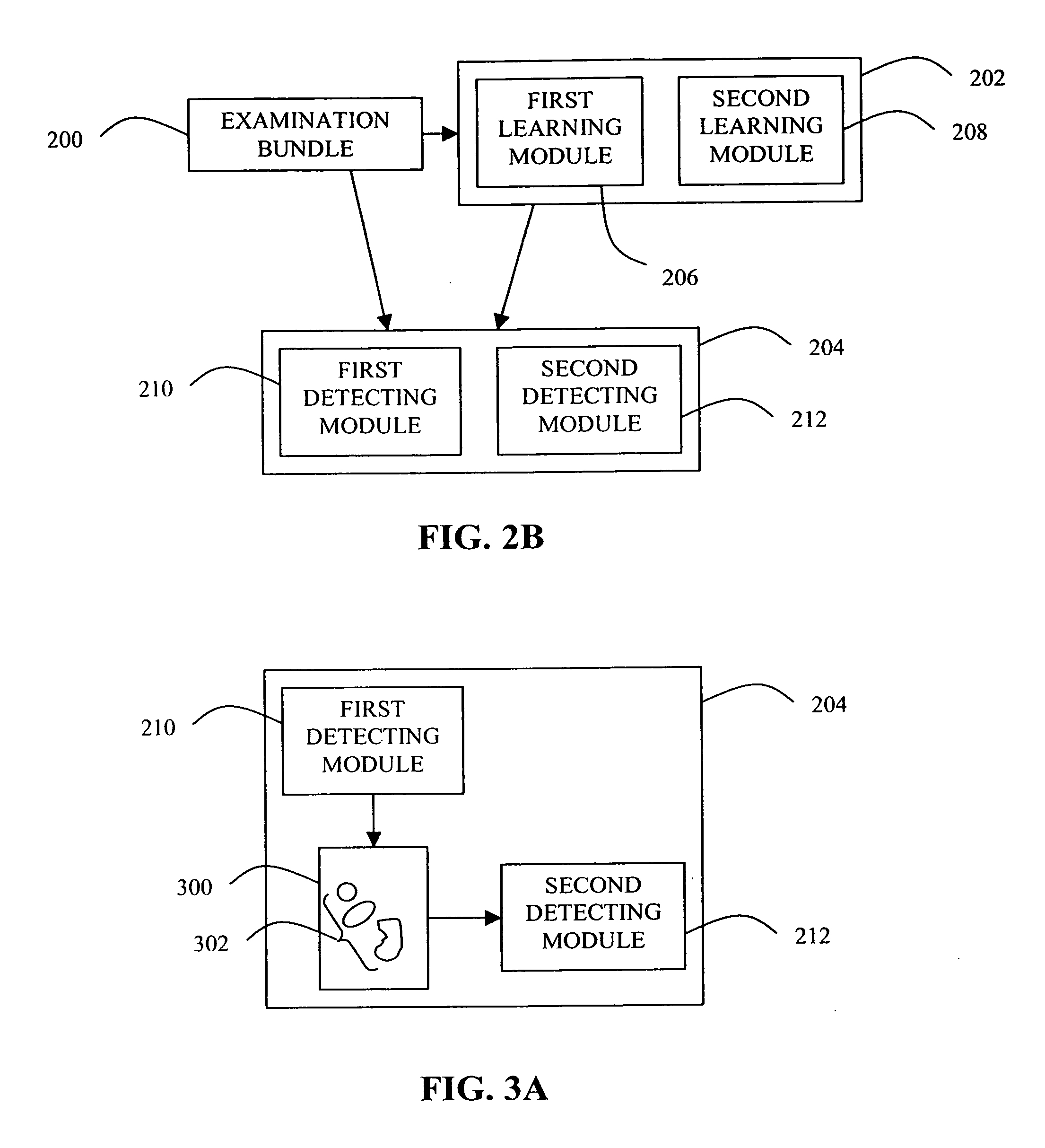
[0044] During a typical imaging examination of a patient by at least two modalities, which will hereinafter be referred to as a multimodal examination, one or more images from each modality can be captured or reconstructed.

[0045] The images captured...


PUM


Abstract

A system for the detection of abnormalities in a medical image of a subject. The system includes an examination bundle, a learning engine, and a detecting engine. The examination bundle includes at least one medical image of the subject from a first modality and at least one medical image of the subject from a second modality. The learning engine is employed for determining the characteristics of abnormalities within the at least one medical image from the first modality and the at least one medical image from the second modality. The detecting engine is employed for detecting abnormalities within at least one of the medical images comprising the examination bundle.

Description

FIELD OF THE INVENTION [0001] The invention relates generally to the field of digital image processing, and in particular to the detection of abnormalities in a medical image. BACKGROUND OF THE INVENTION [0002] Medical imaging plays an important role in the screening, diagnosis, and / or treatment of many diseases because medical images enable a physician to view the internal anatomical structure of a patient or to visualize physiological or metabolic information. A variety of different imaging techniques or modalities can be used in clinical medicine. Some well known techniques / modalities include X-ray and computed tomography (CT), ultrasound, nuclear medicine, ultrasonic imaging, and magnetic resonance imaging (MRI). X-ray and CT, ultrasound, and MRI produce images of anatomical structure, whereas nuclear medicine produces images depicting metabolic uptake or biodistribution of radioactive compounds in various tissues (organs or tumors). Other modalities for imaging functional chara...

Claims


Application Information

Patent Timeline
Patent Type & Authority Applications(United States)
IPC IPC(8): G06K9/00
CPCG06T2207/30061G06T7/0012
Inventor CAHILL, NATHAN D.CHEN, SHOUPUSUN, ZHAOHUIRAY, LAWRENCE A.
Owner CARESTREAM HEALTH INC
Why Eureka
Try Eureka