Vacuum coagulation probes

a coagulation probe and vacuum technology, applied in the field of vacuum coagulation probes, can solve the problems of inability to direct the coagulation of precise regions of soft tissue, inability to selectively ablate desired soft tissue structures, and inability to isolate preserved tissue structures from targeted regions, etc., to achieve consistent and reliable lesion, shorten wall thickness, and increase thermal conduction efficiency

Inactive Publication Date: 2007-02-22
ATRICURE
View PDF98 Cites 90 Cited by
  • Summary
  • Abstract
  • Description
  • Claims
  • Application Information

AI Technical Summary

Benefits of technology

[0007] Increased tissue contact relative to that which can be achieved with known devices is capable of reversing convective cooling effects effect noted above by evoking a compression of the tissue that shortens the wall thickness (e.g., of the atria), ensuring consistent contact throughout the length of the electrode(s), and increasing the efficiency of thermal conduction from the epicardium to the endocardium. As such a more consistent and reliable lesion is created.
[0008] As such, the integrated vacuum-assisted coagulation probes of the subject invention are capable of reliably creating transmural, curvilinear lesions capable of preventing the propagation of wavelets that initiate and sustain atrial fibrillation, atrial flutter, ventricular tachycardia, or other arrhythmia substrate.

Problems solved by technology

Atrial fibrillation surgery involving radiofrequency, D.C., microwave, or other thermal ablation of atrial tissue has a limitation in that tissue contact throughout the length of the electrode(s) is / are not consistent causing variability in the transmission of energy throughout the target length of ablated / coagulated tissue.
Another deficiency of current approaches is the inability to direct the coagulation of precise regions of soft tissue while avoiding underlying or nearby tissue structures.
Conventional approaches are unable to selectively ablate desired soft tissue structures and isolate preserved tissue structures from targeted regions.

Method used


Image

  • Vacuum coagulation probes
  • Vacuum coagulation probes
  • Vacuum coagulation probes

Examples


Embodiment Construction

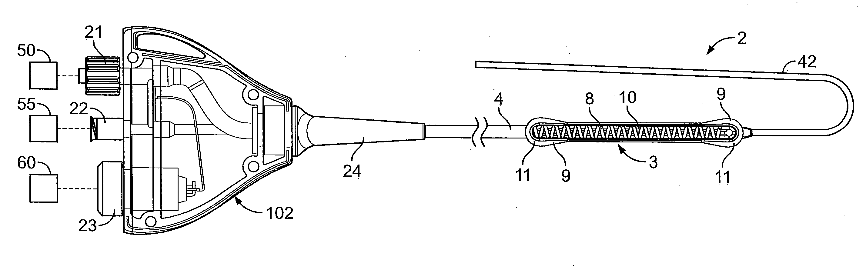
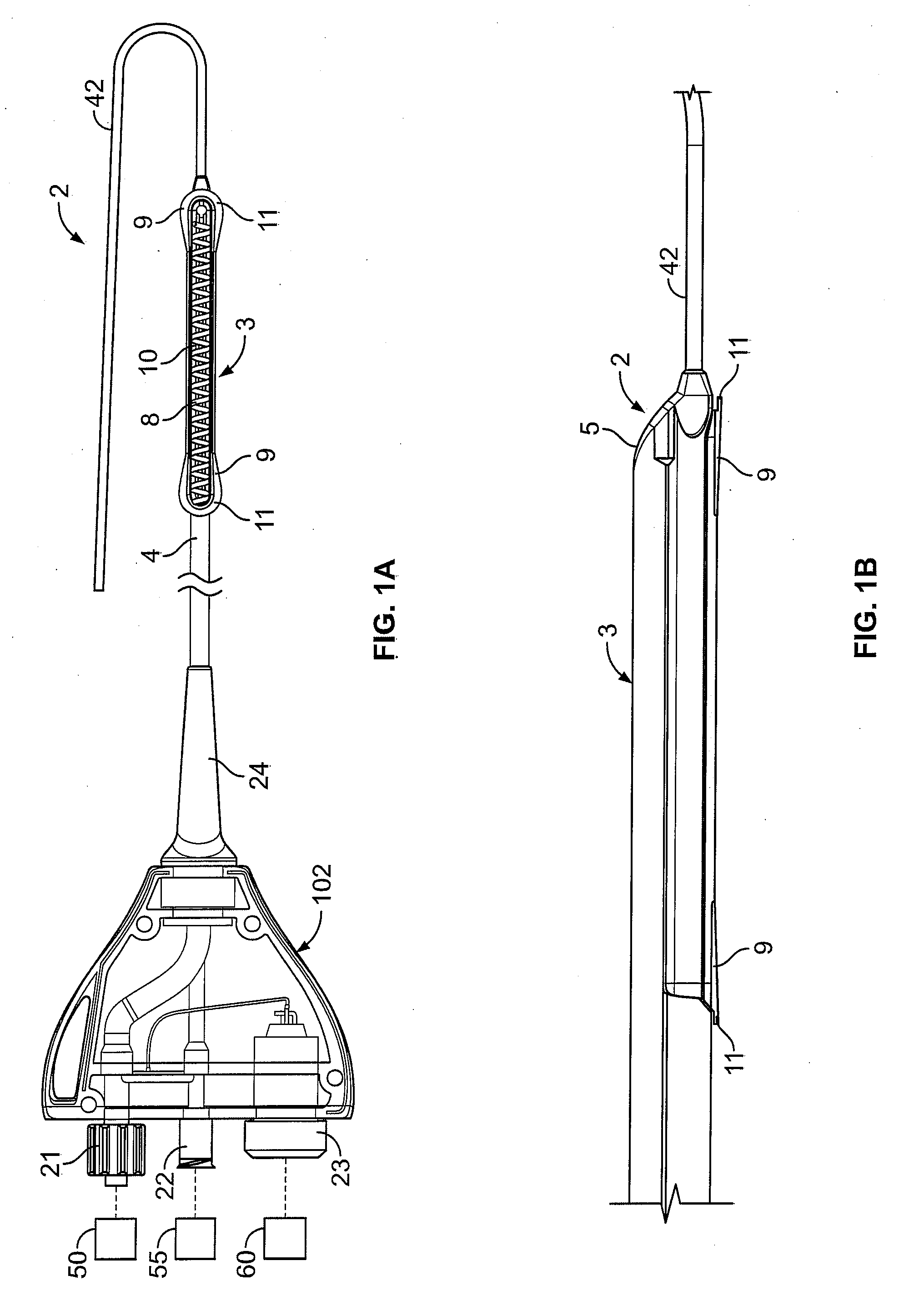
[0058] In light of this framework, a number of exemplary variations of the invention are disclosed—mainly in the context of soft tissue coagulation accomplished through less invasive approaches (e.g., thoracoscopic, arthroscopic, laparoscopic, percutaneous, or other minimally invasive procedures). The integrated vacuum coagulation probe variations disclosed herein produce intimate contact specifically between a soft tissue surface and the electrode(s) or vibration elements used to transmit energy (e.g., radiofrequency or ultrasonic) capable of heating the soft tissue until irreversible injury is achieved making the soft tissue non-viable and unable to propagate electrical impulses, mutate, or reproduce.

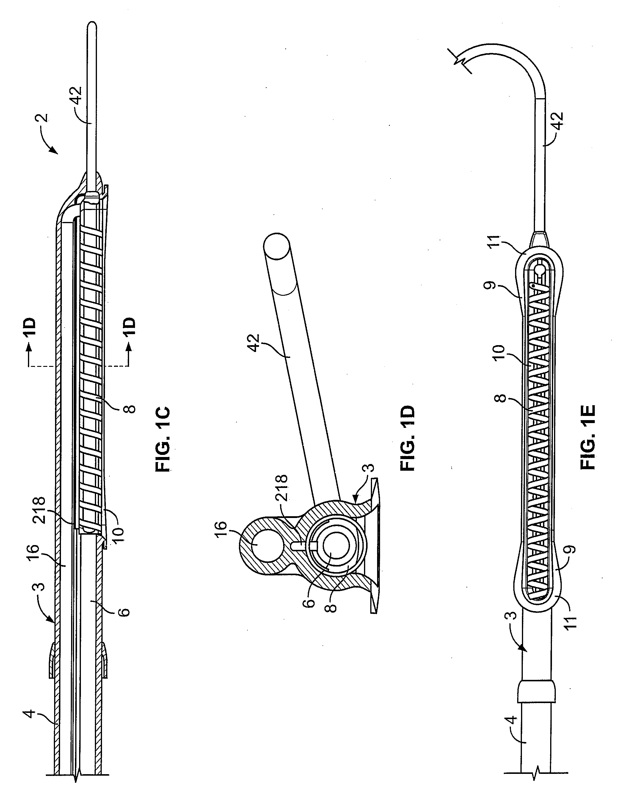
[0059] The integrated vacuum coagulation probe variations may also enable supporting and / or repositioning the soft tissue during coagulation to prevent or minimize shrinking or other change in the shape of the soft tissue associated with heat causing the collagen in the soft tissue t...


PUM


Abstract

A surgical device integrating a suction mechanism with a coagulation mechanism is provided for improving lesion creation capabilities. The device comprises an elongate member having an insulative covering attached about means for coagulating soft tissue. Openings through the covering expose regions of the coagulation-causing elements and are coupled to lumens in the elongate member which are routed to a vacuum source. A fluid source to passively transport fluid along the contacted soft tissue surface may be provided in order to push the maximum temperature deeper into tissue.

Description

CROSS-REFERENCE [0001] This filing is a continuation-in-part of co-pending U.S. patent application Ser. No. 11 / 208,465 entitled “Vacuum Coagulation Probes” filed: Aug. 18, 2005 which is hereby incorporated by reference in its entirety.BACKGROUND [0002] Atrial fibrillation surgery involving radiofrequency, D.C., microwave, or other thermal ablation of atrial tissue has a limitation in that tissue contact throughout the length of the electrode(s) is / are not consistent causing variability in the transmission of energy throughout the target length of ablated / coagulated tissue. This produces gaps of viable tissue that promote propagation of wavelets that sustain atrial fibrillation, or produce atrial flutter, atrial tachycardia, or other arrhythmia substrate. [0003] Another influence in the inability of existing thermal ablation probes to create complete curvilinear, transmural lesions is the presence of convective cooling on the opposite surface of the atrium producing a heat sink that ...

Claims


Application Information

Patent Timeline
Patent Type & Authority Applications(United States)
IPC IPC(8): A61B18/14A61B18/00
CPCA61B18/148A61B18/1492A61B2018/00029A61B2018/00083A61B2018/00291A61B2018/00351A61B2018/1437A61B2018/1472A61B2018/1497A61B2218/002A61B2218/007A61B2018/00577A61B2018/00589A61B2018/1435
Inventor FLEISCHMAN, SIDNEY D.WHAYNE, JAMES G.ROGERS, EARL W.
Owner ATRICURE
Why Eureka
Try Eureka