Administration of dipeptidyl peptidase inhibitors
a technology of dipeptidyl peptidase and inhibitor, applied in the field of medicine, can solve the problems that the membrane-bound proteases active at the site of angiogenesis have not yet been identified, and achieve the effect of increasing the blood supply to the affected tissue and increasing the blood supply
- Summary
- Abstract
- Description
- Claims
- Application Information
AI Technical Summary
Benefits of technology
Problems solved by technology
Method used
Image
Examples
example 1
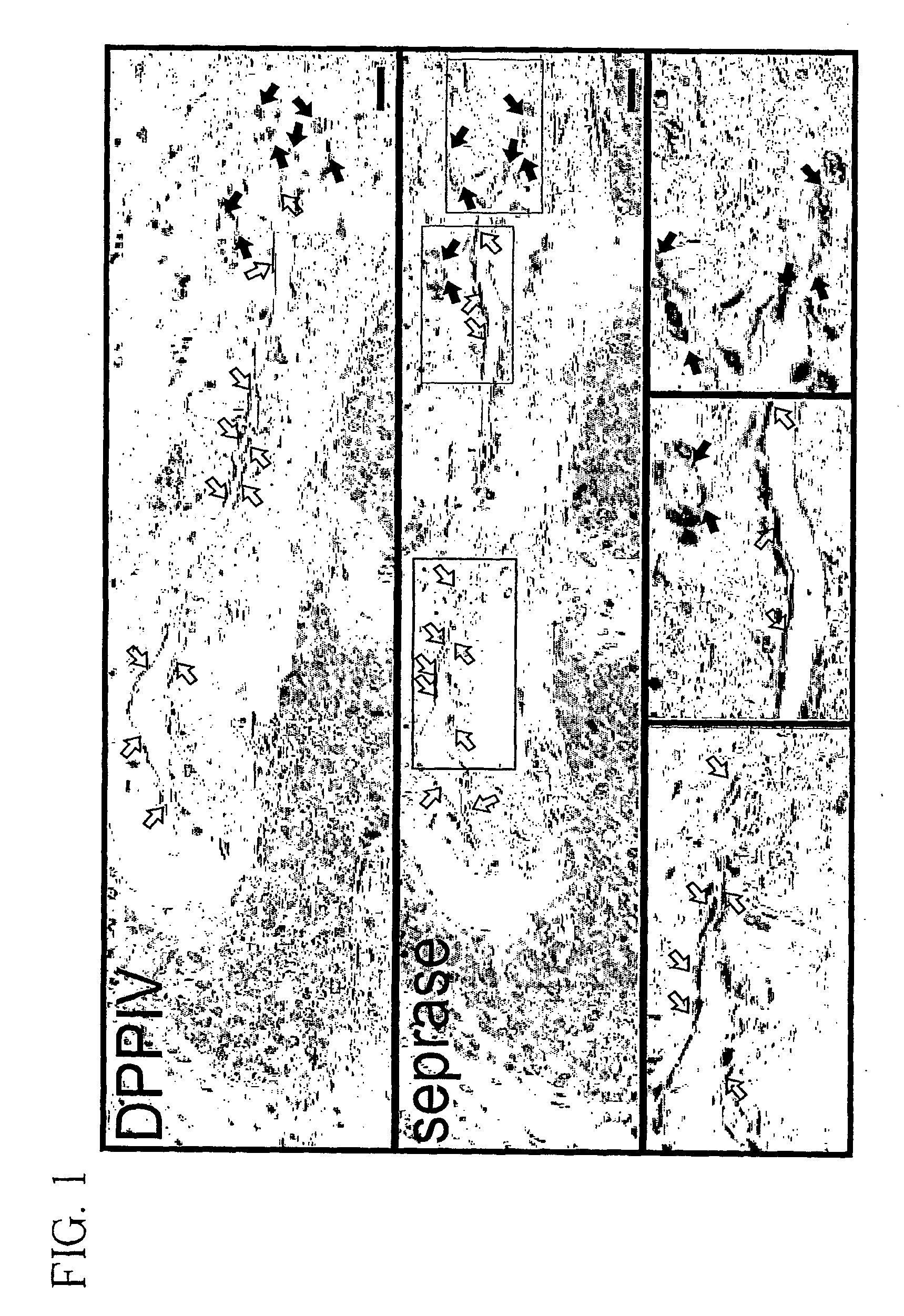
Cytoimmunohistochemical Staining for Seprase and DPPIV in Endothelial Cells Sprouting Vessels and in Normal Tissue.
[0080] To investigate the expression of seprase and DPPIV during angiogenesis, human malignant breast carcinoma tissue or adjacent normal skin were stained with antibodies specific for either seprase or DPPIV. Both seprase and DPPIV were abundantly expressed on the endothelial cells of sprouting vessels (FIG. 1, solid arrows) but were not detectable in other tumor vessels (FIG. 1, open arrows) or in adjacent normal skin from the same donor. These findings indicate that only sprouting sites of blood vessels involved in tumor angiogenesis have enhanced expression of seprase and DPPIV. Consistent with this result, expression of seprase and DPPIV on cultured endothelial cells can be induced by means or factors that enhance cell migration and vessel sprouting (see below).
example 2
Cytoimmunohistochemical Staining for Seprase and DPPIV in Human Primary Cell Culture Monolayers of Different Cell Densities.
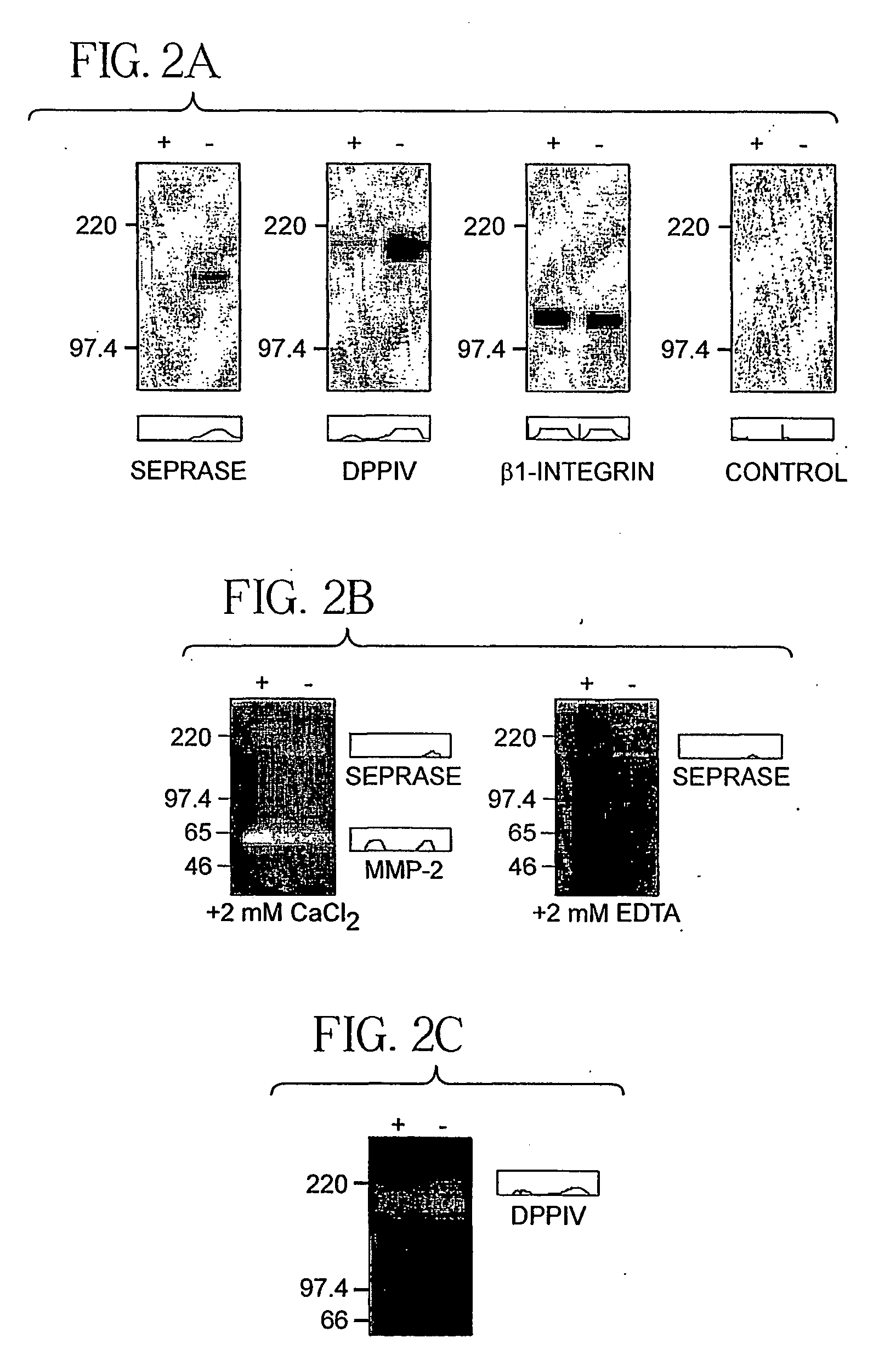
[0081] Endothelial cells of sprouting vessels are migratory and exhibit a lack contact inhibition (Pepper et al., 1993). Monolayer cultures of human umbilical vein endothelial cells (HUVEC) can be induced to migrate by wounding or passage to low cell density (Pepper et al., 1996). This assay was used to examine the expression of seprase and DPPIV in migratory endothelial cells. The confluent HUVEC monolayers were found to contain low levels of seprase and DPPIV and their proteolytic activities were also low (FIG. 2a-c). Passage of monolayers into a sparse culture within 24 hours induced the expression of functional seprase protein; it also caused an increase in DPPIV protein and their proteolytic activities (FIG. 2a-c).
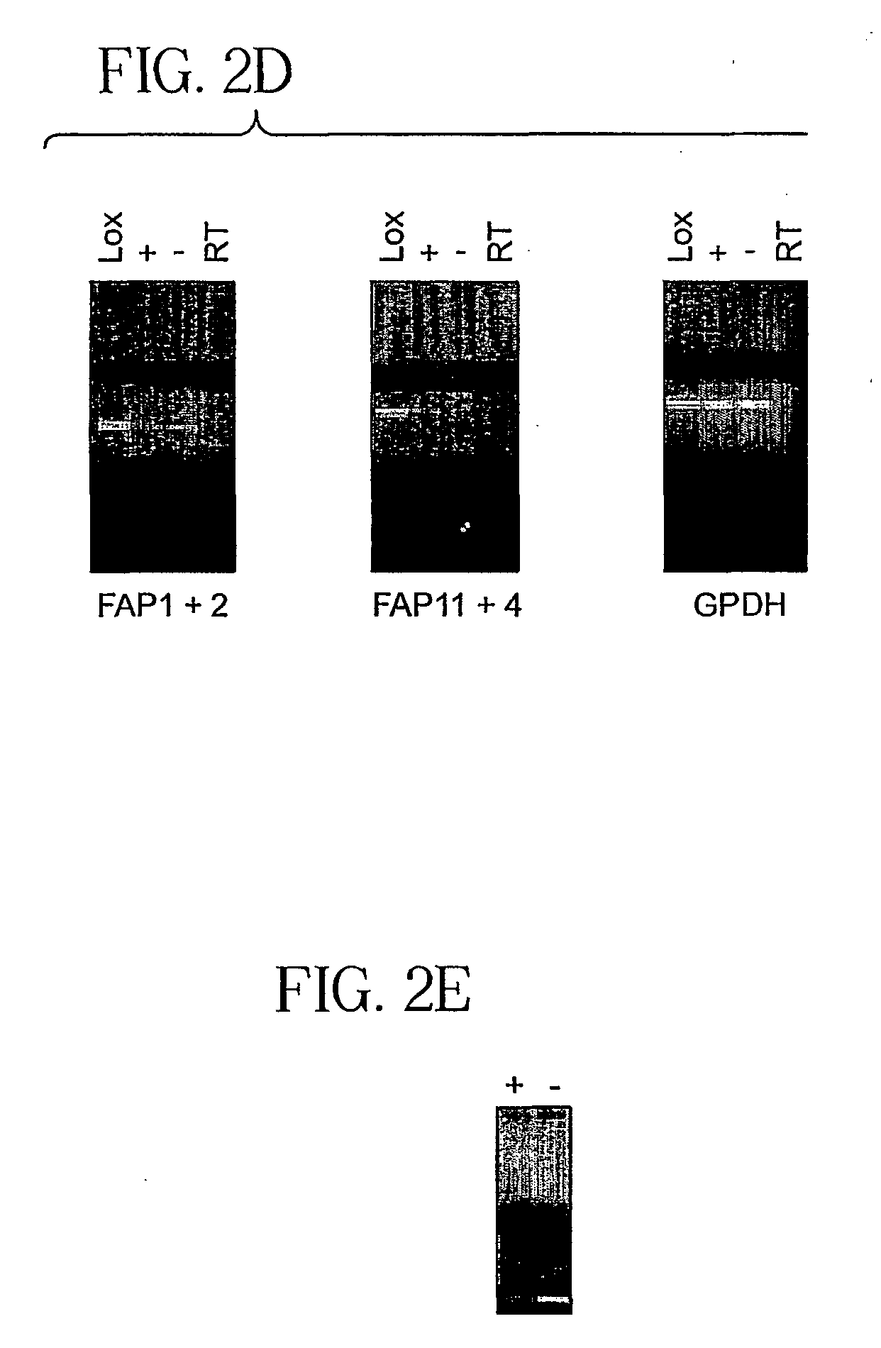
[0082] Both HUVEC cultures had detectable seprase and DPPIV mRNA (FIG. 2d-e). As β1-integrin (Bloch et al., 1997) and membrane-bound MMP-2 (H...
example 3
Expression of Seprase / DPPIV and β1-Integrins During Wound-Induced Endothelial Migration.
[0083] The relative expression of seprase / DPPIV and β1-integrins during wound-induced endothelial migration was examined by double-labeled immunofluoresence. The monolayer wound model consists of a 300 μm width wound on the HUVEC monolayer; migratory activity of HUVEC was visible one hour after wounding (FIG. 2f-g). Expression of p1-integrins was high in endothelial cells at both the wound edge (wounded) and in the monolayer (stationary); in contrast, seprase and DPPIV expression was restricted to migratory cells at the wound edge (FIG. 2f-g, arrows).
[0084] In addition, seprase and DPPIV expression was found on invadopodia (FIG. 2f-g, arrows) and on the perinuclear region (Golgi apparatus) of migratory cells but not in confluent cells. In phase contrast images (FIG. 2f-g), β1-integrins were distributed widely on the surface of migratory cells (FIG. 2f-g, solid arrows) and particularly concentr...
PUM
| Property | Measurement | Unit |
|---|---|---|
| Dimensionless property | aaaaa | aaaaa |
| Dimensionless property | aaaaa | aaaaa |
| Weight | aaaaa | aaaaa |
Abstract
Description
Claims
Application Information
 - R&D
- Intellectual Property
- Life Sciences
- Materials
- Tech Scout
- Unparalleled Data Quality
- Higher Quality Content
- 60% Fewer Hallucinations
Browse by: Latest US Patents, China's latest patents, Technical Efficacy Thesaurus, Application Domain, Technology Topic, Popular Technical Reports.
© 2025 PatSnap. All rights reserved.Legal|Privacy policy|Modern Slavery Act Transparency Statement|Sitemap|About US| Contact US: help@patsnap.com


