Method for localizing a medical instrument introduced into the body of an examination object

a medical instrument and body technology, applied in the field of medical instruments localization, can solve the problems of high radiation dose, patient exposure to considerable x-ray dose, and inability to obtain three-dimensional information about the position, and achieve the effect of reducing costs

Inactive Publication Date: 2007-03-22
SIEMENS HEALTHCARE GMBH
View PDF10 Cites 57 Cited by
  • Summary
  • Abstract
  • Description
  • Claims
  • Application Information

AI Technical Summary

Benefits of technology

[0040] The use of special catheters is not necessary in the method according to the invention, but all electrophysiology catheters, for example, can be detected, as a result of which lower costs are incurred and, moreover, the locating of almost any number of catheters is possible.
[0041] A medical instrument comprising more than one electrode can be used for calibrating. Such a calibration offers the advantage that an absolute scaling of the voltage is obtained. For this purpose, a first electrode, then a second electrode are arranged, for example, in a front area of a catheter at a specified distance of, for example, 3 cm. In a non-flexible catheter, the distance is known so an absolute value of the voltage can thus be derived.

Problems solved by technology

However, a problem with localization by means of an X-ray system is that as a rule no three-dimensional information can be obtained about the position of the catheter, only projection images without depth information.
Furthermore, the patient is often exposed to a considerable X-ray dose.
To achieve this, medical instruments such as catheters have to be navigated in the three-dimensional cavities of the heart, while on the other hand the interventions are often prolonged and in the case of X-ray localization the long duration means that high radiation doses have to be applied.
In contrast to methods which are based on electromagnetic locating, no special and thus expensive catheters are required for this.
In the method of measuring the resistance of the body, there is, however, the problem that in reality the voltage drop does not take place in a linear manner but depends on the individual geometry and conductivity distribution in the body.
Localization which is achieved through measurement of the resistance of the body as outlined above is therefore not exact with deviations in the heart, for example, of several millimeters, which can give rise to problems for examinations or treatments.
In addition, a total of at least six additional electrodes have to be stuck onto the patient, which increases the time expended on conducting the examination.

Method used


Image

  • Method for localizing a medical instrument introduced into the body of an examination object
  • Method for localizing a medical instrument introduced into the body of an examination object
  • Method for localizing a medical instrument introduced into the body of an examination object

Examples


Embodiment Construction

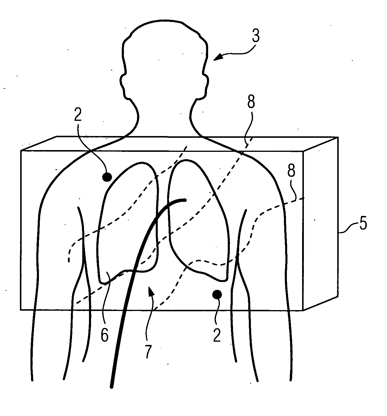
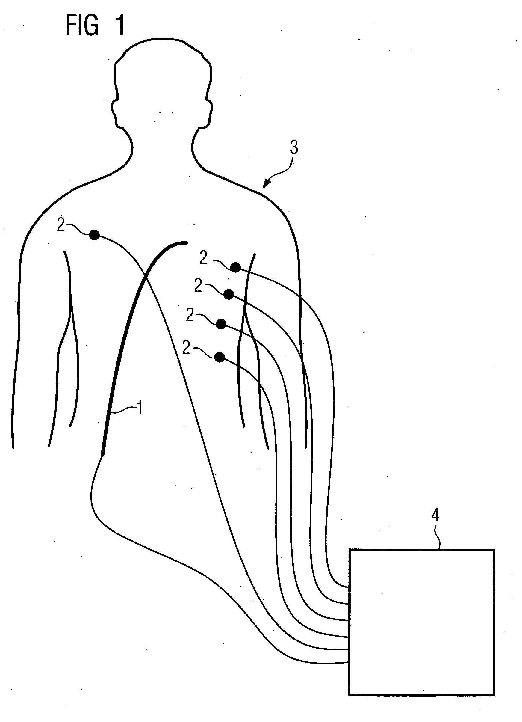
[0050]FIG. 1 shows a diagrammatic sketch of the arrangement of a catheter 1 and of electrodes 2 in a method according to the invention. The catheter 1 is introduced into the body of a patient 3 in order to be used there within the scope of an interventional procedure. Electrodes 2 are arranged on the patient 3, in this case five different skin electrodes. The electrodes 2 are arranged such that they do not all lie in one plane. The electrodes 2 and an electrode, not shown, of the catheter 1 are connected to a device 4 for generating and detecting signals, via which device voltages and / or currents can be applied and measured. In addition, by means of an imaging medical examination device, not shown here, for example for conducting a rotational angiography, three-dimensional image data of a region of interest of the body of the examination object is produced.

[0051]FIG. 2 shows a sketch of an image-recording volume 5 of the patient 3 comprising different tissue types. Besides the five...


PUM


Abstract

A method for localizing a medical instrument, in particular a catheter or guide wire, introduced into the body of an examination object, comprising the steps: arrangement of at least four electrodes on the body of the examination object, recording of three-dimensional image data of a body region of the examination object that is of interest by means of an imaging medical examination device, determination of a model of the electrical conductivity of the examination object as a function of the recorded three-dimensional image data, determination of the electric field distribution when a voltage and/or current is/are applied for at least three different electrode pairs formed from the four electrodes as a function of the conductivity model and the spatial positions of the electrodes, not all the electrodes lying in one plane, application of a voltage and/or a current at each electrode pair and determination of a voltage value at an electrode of the medical instrument for the at least three electrode pairs and determination of the spatial position of the medical instrument as the point of intersection of the equipotential surfaces assigned to the at least three voltage values at the electrode of the medical instrument.

Description

CROSS REFERENCE TO RELATED APPLICATIONS [0001] This application claims priority of German application No. 10 2005 045 093.8 filed Sep. 21, 2005, which is incorporated by reference herein in its entirety. FIELD OF THE INVENTION [0002] The invention relates to a method for localizing a medical instrument, in particular a catheter or guide wire, introduced into the body of an examination object. BACKGROUND OF THE INVENTION [0003] Minimally invasive interventions in the human body or even in animals are carried out as interventional methods with the aid of medical instruments such as for example catheters or guide wires. The area in which the introduced catheter and / or the introduced guide wire is / are located cannot be viewed directly by the person who is carrying out the examination. Determination of the current spatial position of the medical instrument is, however, necessary in order to convey the instrument optionally to the correct location for a treatment or to obtain information ...

Claims


Application Information

Patent Timeline
Patent Type & Authority Applications(United States)
IPC IPC(8): A61B5/05
CPCA61B5/053A61B5/06A61B2019/5253A61B6/12A61B5/7207A61B5/063A61B2034/2053A61B2562/17
Inventor BOESE, JANSANDKAMP, BERNHARD
Owner SIEMENS HEALTHCARE GMBH
Why Eureka
Try Eureka