Method and apparatus for eliminating abnormal blood flow velocities in a color flow image

Active Publication Date: 2007-04-12
SHENZHEN MINDRAY BIO MEDICAL ELECTRONICS CO LTD
View PDF14 Cites 2 Cited by
  • Summary
  • Abstract
  • Description
  • Claims
  • Application Information

AI Technical Summary

Benefits of technology

[0018] An object of present invention is to provide a method and apparatus for eliminating abnormal blood flow velocities in a color flow im

Problems solved by technology

In that case, it is characterized in that the strength of blood flow in a corresponding resolution cell is relatively high and may keep the same direction duri

Method used


Image

  • Method and apparatus for eliminating abnormal blood flow velocities in a color flow image
  • Method and apparatus for eliminating abnormal blood flow velocities in a color flow image
  • Method and apparatus for eliminating abnormal blood flow velocities in a color flow image

Examples


Embodiment Construction

[0043] The preferred embodiment of the present invention is described thoroughly in conjunction with drawings.

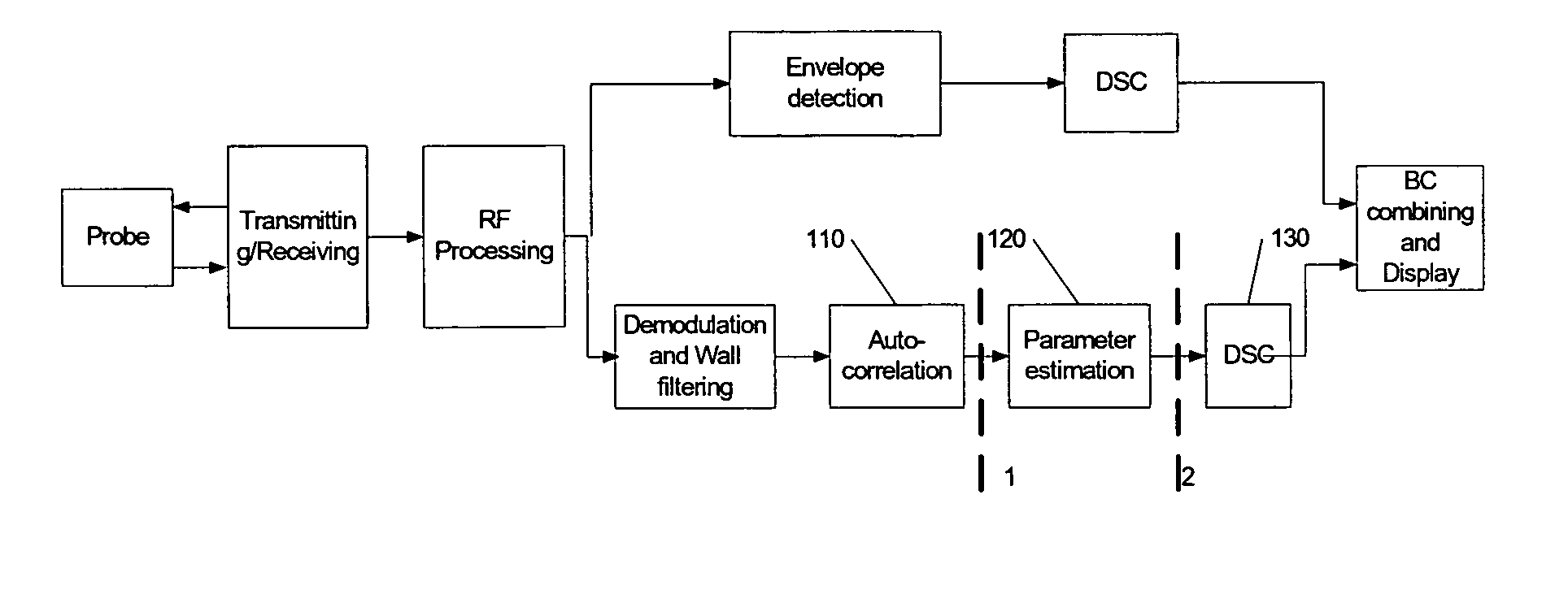
[0044]FIG. 1 is a block diagram showing the structure of a conventional color flow imaging system. As shown in FIG. 1, this system transmits a plurality of coherent pulses towards the interrogation area of a human body via a probe. The backscattered waves of these pulses are received by the system. Then the received signals are divided into two branches after RF processing, which includes amplifying, filtering, analog-digital conversion and beam forming, etc. In one branch, the signals are fed to the processing channel of tissue imaging, and in the other branch, they are provided into the processing channel of color flow imaging. In the processing channel of tissue imaging, the generated data of tissue image are sent to display unit after envelope detection, logarithm compress processing and coordinate conversion, i.e. DSC (Digital Scan Conversion). In the processing channe...


PUM


Abstract

A method for eliminating abnormal blood flow velocities in an ultrasonic imaging system is provided in present invention. The method comprises: receiving signals of blood flow; pre-processing the received signals to generate velocity signs in real time; obtaining a plurality of the velocity signs in a predefined spatial and temporal order, and performing weighted smoothing on the plurality of the velocity signs; determining the direction of blood flow at the pixel to be confirmed according to result of the weighted smoothing. A module for performing above method and a color flow imaging system with such a module are also provided in present invention. The method and apparatus provided by present invention can judge the practical direction of blood flow intelligently by performing spatial-temporal smoothing only on the signs of blood flow velocities, so that the estimation error caused by the system noise,clutter residue, speckle effect and etc. can be corrected.

Description

FIELD OF THE INVENTION [0001] The present invention relates to an ultrasonic diagnostic method and apparatus. Specifically, this invention relates to method and apparatus for ultrasonic color flow imaging, and more particularly relates to a method and apparatus, which is capable of eliminating the pixels indicating abnormal blood flow velocities during the color flow imaging process. BACKGROUND OF THE INVENTION [0002] In ultrasonic diagnostic system, color flow imaging technology is used to measure the moving status and parameters of blood flow or the tissues within a human body, which includes the direction and speed of blood flow, strength of blood flow and the turbulence associated with the blood flow. And the blood flow status will be displayed with pseudo-color on the screen to help doctor make a correct diagnosis. [0003] The basic principle of color flow imaging technology is based on ultrasound Doppler effect to detect the status of the moving subject. When blood flow within ...

Claims


Application Information

Patent Timeline
IPC IPC(8): A61B8/00
CPCA61B8/06
Inventor DONG, YONGQIANG
Owner SHENZHEN MINDRAY BIO MEDICAL ELECTRONICS CO LTD
Why Eureka
Try Eureka