Eureka AIR delivers breakthrough ideas for toughest innovation challenges, trusted by R&D personnel around the world.

A Biomimetic Synthetic Nerve Implant

a biosynthetic nerve and biomimetic technology, applied in the field of biomimetic biosynthetic nerve implants, can solve the problems of long-lasting functional deficits, irreversible injuries to the adult nervous system, and repair strategies that require tissue implantation for bridge repair have not yet matured into clinical practice, so as to improve tissue regeneration capacity, and improve the magnification of regenerated tissue

Inactive Publication Date: 2007-05-03
TEXAS SCOTTISH RITE HOSPITAL FOR CHILDREN
View PDF17 Cites 51 Cited by
  • Summary
  • Abstract
  • Description
  • Claims
  • Application Information

AI Technical Summary

Benefits of technology

[0014] The present disclosure may be described in certain aspects as novel designs for a biosynthetic nerve implant (BNI), which incorporate state of the art biomaterial technology and provide enhanced and directed nerve regeneration both in the peripheral nervous system as well as in the adult injured spinal cord, as compared to other techniques. Advances provided in the disclosure include design of the implant amenable to nanotechnology incorporation, design of a novel scaffold-casting device for medical-grade production, and definition of the cellular and molecular components. The present disclosure includes initial animal evidence demonstrating at the anatomical, behavioral, and electrophysiological levels, that the disclosed BNI better promotes and directs nerve regeneration after sciatic nerve gap repair and dorsal hemisection gap repair of the adult spinal cord.
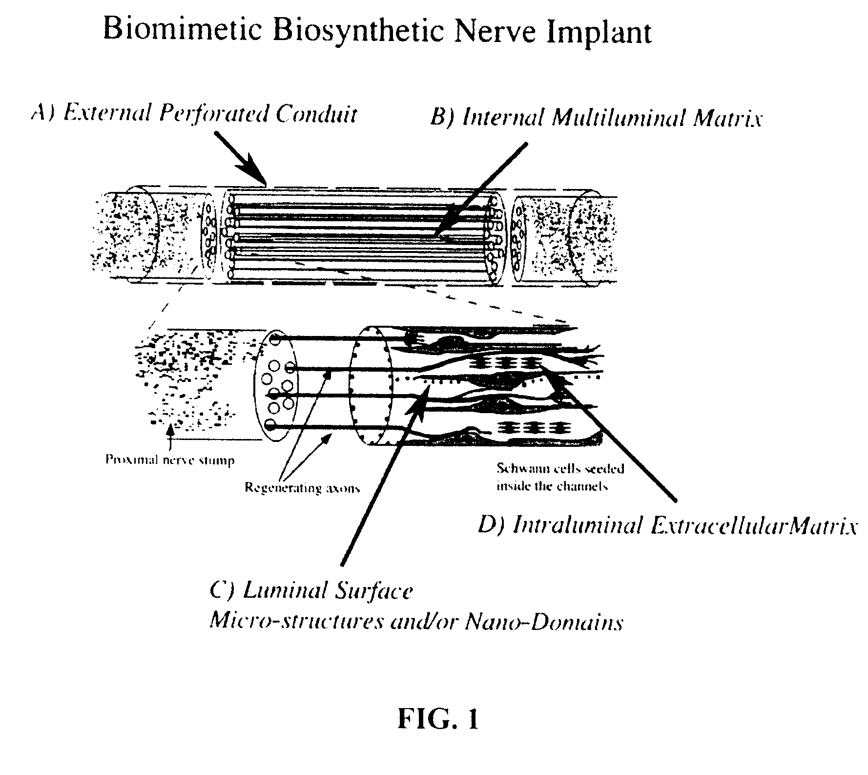
[0015] Preferred embodiments of the disclosure include a biosynthetic nerve scaffold that provides an external, perforated conduit incorporating multiple microchannels within the lumen and including a biodegradable hydrogel matrix. Furthermore, each microchannel may incorporate cells, growth factors and / or extracellular matrix molecules both in the lumen and / or in the walls of the microchannel (FIG. 1). In preferred embodiments, micro- or nanostructures are incorporated in the lumen and / or luminal surface of the microchannels. In some embodiments, a gel-forming matrix is used with the cells in the lumen. When, in certain preferred embodiments, cultured Schwann cells (SCs) are loaded into these channels, the cells are attached to the surface of the microchannels by virtue of a molecularly defined lumen that permits cells to elongate into a three-dimensional viable tissue structure within hours. The early presence and interaction of extracellular matrices components, either natural or synthetic, and / or cellular components, either natural or genetically modified, and the novel incorporation of multiple luminal microdomains within the microchannels, designed for molecular, pharmacological, or electrophysiological manipulations or readings, provide an ideal environment for stimulation and study of the early phases of axon regeneration.
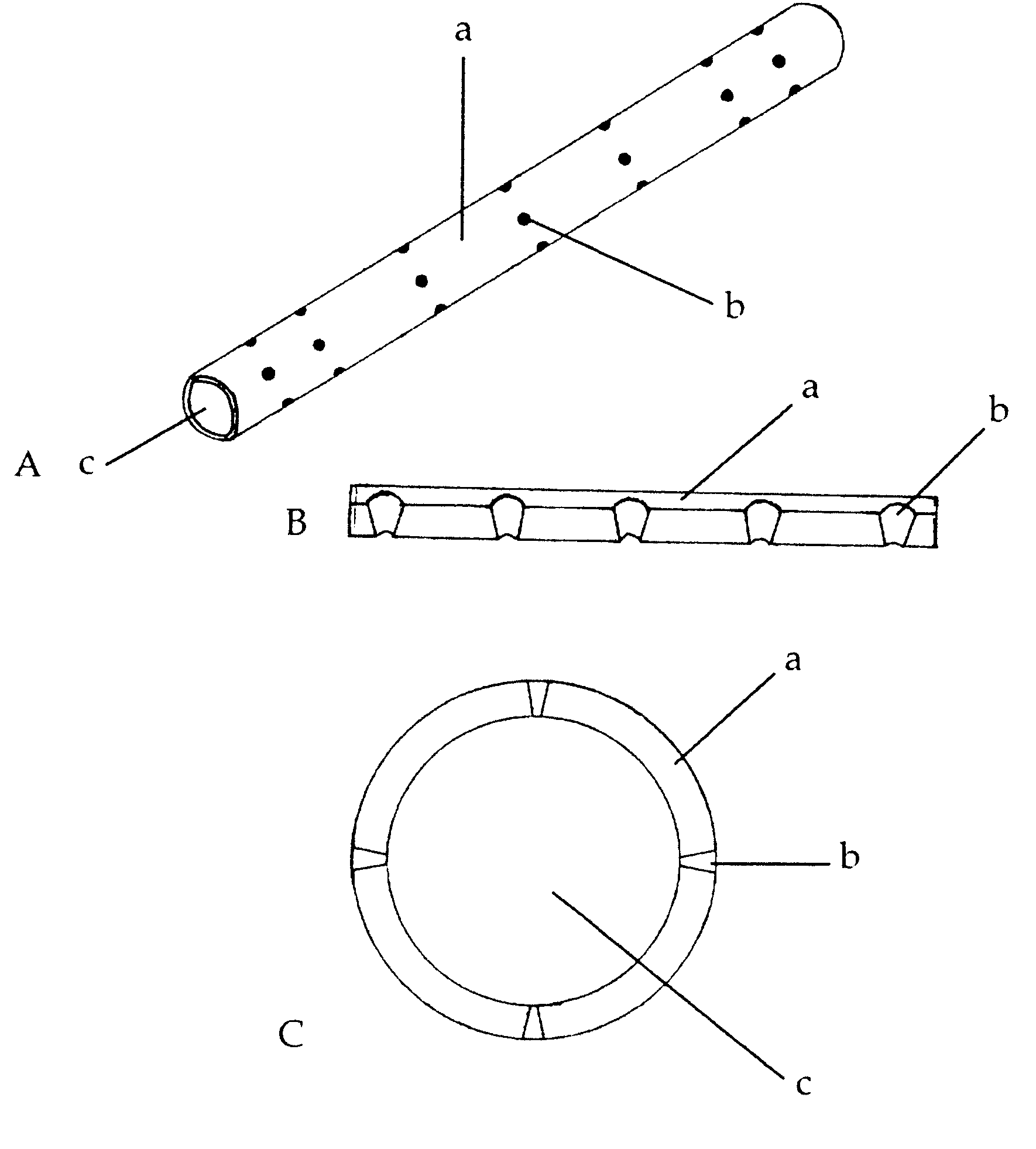
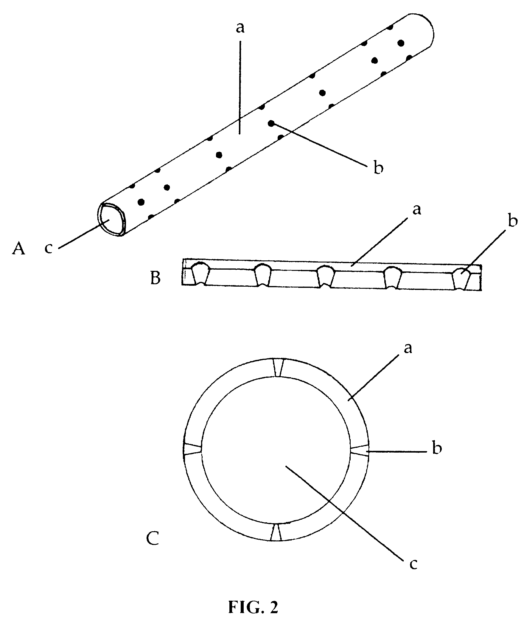
[0016] By forming a permissive substrate for selective neural growth, the initial nerve regeneration events occur faster, and regeneration is accelerated. Although not wishing to be limited to any theory, providing microspheres within the microchannels is contemplated as allowing for the Schwann cells / hydrogel mixture to anchor to the luminal surface of the microchannels. The formed Schwann cell cable is then continuous and somewhat uniform along the microchannels, which is an intuitively better biosynthetic conduit for nerve repair, with a higher potential of improving functional recovery. The present disclosure is not limited to regeneration of nerve cell connections or to nerve tissue of either the central or peripheral nervous systems. The transparent nature of the hydrogel used for casting the nerve scaffold allows for real time observation and dynamic follow up of cellular viability and morphology prior to implantation. Therefore, this disclosure further provides novel methods and compositions for testing the effect(s) of biologically active agents on various cell types.
[0017] The present disclosure also provides a specially designed, three-dimensional scaffold-casting device that is particularly suited for making the tissue scaffolds in a reproducible and sterile manner. The device may function to fabricate a multi-luminal implant scaffold matrix to selectively present molecules or seed cells spatially and temporally in three-dimensions with the required physical, structural, biological and chemical factors to promote cellular development. The disclosed devices are suitable for the production and reproduction of bio-engineered 3-D cellular scaffolds to exact specifications and requirements for basic research and clinical applications in tissue bioengineering, allowing for the effective reproduction and repair of various specialized tissue types and organs by directly addressing the highly complex, three-dimensional, cellular architectural morphology.

Problems solved by technology

Injuries to the adult nervous system are irreversible and bear long lasting functional deficits.
Although numerous approaches have been proposed to repair the injured central (brain and spinal cord) and peripheral (sensory ganglia and sensori-motor nerves) nervous system, repair strategies that require tissue implantation for bridge repairs have not matured yet into clinical practice.
Nerve gaps from segmental tissue loss are routinely repaired by transplanting autogenous nerve grafts; however, this currently accepted “gold-standard” technique results in disappointingly poor (0-67%) functional recovery at the expense of normal donor nerves.
Harvesting of nerve grafts results in co-morbidity that includes scarring, loss of sensation, and possible formation of painful neuroma.
As functional recovery in peripheral nerve reconstruction is poor, clearly, an alternative method for bridging nerve gaps is needed.
Substantial nerve regeneration, however, has never been reported in the reconstruction of human major nerves using silicone tubing.
Despite the fact that the peripheral nerve has an excellent capability of regenerating after a lesion, the main problem is its lack of superior functional recovery compared to autologous nerve repair.
However, there are several limitations.
The manufacture of nerve conduit is rather complicated, it is time consuming, and in most cases requires the use of solvents toxic to the cells.
The dynamic seeding of Schwann cells requires special equipment, involves multiple steps, and the procedure for loading of cells alone can take several hours.
In addition, the material for the conduit is not transparent, and thus not suitable for real time observation and dynamic follow up of cellular and / or tissue morphology and viability.
Thus, despite the recent progress in the engineering of biosynthetic nerve prosthesis, no current design closely resembles the natural morphology of multiple fascicular compartments in the peripheral nerve.
One drawback of current methods of multiluminal nerve repair is that they require rather complicated fabrication techniques.
Several problems still limit the effectiveness of organ bioengineering, and in particular the production of a biomimetic implant.
Unfortunately, the variable availability and degradation of ECM limits cellular growth within the microchannels and thus, their capacity to provide a uniform cellular scaffold for cell growth.
Biodegradable polymers have been used in the surgical repair of peripheral nerves, but their potential for use in the central nervous system has not been exploited adequately.

Method used


Image

  • A Biomimetic Synthetic Nerve Implant
  • A Biomimetic Synthetic Nerve Implant
  • A Biomimetic Synthetic Nerve Implant

Examples


example 1

[0046] Sciatic nerve repair

[0047] Preclinical data on animal models was obtained to evaluate surgical morbidity, immunogenicity, and cellularity of the implants. Using the sciatic nerve gap repair model, two separate cohorts of rats repaired with either seven or fourteen multi-luminal BNIs were examined and compared to animals repaired with empty tubes, tubes filled with collagen, or autologous grafts. Some of the animals were implanted with PTFE Micro-Renathane® tubing that included conical perforations.

[0048] As expected, the recovered implant showed a nerve cable 10 weeks after implantation (FIG. 4). The benefit of the perforations to the polyurethane Micro-Renathane® tubing is also illustrated in FIG. 4. In sharp contrast to the single nerve cable that characterizes the autograft (FIG. 4A) and the simple tubularization repair method (FIG. 4B), multiluminal repair revealed fascicular-like nerve growth throughout the length of the multiluminal BNIs 10-16 weeks after injury (FIG....


PUM


Abstract

A biomimetic biosynthetic nerve implant (BNI) that uses a hydrogel-based, transparent, multi-channel matrix as a 3-D substrate for nerve repair is disclosed. Novel scaffold-casting devices were designed for reproducible fabrication of grafts containing several micro-conduits, and further tested in vivo using a sciatic nerve animal model and repair of the adult hemitransected spinal cord. At 16 weeks post-injury of the sciatic nerve, empty tubes formed a single nerve cable. In sharp contrast, animals that received the multi-luminal BNI showed multiple nerve cables within the available microchannels, better resembling the multi-fascicular anatomy and ultra structure of the normal nerve. In the injured spinal cord, the BNI loaded with genetically engineered Schwann cells were able to demonstrate survival of the grafted cells inside the BNI, and robust axonal regeneration through the implant up to 45 days after repair.

Description

CROSS-REFERENCES TO RELATED APPLICATIONS [0001] The present application is a continuation-in-part of PCT / US04 / 38087, filed Nov. 5, 2004, designating the United States of America and published in English, which claims the benefit of U.S. Provisional Application No. 60 / 517,572, filed Nov. 5, 2003. Each of the above-identified applications is hereby incorporated by reference in its entirety for all purposes.STATEMENT REGARDING FEDERALLY SPONSORED RESEARCH OR DEVELOPMENT [0002] Not applicable. REFERENCE TO A “Microfiche Appendix”[0003] Not applicable. BACKGROUND OF THE INVENTION [0004] 1. Field of the Invention [0005] The present disclosure relates to biomimetic biosynthetic nerve implants for nerve repair, for example spinal cord injury repair. [0006] 2. Description of Related Art [0007] Injuries to the adult nervous system are irreversible and bear long lasting functional deficits. The total costs for the first year of care of paraplegic and quadriplegic patients has been estimated at...

Claims


Application Information

Patent Timeline
Patent Type & Authority Applications(United States)
IPC IPC(8): A61B17/08
CPCA61B17/1128A61L31/005A61L31/129A61L31/145A61L31/146A61L31/16A61L2300/414A61L2430/32A61N1/326
Inventor ROMERO-ORTEGA, MARIOGALVAN-GARCIA, PEDRO
Owner TEXAS SCOTTISH RITE HOSPITAL FOR CHILDREN
Why Eureka
Eureka Blog
Learn More