

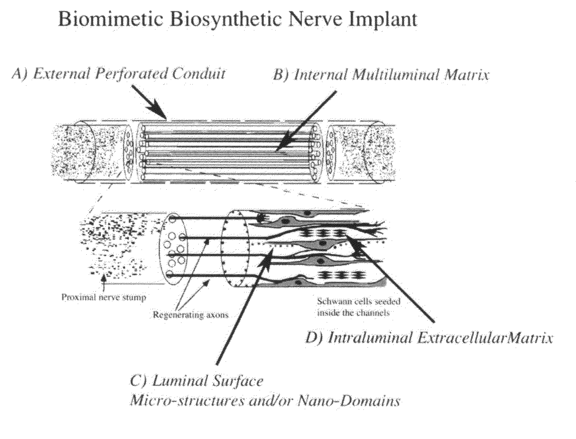
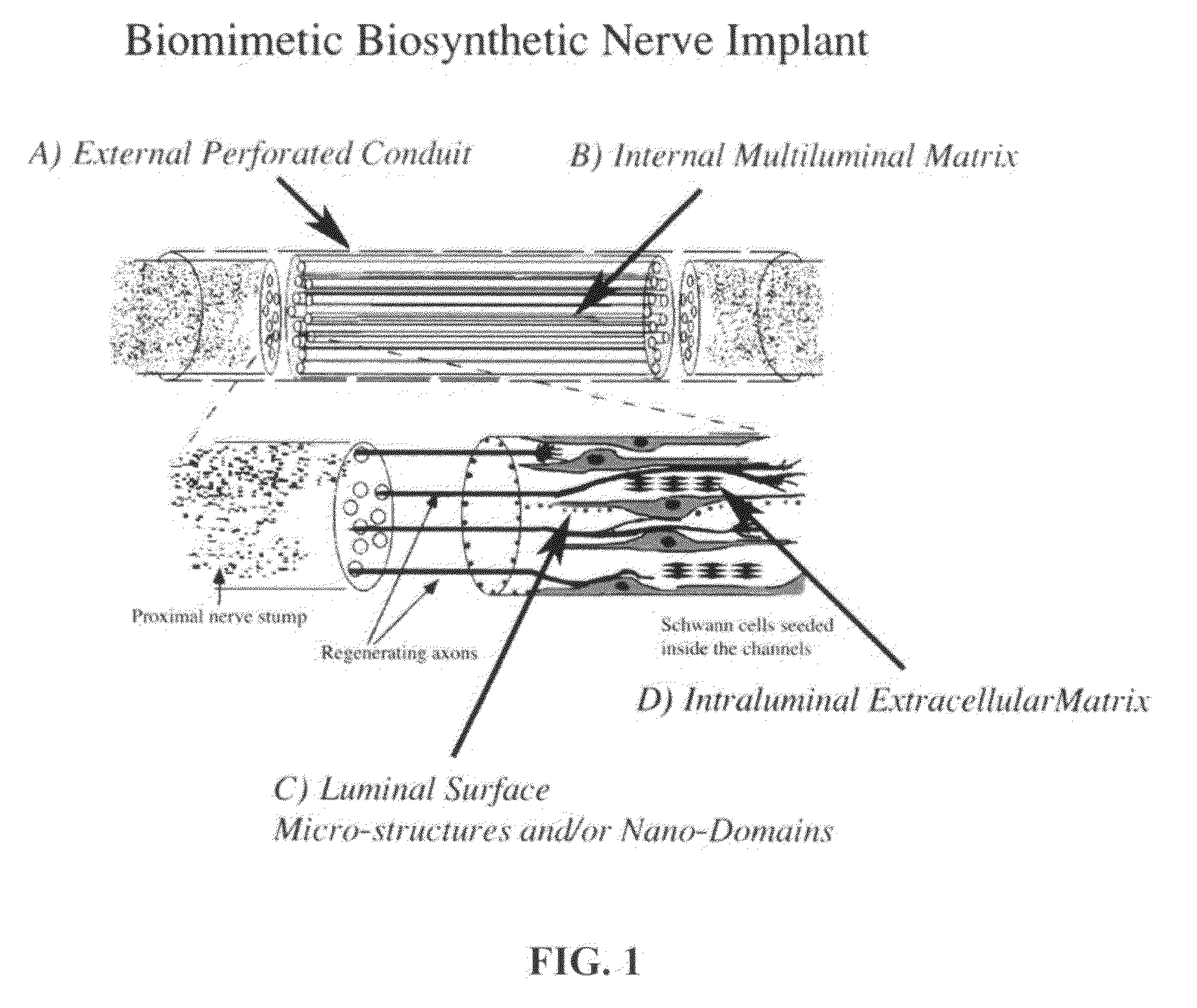
Biomimetic Synthetic Nerve Implant Casting Device
a biosynthetic nerve and casting device technology, applied in the field of biomimetic biosynthetic nerve implants, can solve the problems of long-lasting functional deficits, irreversible injuries to the adult nervous system, and the repair strategies that require tissue implantation for bridge repair have not yet matured into clinical practice, so as to improve tissue regeneration capacity, improve the magnification of regenerated tissue, and achieve the effect of successful guided nerve regeneration
- Summary
- Abstract
- Description
- Claims
- Application Information
AI Technical Summary
Benefits of technology
Problems solved by technology
Method used
Image

Examples
example 1
[0046]Sciatic Nerve Repair
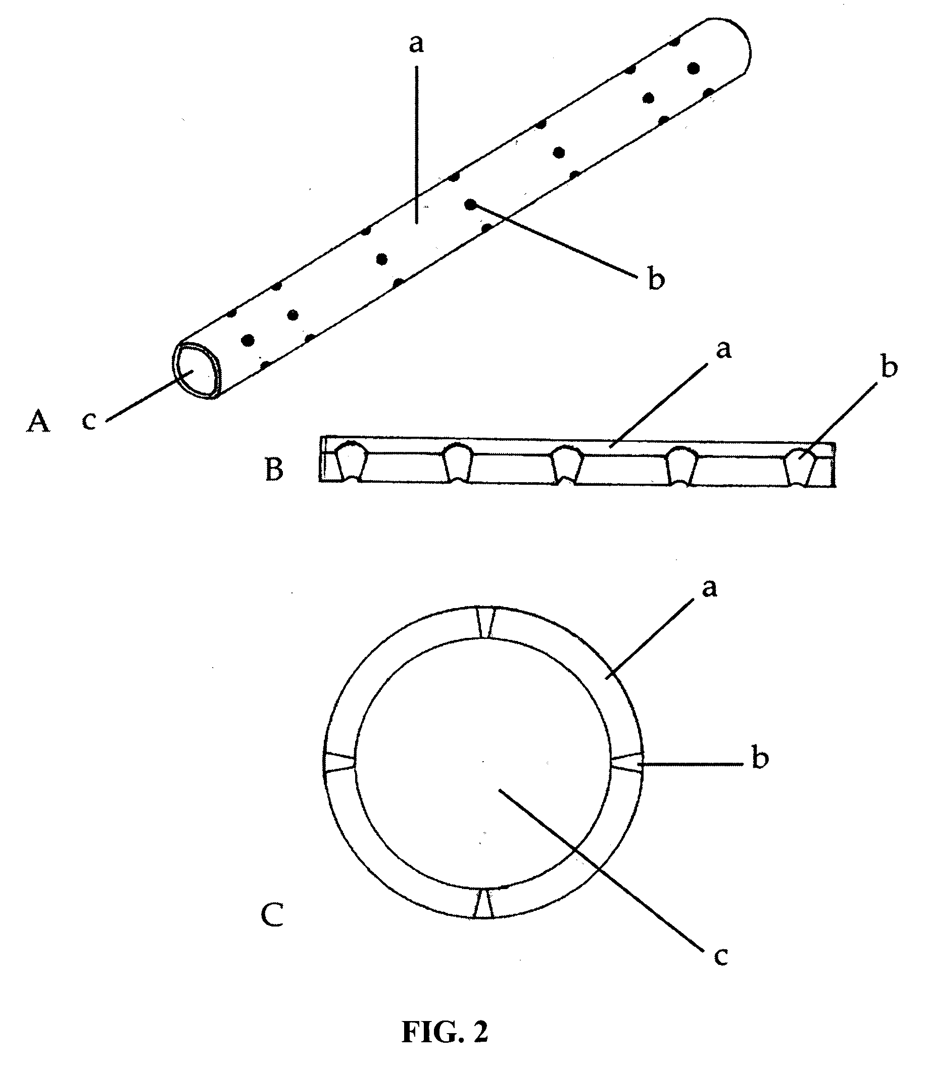
[0047]Preclinical data on animal models was obtained to evaluate surgical morbidity, immunogenicity, and cellularity of the implants. Using the sciatic nerve gap repair model, two separate cohorts of rats repaired with either seven or fourteen multi-luminal BNIs were examined and compared to animals repaired with empty tubes, tubes filled with collagen, or autologous grafts. Some of the animals were implanted with PTFE Micro-Renathane® tubing that included conical perforations.
[0048]As expected, the recovered implant showed a nerve cable 10 weeks after implantation (FIG. 4). The benefit of the perforations to the polyurethane Micro-Renathane® tubing is also illustrated in FIG. 4. In sharp contrast to the single nerve cable that characterizes the autograft (FIG. 4A) and the simple tubularization repair method (FIG. 4B), multiluminal repair revealed fascicular-like nerve growth throughout the length of the multiluminal BNIs 10-16 weeks after injury (FIG. 4C-H...
PUM
| Property | Measurement | Unit |
|---|---|---|
| diameter | aaaaa | aaaaa |
| length | aaaaa | aaaaa |
| diameter | aaaaa | aaaaa |
Abstract
Description
Claims
Application Information