
Interferon assay

a reporter cell and assay technology, applied in the field of methods and reporter cell assays, can solve the problems of difficult diagnosis of diseases, no single laboratory test that can definitively detect lupus, and serious and life-threatening diseases

Inactive Publication Date: 2007-05-24
NEW YORK SOC FOR THE RUPTURED & CRIPPLED MAINTAINING THE HOSPITAL FOR SPECIAL SURGERY
View PDF3 Cites 9 Cited by
  • Summary
  • Abstract
  • Description
  • Claims
  • Application Information

AI Technical Summary

Benefits of technology

The present invention provides methods for detecting mediators that stimulate expression of interferon-inducible genes in a subject, diagnosing systemic lupus erythematosus (SLE) in a subject, monitoring a subject with SLE, and assessing the efficacy of a test compound in treating SLE. These methods involve contacting interferon responsive cells in vitro with a body fluid sample obtained from the subject and detecting the expression level of interferon-inducible genes. The methods can help in identifying individuals with SLE and monitoring their disease progression, as well as assessing the efficacy of a treatment.

Problems solved by technology

In some patients, SLE may be a mild disease, however, in other patients is may be a serious and life-threatening disease.
However, there may be stages of the disease when few symptoms are evident, and patients with SLE may not necessarily exhibit identical symptoms.
In view of the complexities of autoimmune disease symptoms in general, and for lupus in particular, these diseases are difficult to diagnose.
To date there is no single laboratory test that can definitively detect lupus.
The immuno-fluorescent antinuclear antibody (ANA) test is more sensitive, however, positive results are inconclusive because they may be indicative of other diseases.
Skin and kidney biopsies may also be performed in an attempt to diagnose SLE, but these are invasive, costly and often not definitive.
However, these tests have the drawback of detecting only specifically known genes and certain methods require the use of fresh patient cells.
Additionally, there are currently no good biomarkers for monitoring disease progression in patients with SLE.
In some instances the titer of anti-double stranded DNA antibodies can be used to monitor disease state or progression in certain patients; however, this method has limited application and is difficult to interpret.

Method used


Image

  • Interferon assay
  • Interferon assay
  • Interferon assay

Examples


example 1

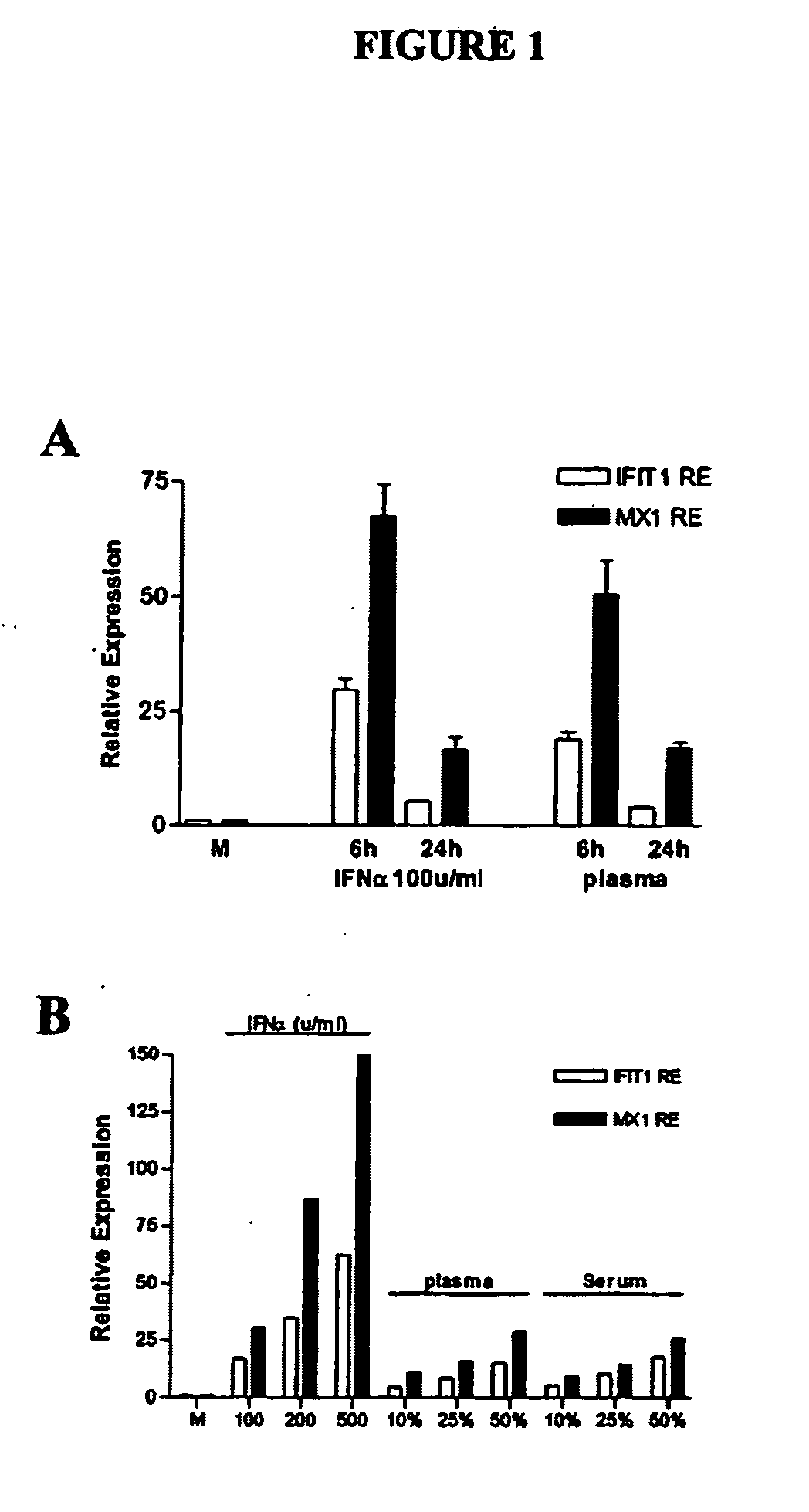
[0097] Induction of IFIG in an in vitro WISH cell system by recombinant human interferon: time course and dose response. To develop an in vitro assay for quantification of IFIG-inducing activity in patient plasma or serum, WISH cell line cells, previously demonstrated to be IFN-responsive (31), were cultured with rhIFNα, rhIFNγ or SLE plasma. Expression of five genes, interferon-induced protein with tetratricopeptide repeats 1 (IFIT1, SEQ ID NO:15,), interferon-induced protein 44 (IFI44, SEQ ID NO:16, deduced amino acid sequence SEQ ID NO:17), protein kinase, interferon-inducible double stranded RNA dependent (PRKR, SEQ ID NO:18, deduced amino acid sequence SEQ ID NO:19) also known as eukaryotic translation initiation factor 2-alpha kinase 2 (EIF2AK2)), C1orf29 (SEQ ID NO:20, deduced amino acid sequence SEQ ID NO:21) and myxovirus (influenza virus) resistance 1 (MX1, SEQ ID NO:22, deduced amino acid sequence SEQ ID NO:23) was measured in cultured WISH cells. These five genes were pr...

example 2

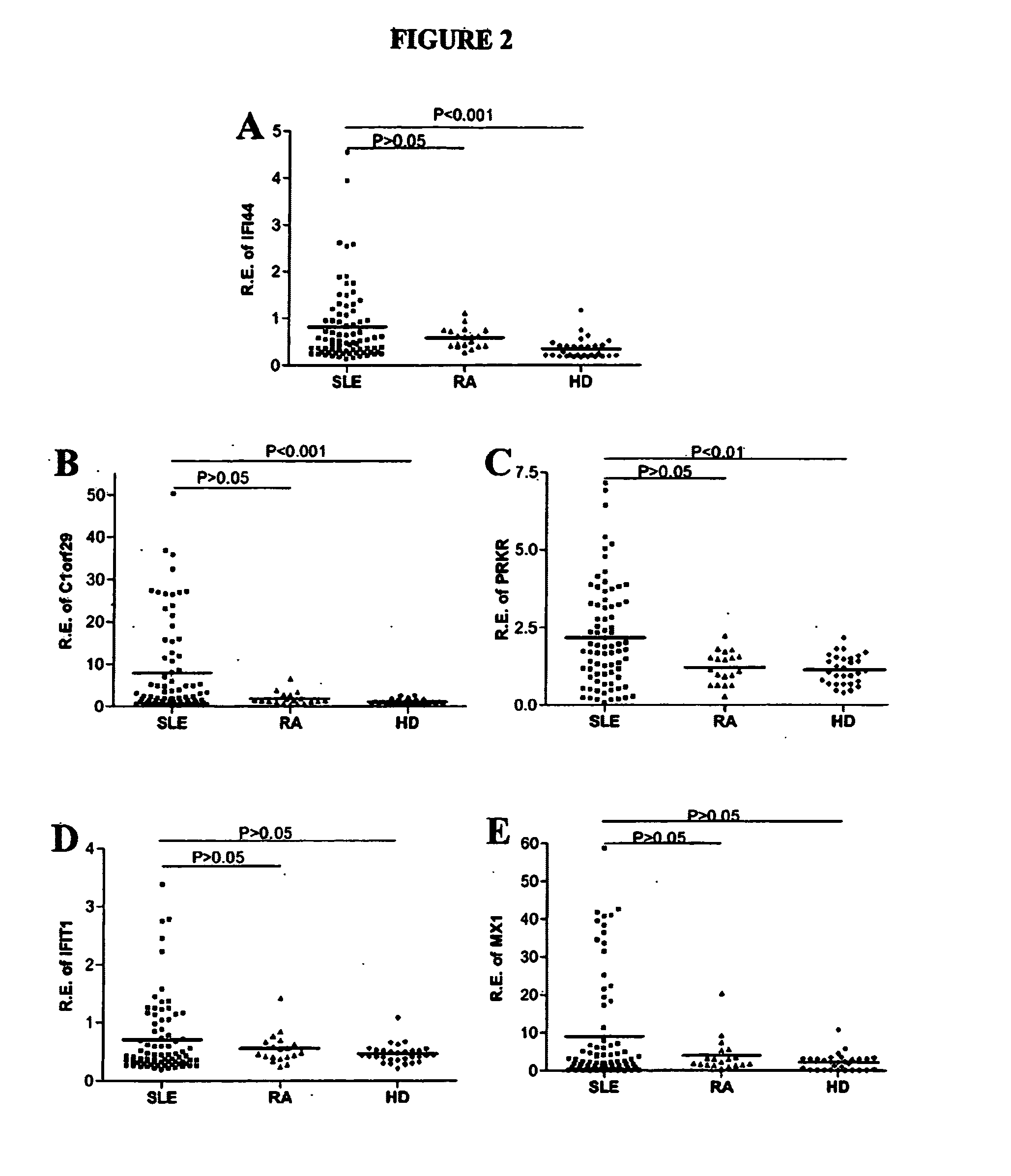
[0101] Correlation of lupus plasma activity with expression of IFIG in SLE PBMC. To assess the level of IFIG-inducing activity in plasmas from a diverse population of SLE patients, as well as in plasmas from disease controls with RA and from healthy subjects, WISH cells were cultured with medium or with plasma from seventy-three SLE patients, nineteen RA patients and thirty healthy donors and IFIG expression was measured by real-time PCR. Five IFNα-inducible genes and one IFNγ-inducible gene were quantified. Plasma samples from patients with SLE (n=73) or RA (n=19), or from healthy donors (n=30), were incubated with WISH cells at a 50% volume / volume concentration for 20 hrs. WISH cells were then lysed and used for RNA isolation, reverse transcription, and amplification by quantitative real-time PCR. Relative expression of five IFNα-induced genes is shown. Mean values for each group are indicated by the horizontal line shown in FIG. 2. P values for differences between SLE patients an...

example 3

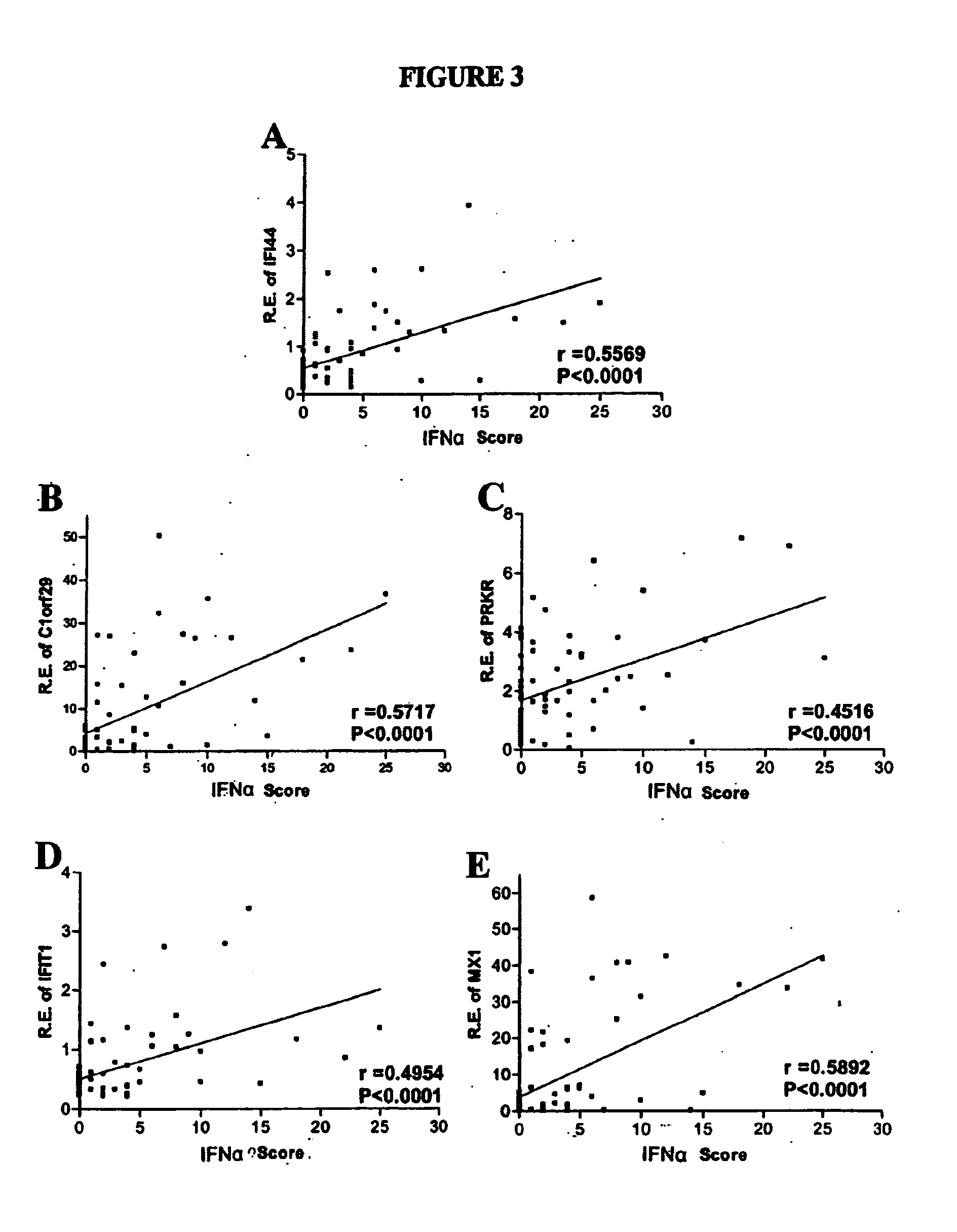
[0103] Correlation of IFNα-inducible genes in WISH cells cultured with SLE plasma with the level of mRNA encoded by IFNα-inducible genes in PBMC cells from patients. The expression of each of these five IFNα-inducible genes in WISH cells cultured with SLE plasma was correlated with the level of mRNA encoded by IFNα-inducible genes in the PBMC of those same patients collected on the same day, as expressed as an IFNα score (FIG. 3). Relative expression of 5 IFNα-induced genes in WISH cells cultured with 50% lupus plasma was plotted against the IFNα score, previously assayed based on real-time PCR analysis of patient PBMC cells. The IFNαscore was calculated based upon previous real-time PCR analysis of expression of three IFNα-inducible genes (IFIT1, IFI44, PRKR) in patient PBMC. The expression of IFIT1, IF44, and PRKR in patient PBMC was described in detail by Kirou et al. 2005 (11). The Spearman rho(r) and p correlation values are shown. The p value for each of the five IFNα-inducibl...


PUM

PropertyMeasurementUnit
temperaturesaaaaaaaaaa

Abstract

The present invention relates to methods and reporter cell assays for determining the ability of a patient sample to induce interferon target gene expression in interferon responsive cells. These methods will be useful for detecting, diagnosing, and monitoring those who have or are at risk of various autoimmune disorders or diseases, including systemic lupus erythematosus (SLE) and Sjogren's syndrome.

Description

CROSS REFERENCE TO RELATED APPLICATIONS [0001] This application claims priority from U.S. Provisional Application Ser. No. 60 / 680,931, filed May 12, 2005, which is hereby incorporated by reference in its entirety.FEDERALLY SPONSORED RESEARCH OR DEVELOPMENT [0002] This invention was made in part in the course of research sponsored by the National Institutes of Health (NIH) Grants AR-050829 and AI-052422, the Alliance for Lupus Research, the Lupus Research Institute, and the Mary Kirkland Center for Lupus Research. The U.S. government may have certain rights in this invention.FIELD OF THE INVENTION [0003] The present invention relates to methods and reporter cell assays for determining the ability of a patient sample to induce interferon target gene expression in interferon responsive cells. BACKGROUND OF THE INVENTION [0004] Prototypical systemic autoimmune diseases include systemic lupus erythematosus (SLE), scleroderma, mixed connective tissue disease, Sjogren's syndrome and other ...

Claims


Application Information

Patent Timeline
Patent Type & Authority Applications(United States)
IPC IPC(8): C12Q1/68G01N33/567
CPCC12Q1/6883G01N33/564G01N2500/10C12Q2600/136C12Q2600/158
Inventor CROW, MARY K.KIROU, KYRIAKOSHUA, JING
Owner NEW YORK SOC FOR THE RUPTURED & CRIPPLED MAINTAINING THE HOSPITAL FOR SPECIAL SURGERY
Learn More