Methods and systems for segmental lung diagnostics

a segmental lung and diagnostic technology, applied in the field of respiratory medicine, can solve the problems of less accurate method for directly determining the extent of hyperinflation, unlikely success of such treatments, etc., and achieve the effect of reducing the risk

Inactive Publication Date: 2007-06-21
PULMONX
View PDF65 Cites 101 Cited by
  • Summary
  • Abstract
  • Description
  • Claims
  • Application Information

AI Technical Summary

Benefits of technology

[0015] Minimally invasive methods, systems and devices are provided for qualitatively and quantitatively assessing the condition and function of individual lung compartments, including the extent of hyperinflation of a lung compartment, compliance of a lung compartment, efficiency of gas exchange within a lung compartment such as the value of oxygen uptake within a lung compartment, the directionality of collateral flow channels between adjacent lung compartments, and the rate or degree of blood flow and / or blood flow / unit of volume within a lung compartment. The methods, systems, and devices generally rely on accessing, isolating, and at least partially occluding a target lung compartment within the lung of a living patient in order to perform the diagnostic protocol. Typically, a lung of the patent is accessed by advancement of a catheter through the tracheobronchial tree to an airway, typically referred to as feeding bronchus, which feeds the target lung compartment. The airway is usually occluded by an expansible occlusion member, typically a balloon on the catheter, and a variety of measurements may be taken with or through the catheter in a manner which presents a minimum risk to the patient.

Problems solved by technology

If flow through the collateral channels allow air to collectively enter the target lung compartment when occluded, the success of such treatments is unlikely.
It will appreciated that this method for directly determining the extent of hyperinflation will usually be less accurate in lung compartments having collateral flow channels which allow airflow into the lung compartment from adjacent lung compartments.

Method used


Image

  • Methods and systems for segmental lung diagnostics
  • Methods and systems for segmental lung diagnostics
  • Methods and systems for segmental lung diagnostics

Examples


Embodiment Construction

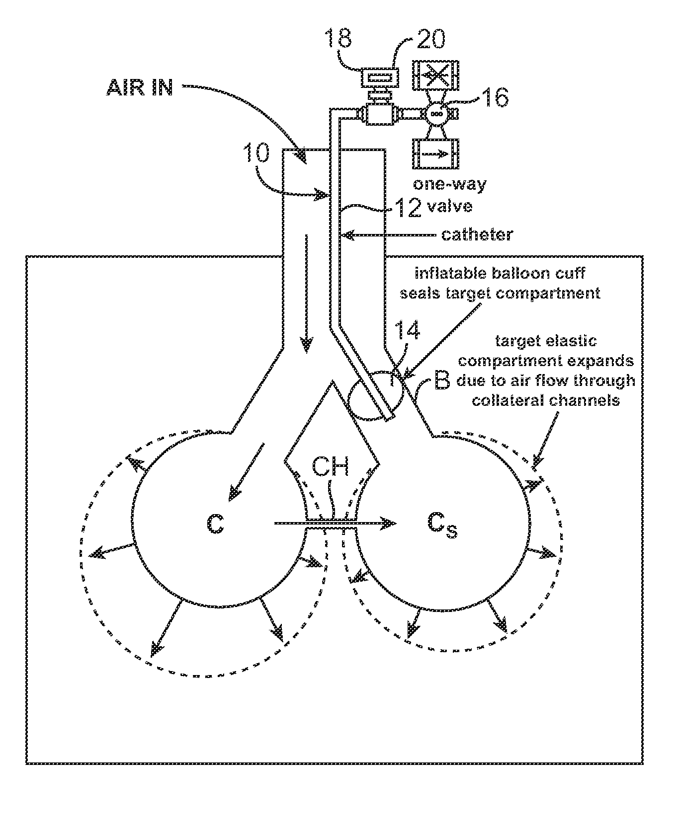
[0042] Minimally invasive methods, systems and devices are provided for qualitatively and quantitatively assessing lung condition and function, particularly in target lung compartments or segments which have been isolated from the remainder of the lung. FIGS. 4A-4D illustrate a system which can be utilized for performing the various diagnostic protocols of the present invention and includes a catheter 10 which may be advanced through a tracheobronchial tree to the feeding bronchus B (upper airway) of the target area Cs, the lung compartment targeted for treatment or isolation. The catheter 10 comprises a shaft 12 having at least one lumen therethrough and an occlusion member 14 mounted near its distal end. The occlusion member 14 of the catheter 10 is adapted to seal the area between the catheter shaft 12 and the bronchial wall such that only a lumen inside the catheter which extends the entire length of the catheter is communicating with the airways distal to the seal. The seal, or...


PUM


Abstract

Minimally invasive systems and methods are provided for diagnosing conditions in target lung compartments. Using catheters capable of isolating the target lung compartments and measuring one or more of collateral ventilation, pressure, flow rate, and volume, conditions such as hyperinflation, compliance, gas exchange including oxygen uptake, directionality of collateral channels, blood flow, and blood flow per unit lung volume may be assessed.

Description

CROSS-REFERENCES TO RELATED APPLICATIONS [0001] This application is a continuation of PCT / US 06 / 27478 (Attorney Docket No. 017534-003010PC) filed Jul. 13, 2006, which claimed the benefit of U.S. Provisional No. 60 / 699,289 (Attorney Docket No. 017534-003000US), filed on Jul. 13, 2005, and is a continuation-in-part of U.S. application Ser. No. 11 / 296,951 (Attorney Docket No. 017534-002820US), filed on Dec. 7, 2005, the full disclosures of which are incorporated herein by reference.BACKGROUND OF THE INVENTION [0002] 1. Field of the Invention [0003] The present invention relates generally to respiratory medicine and more specifically to the field of assessing lung condition and function in isolated lung compartments. [0004] The lungs comprise a plurality of bronchopulmonary compartments, referred to hereinafter as “lung compartments,” which are separated from one another by a double layer of infolded reflections of visceral pleura called the “fissures.” The fissures which separate the l...

Claims


Application Information

Patent Timeline
Patent Type & Authority Applications(United States)
IPC IPC(8): A61B5/08
CPCA61B5/0813A61B5/085A61B5/087A61B5/093
Inventor ALJURI, NIKOLAIVENEGAS, JOSE G.PERKINS, RODNEY C.
Owner PULMONX
Why Eureka
Try Eureka