Fluid sampling, analysis and delivery system

a technology of fluid sampling and analysis, applied in the field of fluid sampling, analysis and delivery system, can solve the problems of foreign body reactions related to scar tissue buildup around the sensor, inability to wide-spread and routine use, repeatability and associated errors of non-invasive blood sampling techniques, etc., to achieve smooth penetration, small volume of blood, and minimal skin irritation

Inactive Publication Date: 2005-10-13
UNIV TECH INT
View PDF9 Cites 64 Cited by
  • Summary
  • Abstract
  • Description
  • Claims
  • Application Information

AI Technical Summary

Benefits of technology

[0009] The principle of semi-invasive blood sampling could be easily related to the operation of a mosquito, which penetrates the skin to collect a very small volume of blood, with minimal amount of skin irritation. Smooth penetration is ensured because of the jagged shape of the maxilla, and blood transport over relatively long distance is made possible by a feeding pump. Anesthetizing saliva provides for a painless penetration, while the minimal irritation is associated with the anticoagulant used [35] (FIG. 3.1a). The electronic cousin of the mosquito, the e-Mosquito™, somewhat differs from the approach utilized by the real mosquito, although the end result, the acquisition of a blood sample, remains the same (FIG. 3.1b).
[0010] The total size of one single e-Mosquito™ may be in the range of a few millimeters and a set of these miniature cells can be applied as a patch on to the skin, similarly to a band-aid. The short microneedle provides completely painless blood sampling. The electrochemical sensors need only a very small blood sample for reliable assessment of blood glucose or other blood parameters, utilizing the collected static blood volume. An antiseptic layer provides minimal risk of infection and skin irritation. The e-Mosquito™ is a single-use device. The device features a real-time monitoring of blood parameters and is capable of sending wirelessly the results either to a remote device with a display (i.e. wrist watch, cell phone, personal digital assistant, etc.) or directly to a medical authority (i.e. hospital, clinic, medical doctor, etc.)
[0011] Taking into account the technological complications related to designing a microelectronic “horizontal drilling” setup to mimic the operation performed by the maxilla of the real mosquito (see FIG. 3.1a), a simple vertical penetration was considered. However, the “horizontal drilling” of the mosquito has several important biological advantages, including the possibility to search for arteries or capillaries, and the capability to directly penetrate a vessel, thus extracting maximal blood volume from it. In the case of the e-Mosquito™, however, both of these advantages can be bypassed by (a) introducing a matrix of single-use e-Mosquito™ cells (FIG. 3.2), with the individual actuation of which a direct vertical hit on a capillary by one of the needles becomes inevitable; and (b) reducing the blood volume needed for reliable analysis.
[0012] The second major difference between an actual mosquito and its “electronic cousin” is the penetrating depth, which in the latter case is about 400 um, thus ensuring the reach of capillaries but not neural endings, thus providing for a painless operation (FIG. 4.1), and also avoiding the need for accompanying anesthetizing liquid during the bite.
[0013] Thirdly, the e-Mosquito™ does not have a feeding pump, but relies on the intricate balance between capillary forces and pressure differences considered during the design process. This intricate balance, combined with the fact that each e-Mosquito™ cell in the matrix is a single-use device, also avoids the use of anticoagulant and alleviates the need to clean individual cells after use, thus keeping the microneedle actuation a sterile one-time event. In other words, each individual e-mosquito cell within the framework of the matrix dies immediately after fulfilling its primary mission, which is to extract a given volume of blood required for reliable stationary blood analysis associated with the particular application of the device. Should a needle from a given cell of the matrix fail to hit a capillary and fulfill its mission, the entire cell is not reused—another cell picks up the mission at a slightly different location.
[0014] In addition, penetration of the skin and microneedle withdrawal in the e-Mosquito™ is performed through an antiseptic layer adhesive to the skin, thus avoiding any potential skin irritations or infections.

Problems solved by technology

However, the impact of non-quantifiable external factors (e.g., variable capillary concentration in the interrogated tissue volume, changeable capillary volumes associated with blood pressure dynamics, dynamic absorption capabilities of the surrounding living tissue, etc.), make the reliability, the repeatability and the associated errors of non-invasive blood sampling techniques inadequate to warrant wide-spread and routine utilization [6].
A variety of electrochemical techniques have been suggested, but the limitations of this approach include foreign-body reactions related to scar tissue buildup around the sensor, and the complexities associated with data transmission from the implant to external data loggers.
In important studies, inconsistently decreasing sensor activity has been found in less than 24 hours, questioning the repeatability of this approach even if the signal transmission issues were to be completely resolved [2, 7-9].
Nevertheless, needle based blood collection is necessary in order to harvest an adequate volume of blood for analysis and electronic reporting, which presents a significant inconvenience for wide groups of patients and medical professionals alike, such as diabetic patients and laboratory nurses.
Many intellectual property instruments (patents and patent applications) have been released on invasive blood sampling [21, 23-29] and on transdermal drug delivery through microneedles [23, 30-32], however these approaches are deficient in automated real-time actuation and control of the microneedle.
These approaches do not present a self-contained, real-time, closed-loop control device, utilizing an integrated microsystem mountable on a disposable adhesive patch, and containing a matrix of individually, but sequentially actuated, single-use elementary cells mounted in a disposable platform such as a self-adhesive patch.

Method used


Image

  • Fluid sampling, analysis and delivery system
  • Fluid sampling, analysis and delivery system
  • Fluid sampling, analysis and delivery system

Examples


Embodiment Construction

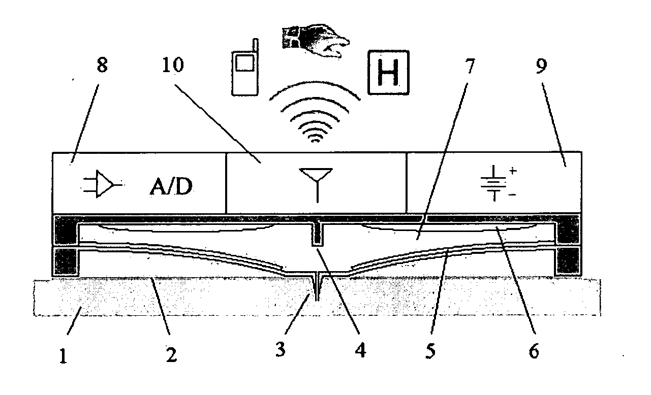
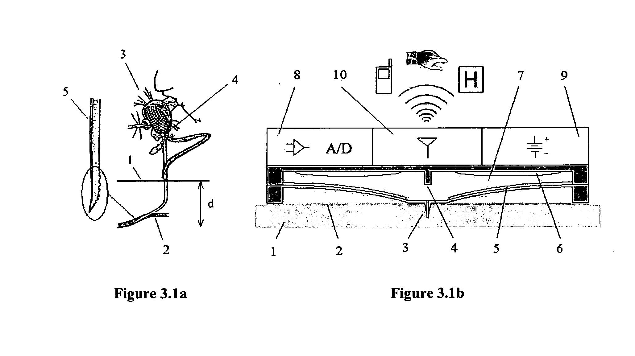
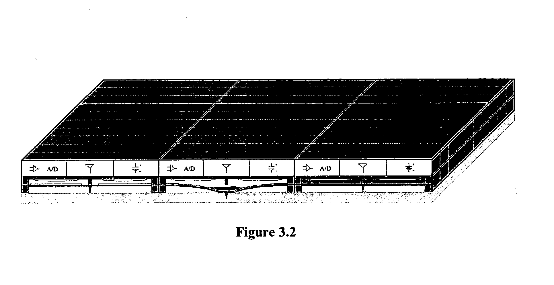
[0052] Design of the Building Blocks

[0053] Microneedle

[0054] A variety of different approaches have been put forward in designing the microneedles. In terms of fabrication techniques, in-plane [13, 17, 20, 36, 37] and out-of-plane [11, 12, 15, 16, 38-46] solutions have been suggested. Single microneedles are normally produced in-plane, and are much larger in size, while out-of-plane designs are smaller and are routinely fabricated in a matrix. A clear distinction is made between microneedles for drug delivery, and for blood sampling. In-plane needles are efficiently used for blood sampling because of their longer size [13, 20, 36]. With the recognition of the efficacy of subcutaneous drug delivery, out-of-plane microneedles are routinely preferred for that purpose [12, 15, 38, 47].

[0055] The human skin is composed of three layers: (1) the stratum corneum is the outermost layer and is made of dead tissues; (2) the epidermis, a tissue of living cells and interstitial plasma, which ...


PUM


Abstract

The lack of safe, reliable, automated and clinically acceptable blood sampling has been the main problem precluding the development of real-time systems for blood analysis and subsequent closed-loop physiological function control. While the analysis of a static blood sample in laboratory conditions has been rapidly advancing in reliability and blood volume reduction, non-invasive real-time blood analysis performed in vivo (while the blood is circulating in the body) has been elusive and unreliable. In this study we propose an innovative idea for semi-invasive blood sampling and analysis, which resembles the operation of a mosquito. At a miniature scale the proposed system does penetrate the skin to extract a static blood sample for further analysis, but the extent of this penetration, and the fact that it can be made painless, is particularly attractive for such applications as automated glucose analysis for closed-loop control of insulin infusion (artificial pancreas), continuous drug monitoring, or even periodic DNA analysis for security and identification purposes. These design aspects are described, and a specific implementation, applying MEMS (Micro Electro Mechanical Systems) technology, is suggested. The proposed microsystem is a matrix of individually controllable e-Mosquito™ cells, packaged in a disposable patch and attached to the skin, could be an avenue for real-time semi-invasive blood analysis and diagnostics.

Description

CROSS-REFERENCE TO RELATED APPLICATIONS [0001] This application claims the benefit of priority under 35 U.S.C. 119(e) of U.S. provisional application No. 60 / 526,595 filed Dec. 4, 2003.BACKGROUND OF THE INVENTION [0002] Blood sampling is essential for diagnosing and testing a wide variety of disorders and medical conditions, as well as for DNA testing, and blood donation screening [1]. Contemporary blood sampling techniques can be generally classified as invasive, implanted and non-invasive [2]. All references in square brackets are listed at the end of the patent disclosure and incorporated by reference herein. [0003] Non-invasive blood sampling and subsequent analysis usually involves optical or ultrasonic tissue interrogation techniques [3], which generally depend on the location and the characteristics of the tissue volume studied. For example, it has been shown that glucose concentration in a given blood volume does have specific optical and ultrasonic signatures [4], and claims...

Claims


Application Information

Patent Timeline
Patent Type & Authority Applications(United States)
IPC IPC(8): A61B5/00A61M1/00A61M5/142B65D81/00
CPCA61B5/14514A61B5/14532A61B5/14865A61M2205/3576A61M2205/0244A61M2205/3546A61M5/14244
Inventor KALER, KARAN V.I.S.GATTIKER, GIORGIOMINTCHEV, MARTIN P.
Owner UNIV TECH INT
Why Eureka
Try Eureka