Foam electrode and method of use thereof during tissue resection

a foam electrode and tissue resection technology, applied in the field of tissue ablation devices and methods, can solve the problems of increasing the complexity, time and expense of the resection procedure, affecting the safety of patients, so as to prevent tissue charring and prevent blood or fluid loss.

Inactive Publication Date: 2007-07-05
BOSTON SCI SCIMED INC
View PDF39 Cites 77 Cited by
  • Summary
  • Abstract
  • Description
  • Claims
  • Application Information

AI Technical Summary

Benefits of technology

[0018] Although the present inventions should not necessarily be limited in their broadest aspects, the use of an electrically insulative hydrophilic material is advantageous in that the hydrophilic electrode cannot be activated until it absorbs a sufficient amount of electrically conductive fluid. Thus, unlike other electrosurgical systems that utilize saline, the inactivation of the hydrophilic electrode will prevent tissue charring from occurring if the flow of saline is cutoff or is otherwise insufficient. In addition, because the absorbent nature of the hydrophilic electrode provides only the necessary amount of electrically conductive fluid to the tissue site by limiting the electrically conductive fluid to the tissue-electrode interface, the electrical energy conveyed to or from the hydrophilic electrode is focused at the desired location, and the need for aspirating electrically conductive fluid, which may otherwise accumulate at the tissue site using other saline systems, is obviated.
[0019] The method further comprises separating the tissue along the resection line to resect the tissue portion to be removed from the tissue portion to be retained. For example, tissue separation may comprise mechanically or electrically separating the coagulated tissue along the resection line. Thus, it can be appreciated that, because the tissue has been coagulated, blood or fluid loss is prevented, or at least minimized during the resection process. Electrical separation of the tissue may be accomplished with the hydrophilic electrode or another electrode separate from the hydrophilic electrode. In the later case, the hydrophilic electrode and other electrode may be conveniently mounted on a single probe, and / or electrical energy may be conveyed to or from the hydrophilic electrode and the other electrode, wherein tissue coagulation and tissue separation is simultaneously achieved. The tissue coagulation may be performed prior to the tissue separation, whereby the coagulated tissue is separated. Optionally, electrical energy may be conveyed to or from the hydrophilic electrode to seal an anatomical vessel (e.g., a blood vessel) that traverses the resection line, and the sealed blood vessel can then be transected.
[0020] In accordance with a second aspect of the present inventions, an electrosurgical probe is provided. The electrosurgical probe comprises an elongated probe shaft, which may be rigid, and a tissue coagulation electrode mounted to the distal end of the probe shaft. The coagulation electrode is configured for absorbing an electrically conductive fluid, and has a leading surface that can be placed in contact with tissue. The leading surface of the coagulation electrode may be straight, such that it can be placed along a straight resection line. The coagulation electrode may be composed of a hydrophilic material having the characteristics and accompanying advantages previously described above. In an optional embodiment, the electrode surgical probe may further comprising a fluid delivery conduit extending through the probe shaft in fluid communication with the coagulation electrode. In this manner, electrically conductive fluid may be readily provided to the coagulation electrode on demand without displacing the electrosurgical probe from the tissue site. Alternatively, the coagulation electrode may be repeatedly dipped in a source of electrically conductive fluid.

Problems solved by technology

When used to cut tissue, the electrical arcing and corresponding current flow results in a highly intense, but localized heating, sufficient enough to break intercellular bonds, resulting in tissue severance.
Whichever modality is used, extensive bleeding can occur, which can obstruct the surgeon's view and lead to dangerous blood loss levels, requiring transfusion of blood, which increases the complexity, time, and expense of the resection procedure.
In the case where an organ is resected, application of RF energy divides the parenchyma, thereby skeletalizing the organ, i.e., leaving vascular tissue that is typically more difficult to cut or dissect relative to the parenchyma.
When electrosurgically resecting tissue, care must be taken to prevent the heat generated by the electrode from charring the tissue, which generates an undesirable odor, results in tissue becoming stuck on the electrosurgical probe, and most importantly, increases tissue resistance, thereby reducing the efficiency of the procedure.
As a result, if the fluid and removed tissue is not effectively aspirated from the tissue site, the electrosurgery may either be inadequately carried out, or a greater than necessary amount of energy must be applied to the electrode to perform the surgery.
Increasing the energy used during electrosurgery increases the chance that adjacent healthy tissues may be damaged.
For example, if flow of the fluid is momentarily stopped, e.g., if the tube supplying the fluid is kinked or stepped on, or the port on the fluid delivery device becomes clogged or otherwise occluded, RF energy may continue to be conveyed from the electrode, thereby resulting in a condition where tissue charring may occur.
While electrosurgical resection of tissue reduces the amount of blood loss, as compared to other tissue resection modalities, it still involves a tedious process that includes painstakingly cutting / dissecting through the parenchyma and ligating and cutting though blood vessels.

Method used


Image

  • Foam electrode and method of use thereof during tissue resection
  • Foam electrode and method of use thereof during tissue resection
  • Foam electrode and method of use thereof during tissue resection

Examples


Embodiment Construction

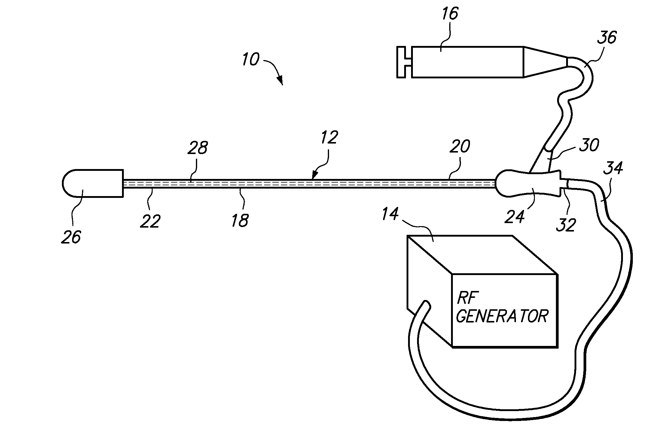
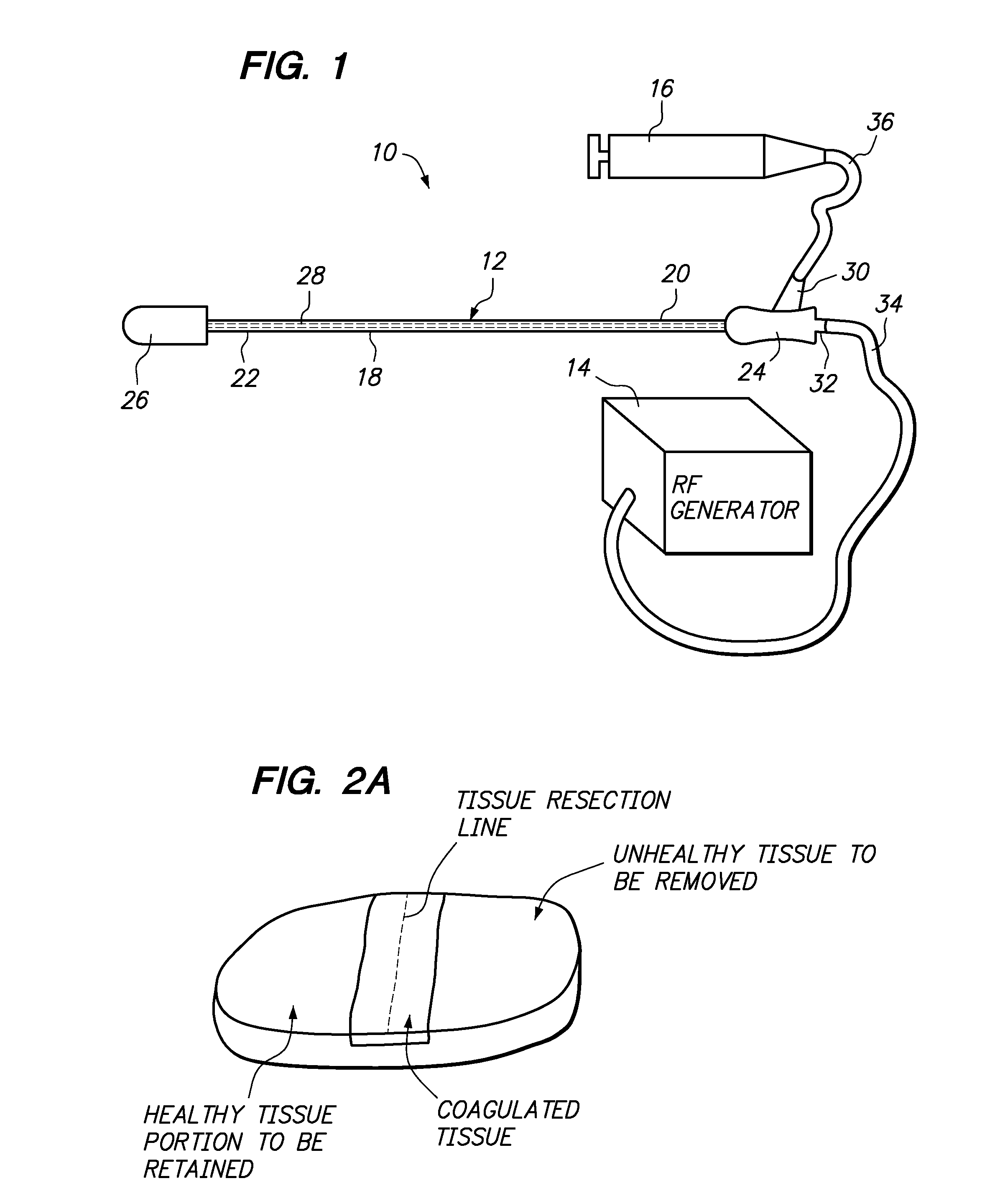
[0073]FIG. 1 illustrates a tissue resection system 10 constructed in accordance with a preferred embodiment of the present inventions. The tissue resection system 10 generally comprises tissue coagulation / resection probe 12 configured for coagulating and resecting tissue, an ablation energy source, and in particular a radio frequency (RF) generator 14, configured for supplying RF energy to the tissue resection probe 12 in a controlled manner, and an electrically conductive fluid source, and in particular a syringe 16 configured for supplying electrically conductive fluid (e.g., saline) to the resection probe 12 to provide an electrically conductive path for the RF energy from the resection probe 12 to the tissue to be coagulated / resected.

[0074] The coagulation / resection probe 12 generally comprises an elongated probe shaft 18 having a proximal end 20, a distal end 22, a handle assembly 24 mounted to the proximal shaft end 20, a tissue coagulation / resection assembly 26 mounted to th...


PUM


Abstract

Assemblies and methods are provided for resecting a portion of tissue to be removed (e.g., unhealthy tissue, such as cancerous tissue) from a portion of the tissue to be retained (e.g., healthy tissue) within a patient is provided. An electrically conductive fluid, such as saline, may be absorbed into a hydrophilic electrode. Electrical energy (e.g., radio frequency (RF) energy) is conveyed to or from the hydrophilic electrode while being moved in proximity to the tissue along a resection line, whereby tissue adjacent to the resection line is coagulated. A resection member, such as a blunt resection member or a resection electrode, which may be on the same device as the hydrophilic electrode, is used to separate the tissue along the resection line to resect the tissue portion to be removed from the tissue portion to be retained.

Description

RELATED APPLICATION DATA [0001] The present application claims the benefit under 35 U.S.C. § 119 to U.S. provisional patent application Ser. No. 60 / 755,713 filed Dec. 29, 2005, which is hereby incorporated by reference.FIELD OF INVENTION [0002] The present inventions generally relate to tissue ablation devices and methods, and particularly to ablation devices and methods for achieving tissue resection. DESCRIPTION OF RELATED ART [0003] Today, electrosurgery is one of the widely used surgical modalities for treating tissue abnormalities. Electrosurgical devices fall into one of two categories, monopolar devices and bipolar devices. Generally, surgeons are trained in the use of both monopolar and bipolar electrosurgical techniques, and essentially all operating rooms will be found equipped with the somewhat ubiquitous instrumentality for performing electrosurgery. [0004] Monopolar electrosurgical devices typically comprise an electrosurgical probe having a first or “active” electrode ...

Claims


Application Information

Patent Timeline
Patent Type & Authority Applications(United States)
IPC IPC(8): A61B18/14
CPCA61B18/1402A61B2018/00065A61B2018/00404A61B2018/1472A61B2018/00601A61B2018/00607A61B2018/00589
Inventor RIOUX, ROBERT F.DICARLO, PAULOYOLA, ARNOLD
Owner BOSTON SCI SCIMED INC
Why Eureka
Try Eureka