Targeting endothelium for tissue-specific delivery of agents

a technology of endothelium and tissue, applied in the direction of immunoglobulins against animals/humans, drug compositions, medical preparations, etc., can solve the problems of caveolae's inability to transport their cargo across cells, and the inability to perform much less effectively

Inactive Publication Date: 2007-07-12
SCHNITZER JAN E
View PDF6 Cites 3 Cited by
  • Summary
  • Abstract
  • Description
  • Claims
  • Application Information

AI Technical Summary

Benefits of technology

[0008] Thus, purified caveolae, G domains (lipid rafts), and co-isolated caveolae and G domains as described herein are useful for the identification of molecules and proteins which are involved in intra- or trans-cellular transport and cell surface signal transduction and communication. They thus make it possible to identify new means by which molecules can be delivered to plasma membranes and, if desired, enter the cell, cross from one side of the cell to the other, or provide a signal to the cell that alters its function. For example, the purified caveolae and the purified G domains (lipid rafts) can be used to make specific probes or antibodies. Antibodies or ligands which are specific to the caveolae, or to the purified G domains, can be used as vectors to target the caveolae or G domains and to influence the transport of molecules into and / or across the plasma membrane. Such vectors can be used to deliver agents into and / or across the cell, such as drugs, genes, or antibodies, and particularly to deliver agents into and / or across the endothelium. The vectors can contain an active component (e.g., the drug, gene, antibody, or other agent) and a transport component (e.g., an antibody or ligand specific to caveolae or to a protein, peptide or ligand within caveolae).

Problems solved by technology

Although such agents are certainly justified by their success in vitro, they frequently perform much less effectively in vivo where the agent must reach its target cells in a tissue in sufficient quantities to be potent while sparing bystander organs (Jain, R. K., Nat Med 4:655-7 (1998)).
Whether caveolae can traffic their cargo across cells (transcytosis) has previously been unproven, primarily because comparative analysis has not been possible using probes capable of targeting caveolae with high affinity and specificity in vivo vs. physically identical, nontargeting control probes.

Method used


Image

  • Targeting endothelium for tissue-specific delivery of agents
  • Targeting endothelium for tissue-specific delivery of agents
  • Targeting endothelium for tissue-specific delivery of agents

Examples


example 1

Isolation of Coated Membrane Pellets (P)

[0069] Isolation of caveolae associated with the endothelial cell surface has many difficulties. The endothelium represents but a small percentage of a diverse population of cells in any organ. Unfortunately, isolating endothelial cells from tissues as a primary source or even for growth in culture causes morphological changes, including a very significant loss in cell surface caveolae (Schnitzer, J. E., et al., Biochem. Biophys. Res. Commun. 199:11 (1994)). Also, noncoated vesicles that are very similar in size and density to plasmalemmal caveolae and may even contain the caveolar marker protein caveolin (Dupree, P., et al., EMBO J. 12:1597 (1993); Kurzchalia, T. V., et al., J. Cell Biol. 118:1003 (1992)), may be found in other cellular compartments such as the trans-Golgi network. Moreover, caveolae may vary according to cell type (Izumi, T., et al., J. Electron Microsc. 38:47 (1989)). To overcome the above problems, a strategy of first isol...

example 2

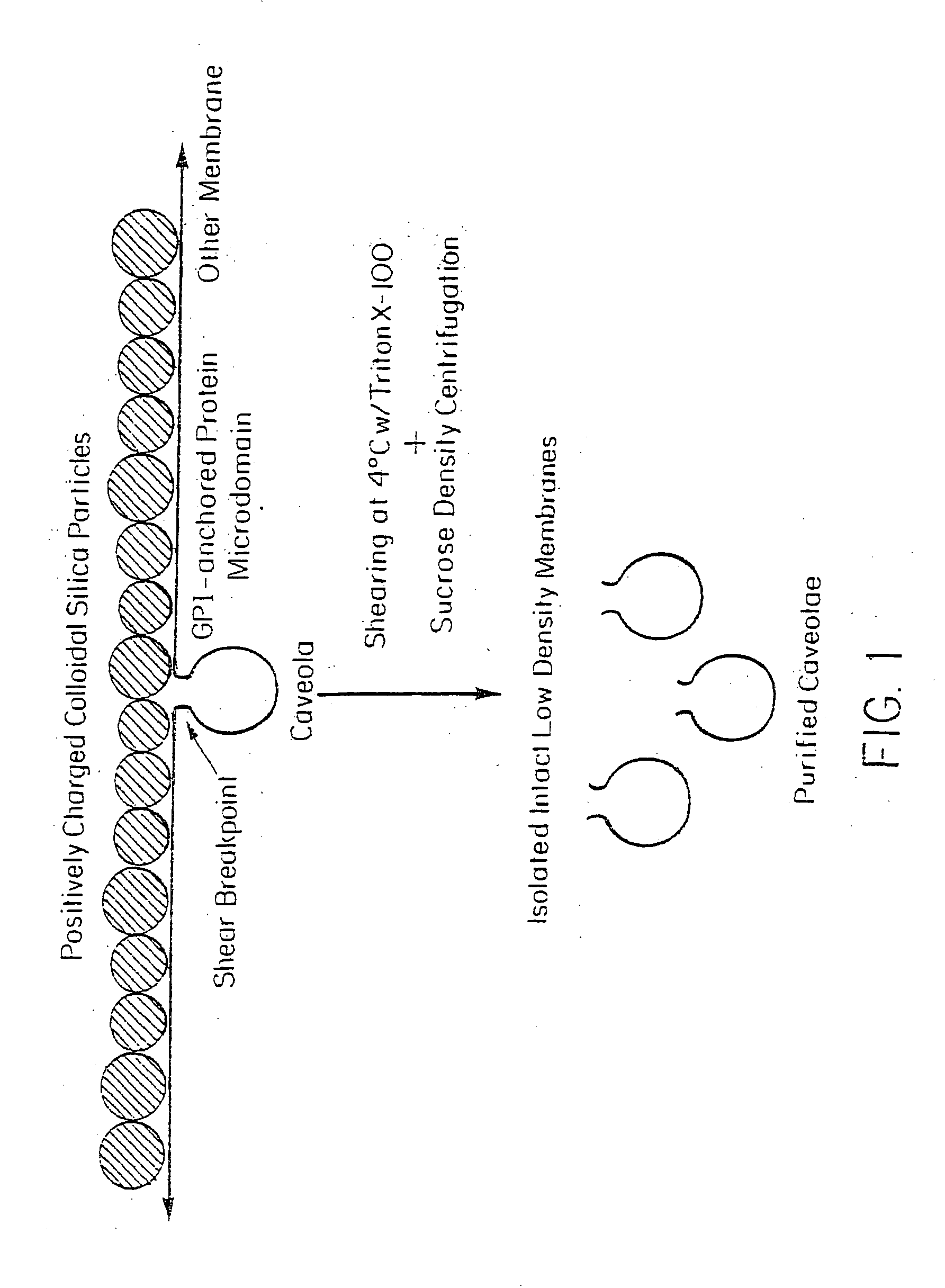
Isolation of Purified Caveolae (V)

[0073] Caveolae attached on the cytoplasmic side of the plasma membranes, opposite to the silica coating in the silica-coated membrane pellets (P), were stripped from the membranes by shearing during homogenization at 4° C. in the presence or absence of Triton X-100. They were then isolated by sucrose density gradient centrifugation to yield a homogenous population of biochemically and morphologically distinct caveolar vesicles (V). This technique is represented schematically in FIG. 1.

[0074] Isolation of Vesicles Stripped from Silica-Coated Membranes.

[0075] The silica-coated membranes (P) in Triton X-100 were stripped of protruding caveolae by shearing in a homogenizer and then subjected to sucrose density centrifugation to isolate the caveolae. Analysis of 34 fractions from the sucrose gradient revealed a peak signal for caveolin well separated from that for ACE. Little caveolin was detected in the silica-coated membrane after removal of the ves...

example 3

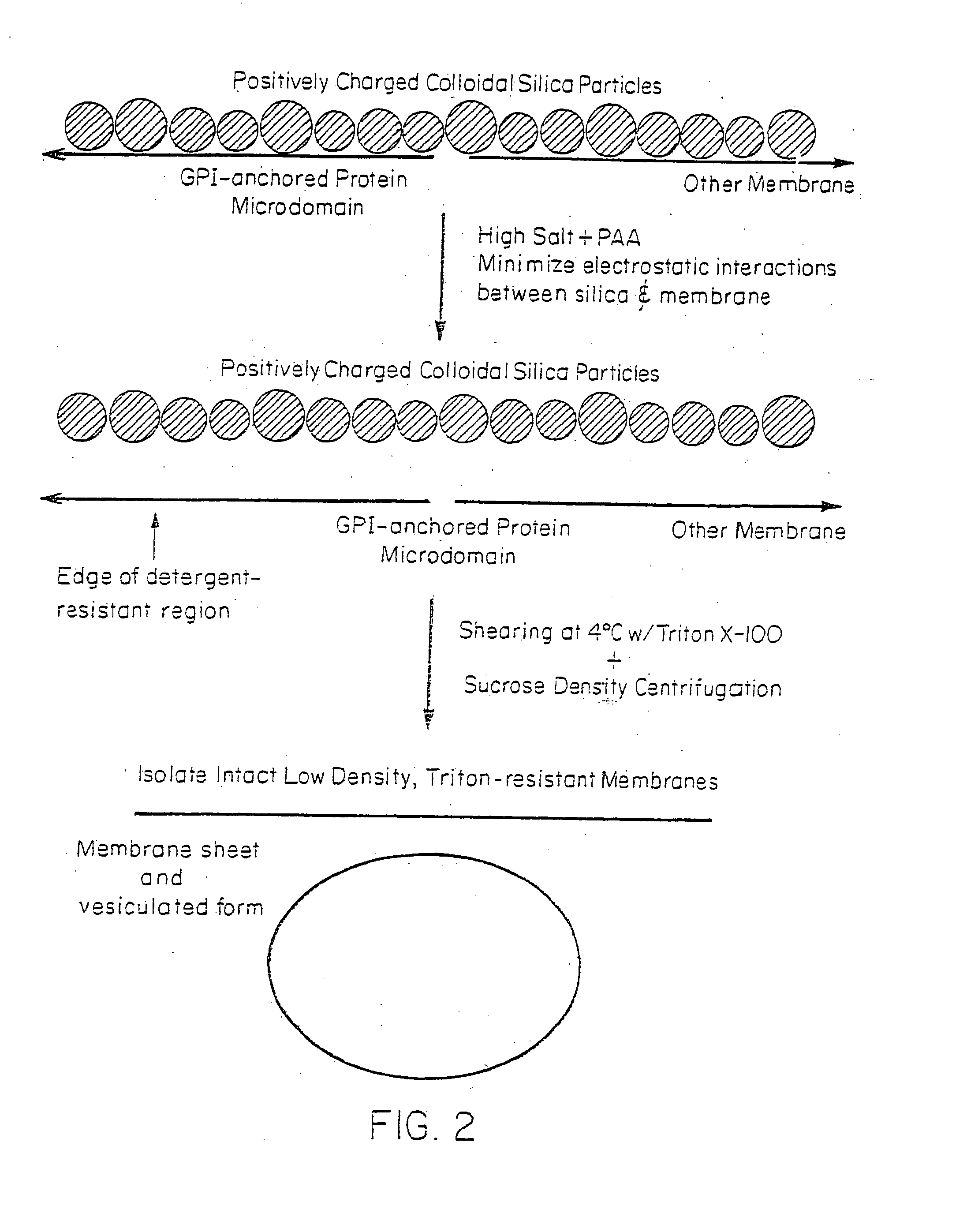
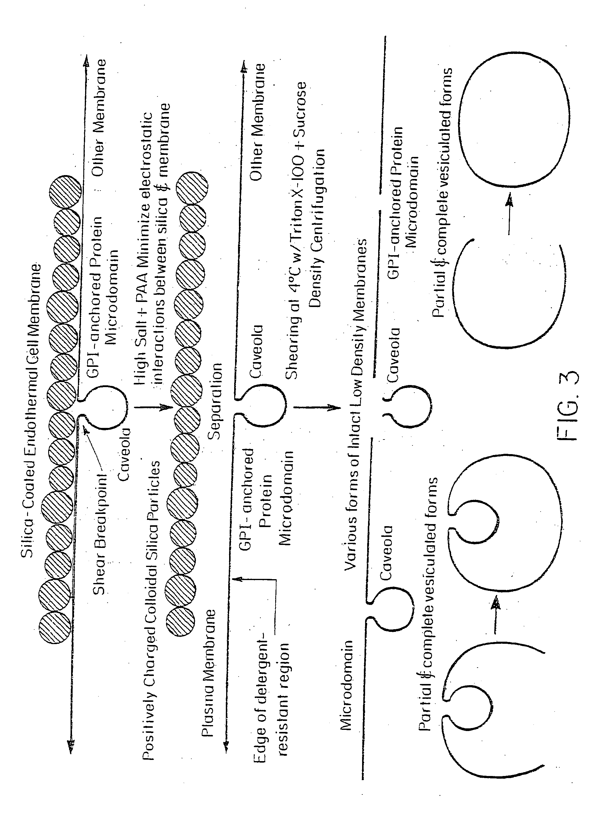
Isolation of Microdomains of GPI-Anchored Proteins (G)

[0082] The silica coating of the outer membrane surface altered the way in which the GPI-anchored proteins interacted with various detergents and thus prevented the separation of noncaveolar, detergent-resistant microdomains from the cell membranes. Cationic silica particles interact with the anionic cell surface to stabilize it against vesiculation or lateral rearrangement by immobilizing membrane molecules (Chaney, C. K. and Jacobson, B. S., J. Biol. Chem. 258:10062 (1983); Patton, W. F., et al., Electrophoresis 11:79 (1990)). Because the silica particles uniformly coated the cell surface but were rarely associated with or present inside the caveolae because of their size, it is likely (Schnitzer, J. E., et al., Proc. Natl. Acad. Sci. USA 92:1759 (1995); Jacobson, B. S., et al., Eur. J. Cell Biol. 58:296 (1992)) that the plasma membrane was stabilized by being firmly attached on one side to most, if not all, nonvesiculated regi...


PUM

PropertyMeasurementUnit
temperatureaaaaaaaaaa
physiologic temperatureaaaaaaaaaa
pHaaaaaaaaaa

Abstract

Mechanisms and routes by which molecules can be delivered into cells, particularly endothelial cells, through the action of caveolae, G domains and other plasma membrane domains and components, are described, as are delivery systems comprising antibodies to caveolar proteins or receptors and also agents conjugated to the antibody or ligand, which can be used to deliver agents to, into, and/or across the endothelium, for example, for imaging purposes.

Description

RELATED APPLICATIONS [0001] This application is a continuation of Ser. No. 10 / 631,481, filed Jul. 31, 2003, which is a Continuation-in-part of U.S. application Ser. No. 10 / 056,230, filed on Jan. 24, 2002, which is a Continuation-in-part of U.S. application Ser. No. 09 / 734,490, filed on Dec. 11, 2000, now abandoned, which is a Continuation of U.S. application Ser. No. 09 / 029,459, filed Jun. 25, 1998, now U.S. Pat. No. 6,255,457, which is the U.S. National stage of International Application No. PCT / US96 / 14177, filed on Sep. 5, 1996, published in English, which is a continuation-in-part (or a continuation of) U.S. application Ser. No. 08 / 582,917, filed Jan. 4, 1996, now U.S. Pat. No. 5,776,770, which claims the benefit of U.S. Provisional Application No. 60 / 003,453, filed Sep. 8, 1995; and the previously identified PCT / US96 / 14177, filed on Sep. 5, 1996 also claims the benefit of U.S. Provisional Application No. 60 / 018,791, filed May 31, 1996, and the benefit of U.S. Provisional Applica...

Claims


Application Information

Patent Timeline
Patent Type & Authority Applications(United States)
IPC IPC(8): A61K51/00A61K49/10A61K48/00A61K9/127A61K9/00A61K38/00A61K51/10C07K14/47C07K14/705C07K16/28C12NC12N9/22C12N9/88
CPCA61K9/0019A61K9/1275A61K38/00A61K51/1027C12N9/88C07K14/705C07K16/28C12N9/22C07K14/47A61P35/00
Inventor SCHNITZER, JAN E.
Owner SCHNITZER JAN E
Why Eureka
Try Eureka