
Endoscope system

a technology which is applied in the field of endoscope and endoscope system, can solve the problems of difficult treatment and difficulty in inserting the probe into the small intestine, and achieve the effect of improving the efficiency of diagnosis

Inactive Publication Date: 2007-07-12
OSAKA UNIV
View PDF11 Cites 90 Cited by
  • Summary
  • Abstract
  • Description
  • Claims
  • Application Information

AI Technical Summary

Benefits of technology

[0053] As described above, according to the present invention, it is possible to provide an endoscope and an endoscope system that are free from a dead area and capable of preventing the physician from overlooking any nidus.
[0054] In addition, it is possible to provide an endoscope and an endoscope system that improve the physician's diagnosis efficiency.
[0055] Thus, it is possible to obtain panoramic images of side surfaces inside the digestive organs, including side surfaces in the forward direction and side surfaces in the backward direction, images of which are conventionally difficult to take, resulting in extremely significant contributions to preventing the physician from overlooking any lesion and the improvement of diagnosis efficiency and immeasurable contributions to the field of medical instruments and the progress of medical science.

Problems solved by technology

However, the small intestine of an adult male is as long as about 3 m, and therefore it is difficult to insert the probe into the small intestine.
The capsule endoscope is expected to be considerably effective in observing the small intestine, although treatment therewith is difficult.

Method used


Image

  • Endoscope system
  • Endoscope system
  • Endoscope system

Examples


first embodiment

[0091] [Configuration of Endoscopes]

[0092] The configuration of endoscopes according to the present embodiment is described with respect to two types of endoscopes: a probe-type endoscope and a capsule endoscope.

[0093] 1. The Probe-Type Endoscope

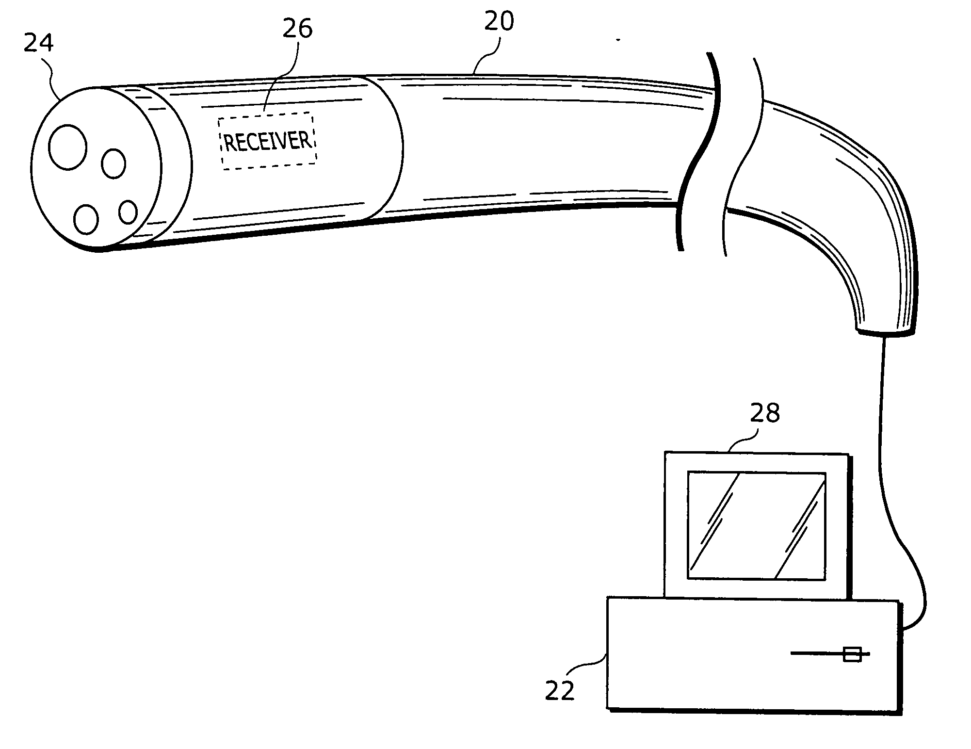
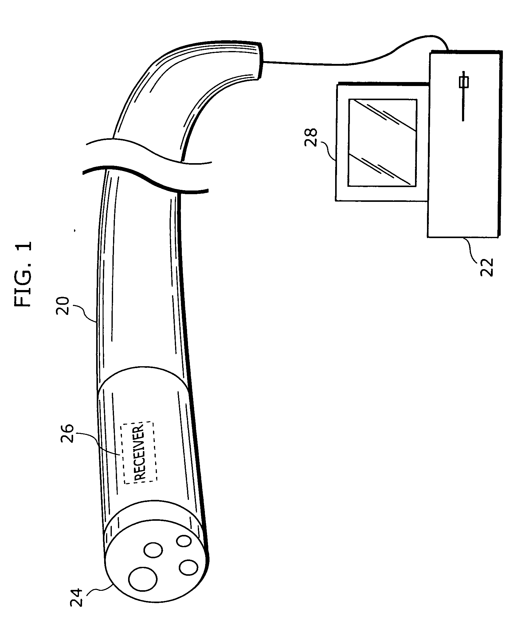
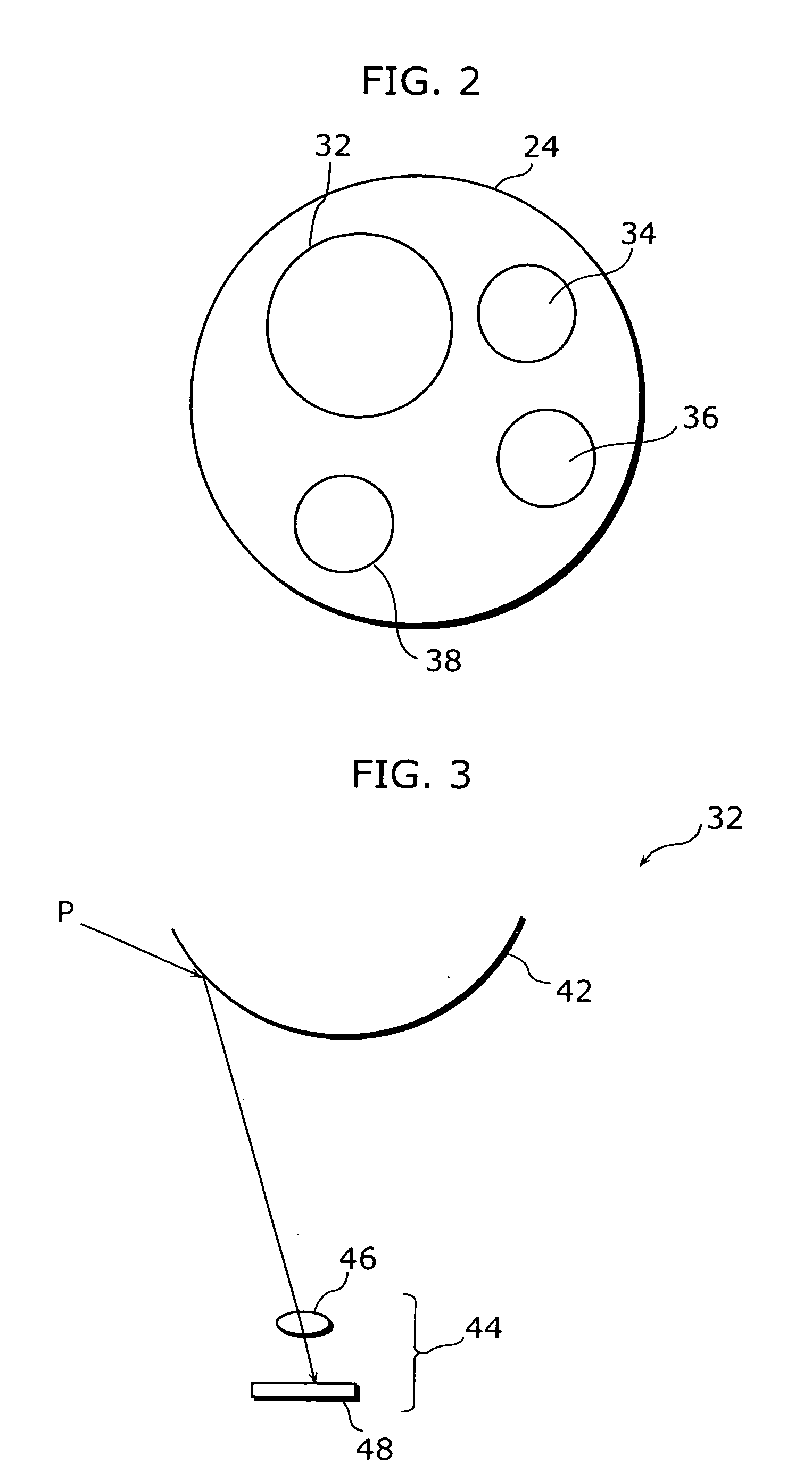
[0094]FIG. 1 is a diagram illustrating the configuration of a probe-type endoscope according to the first embodiment of the present invention. FIG. 2 is an external view of a tip portion 24 of the probe-type endoscope 20 shown in FIG. 1. The tip portion 24 of the probe-type endoscope 20 is provided with an omnidirectional camera 32, a light 34, a forceps 36 and a rinse water injection port 38.

[0095] The omnidirectional camera 32 is a device for taking images the inside of digestive organs, and is able to take 360-degree images of its surroundings. The light 34 is used for lighting up the inside of the digestive organs. The forceps 36 is a tool used for pinching and pressing tissues and nidi inside the digestive organs. The rinse water inj...

second embodiment

[0145] Described next is the configuration of an endoscope according to a second embodiment of the present invention. The configuration of the endoscope according to the second embodiment is similar to that of the probe-type endoscope or the capsule endoscope according to the embodiment. However, it differs from the first embodiment in the following three points.

[0146] (1) In the first embodiment, the motion estimation of the omnidirectional camera 32 is carried out by detection from corresponding image points in a sequence of temporally successive images, whereas in the second embodiment, feature regions in images are obtained to associate the regions.

[0147] (2) Additionally, in the first embodiment, the segmentation movement of the inner wall of a digestive organ is formulated to correct the camera motion, whereas in the second embodiment, in addition to that, the peristalsis movement of the inner wall of the digestive organ is also formulated.

[0148] (3) Further, in the first e...


PUM


Abstract

An endoscope that is free from a dead area and capable of preventing the physician from overlooking any nidus is an endoscope for taking the inside of digestive organs, and the endoscope is provided with an omnidirectional camera (32), a light (34), a forceps (36) and a rinse water injection port (38) at the tip (24). The omnidirectional camera (32) is a device for taking the inside of digestive organs, and is able to take 360-degree images of its surroundings. A probe-type endoscope (20) is provided with a receiver (26) composed of orthogonal coils, and the receiver (26) is used for estimating the position and attitude of the probe-type endoscope (20). An image taken by the omnidirectional camera (32) is presented on a display unit (28) of an image processing device (22) connected to the probe-type endoscope (20). In the image processing device, a video mosaicking process is performed on a plurality of images obtained by the omnidirectional camera (32) to generate a panoramic image of the inside of a digestive organ.

Description

TECHNICAL FIELD [0001] The present invention relates to endoscopes and endoscope systems and, in particular, to an endoscope and an endoscope system for observing the inside of the digestive organs. [0002] 2. Background Art [0003] Conventionally, in the field of medical practice, probe-type endoscopes are used for examining the digestive organs. The endoscopic probe has a camera, a light, a forceps and a rinse water injection port mounted to its tip. The physician inserts the probe through the oral cavity or the anus into a digestive organ, and carries out diagnosis, collection of a lesion and treatment, while monitoring video obtained by the camera at the tip. [0004] With an endoscope inserted from the oral cavity, examination and treatment of the esophagus, stomach and duodenum are carried out, whereas with an endoscope inserted from the anus, examination and treatment of the rectum and large intestine are carried out. However, the small intestine of an adult male is as long as ab...

Claims


Application Information

Patent Timeline
IPC IPC(8): A61B1/04A61B1/018A61B1/12
CPCA61B1/00009A61B1/126A61B1/041A61B1/00177A61B1/000094
Inventor YAGI, YASUSHIECHIGO, TOMIOSAGAWA, RYUSUKE
Owner OSAKA UNIV
Learn More