Dental x-ray apparatus and method of positioning a patient therein

a technology of x-ray apparatus and patient, which is applied in the field of dental x-ray apparatus and the method of positioning a patient therein, can solve the problems of helmets must be worn, and insufficient use of ultrasound sensors for dental radiography, so as to accelerate the alignment procedure and improve the accuracy of alignmen

Active Publication Date: 2007-08-09
DENTAL IMAGING TECH +1
View PDF9 Cites 56 Cited by
  • Summary
  • Abstract
  • Description
  • Claims
  • Application Information

AI Technical Summary

Benefits of technology

[0016] One advantage of the present invention is that it accelerates the alignment procedure and achieves a higher accuracy of the alignment.
[0017] Another advantage of the present invention is that the correct alignment of the patient is monitored during the imaging process and appropriately corrected if necessary.
[0018] Other features and advantages of the present invention will be apparent from the following more detailed description of the preferred embodiment, taken in conjunction with the accompanying drawings which illustrate, by way of example, the principles of the invention.

Problems solved by technology

However, a helmet has to be worn by the patients, which requires a very careful positioning if large positioning errors are to be avoided.
This system seems to transfer the problem of the patient's positioning to the positioning of the helmet.
The active helmets carry magnetic or ultrasound sensors (magnetic sensors are badly distorted by ferromagnetic material, and ultrasound sensors do not appear to be so accurate).
However, the use of a helmet or any similar device is not adequate for dental radiography.
This approach requires additional 2D radiographies, and does not meet the needs of dental radiography.
Although automatic 3D scanners have been on the market for few years (e.g., Minolta), their impact on the costs of an x-ray machine is quite high.
Moreover, these systems are difficult to operate automatically for several reasons.
First, very little information is available for alignment, i.e, the body shape does not have geometrical features (sharp peaks, valleys, etc.), which would allow defining robust error measures, to be used to evaluate alignment.
In the second case, the surface to surface distance has to be determined, which requires a normal to the surface computation that is time consuming.
Second, once this alignment has been achieved and checked, the operator exits from the room where the orthopantomographic apparatus and the patient are located to start the x-ray imaging process.
Unfortunately, during this time period before the operator starts the x-ray imaging process, there is a high likelihood that the patient will move out of the proper alignment position for the x-ray imaging process.
Moreover, the possibilities for an operator of the x-ray apparatus to move within the working space of an orthopantomographic system is very restricted, and a good analysis of the patient alignment can be uncomfortable for the operator.

Method used


Image

  • Dental x-ray apparatus and method of positioning a patient therein
  • Dental x-ray apparatus and method of positioning a patient therein
  • Dental x-ray apparatus and method of positioning a patient therein

Examples


Embodiment Construction

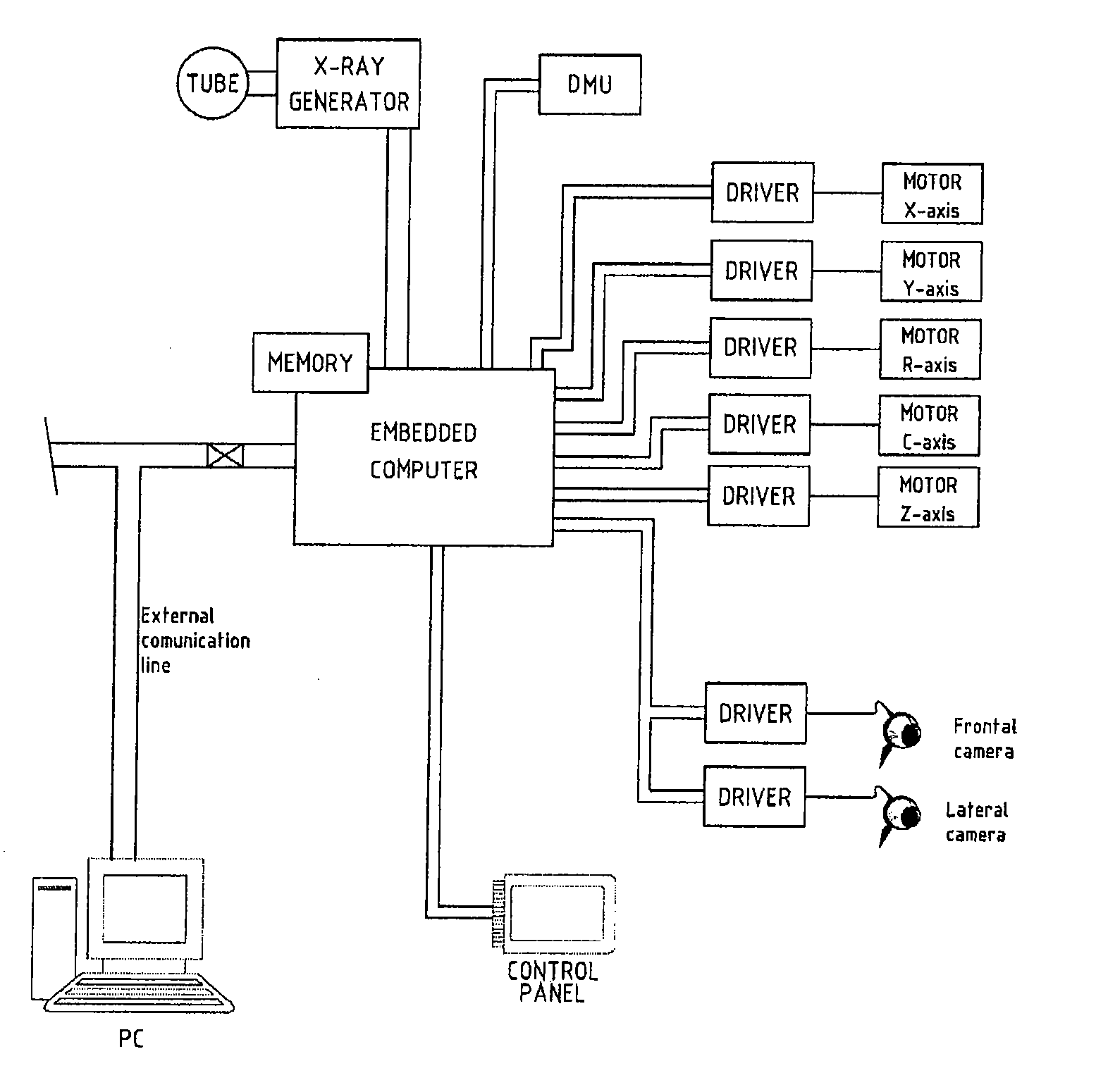
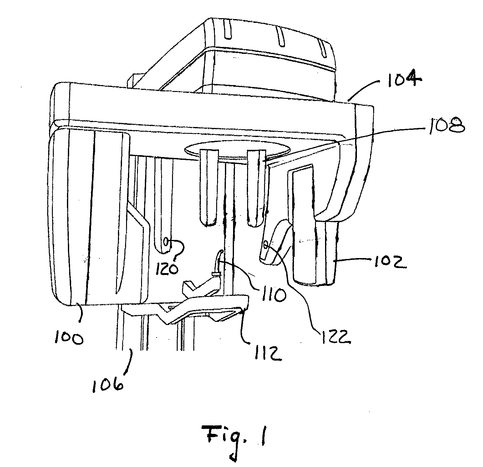
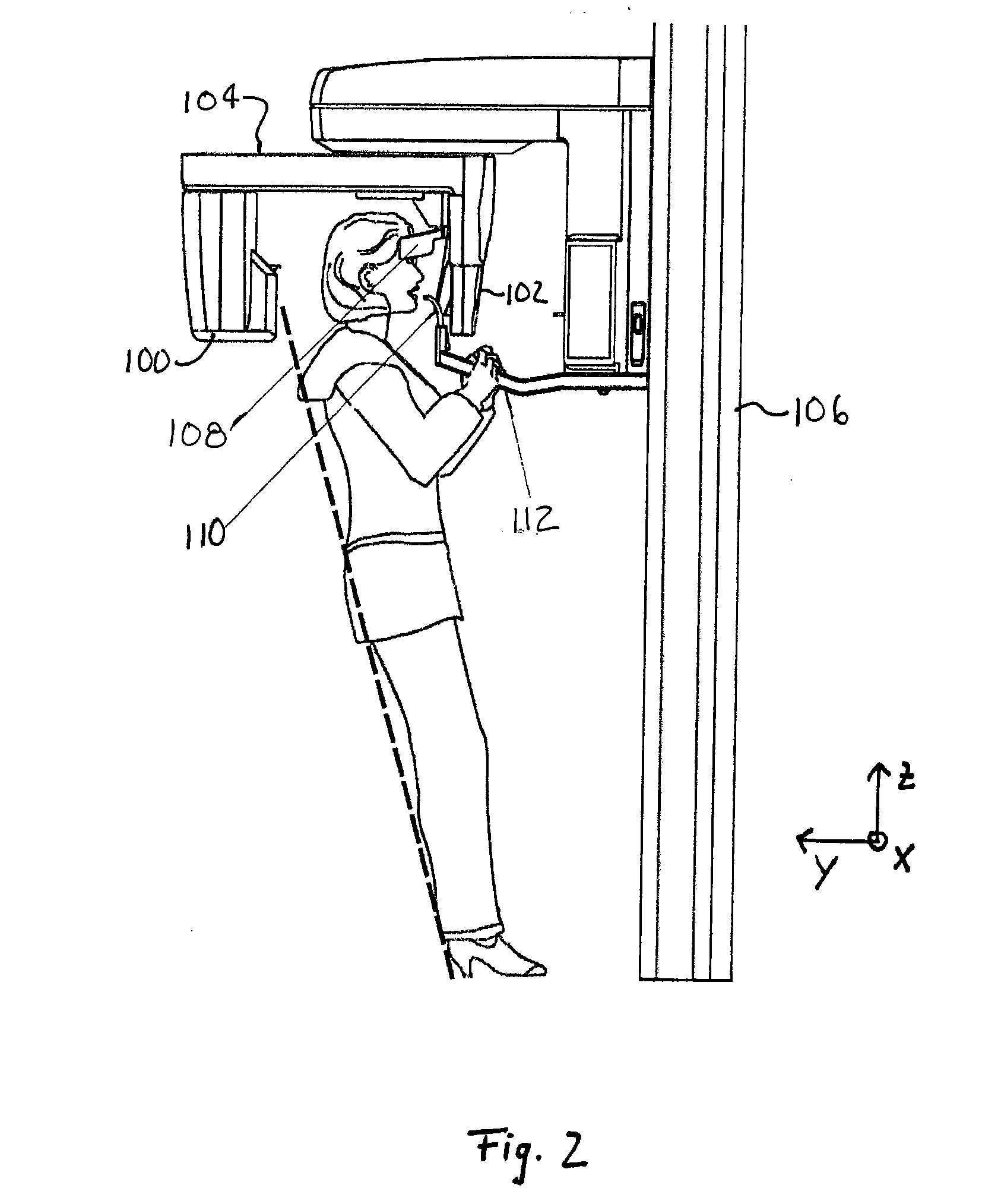
[0026] In one embodiment, the general setup of the x-ray apparatus of the present invention and the kinematic system thereof corresponds to those systems disclosed in U.S. Pat. No. 4,985,907 or US Patent Application Publication No. 2004-0190678 A1 (also published as International Publication No. WO 2004 / 014232), both of which references are hereby incorporated by reference in their entireties. The x-ray apparatus of the present invention differs from these prior apparatuses in that the present invention includes one or more video cameras that allow an improved and simplified apparatus-patient alignment for radiography, which alignment process may be essentially remote controlled.

[0027] The x-ray apparatus of the invention is preferably adapted for the following well-known techniques in dental radiography: orthopantomography, scannography, linear tomography, cephalography, and / or dental volume reconstructions. Preferably, the x-ray apparatus is used with an orthopantomographic, ceph...


PUM


Abstract

A dental x-ray apparatus is provided that includes an x-ray system having an x-ray source and x-ray detection means; positioning means for positioning a patient's head in a predefined position with respect to the x-ray system; and at least one video camera for generating video images of the patient's head.

Description

CROSS REFERENCE TO RELATED APPLICATIONS [0001] This application claims the benefit of U.S. Provisional Application No. 60 / 764,050 filed on Feb. 1, 2006.BACKGROUND OF THE INVENTION [0002] The present invention relates to a method of positioning a patient in an x-ray apparatus for dental x-ray radiography. The invention further relates to a dental x-ray apparatus for implementing the method. [0003] In dental radiography, a precise position of the patient's head with respect to the radiological apparatus is mandatory to achieve the best focusing and best image quality. The need for controlling the position of a patient has first arisen in the radiotherapy domain. In this field, the X-ray dose can be greatly increased, up to 10 times, if the position of the patient is known with high accuracy. The increased X-ray dosage allows limiting the exposure of healthy tissue to radiation. Mechanical Systems and Fixed X-ray Systems [0004] The first system developed for radiotherapy was based on ...

Claims


Application Information

Patent Timeline
Patent Type & Authority Applications(United States)
IPC IPC(8): A61B6/14
CPCA61B6/08A61B6/14A61B6/548A61B6/465A61B6/463
Inventor ROTONDO, GIUSEPPERINALDI, GERARDOBORGHESE, N. ALBERTOFROSIO, IURI
Owner DENTAL IMAGING TECH
Why Eureka
Try Eureka