Method for assessing the contraction synchronization of a heart

a technology of synchronization and heart contraction, applied in image data processing, sensors, catheters, etc., can solve the problems of high user dependence, inconvenient use, and inability to accurately assess the contraction synchronization of the heart, and achieve the effect of minimizing the negative impact of dyskinetic wall segments

Inactive Publication Date: 2007-10-04
INST DE CARDIOLOGIE DE MONTREAL
View PDF0 Cites 60 Cited by
  • Summary
  • Abstract
  • Description
  • Claims
  • Application Information

AI Technical Summary

Benefits of technology

[0045] Results. Overall, 235 patients (79% male; mean age 62±11 years) completed the study. LVEFs were similar by planar (33.5±13.5%) and 3D (34.7±12.7%) methods (r=0.83, P<0.0001). Mean phase angles for planar and tomographic methods were 126.3±29.6° and 124.4±28.7°, respectively (r=0.53; P<0.0001). Phase and amplitude signals were incorporated in the CHI, which was non-normally distributed with a median of 85.9% (IQR 77.7%, 92.2%). This index minimized the negative impact of dyskinetic wall segments with limited regional motion. The planar heterogeneity index (sdΦ) was 28.2° (IQR 17.5°, 46.8°) and correlated inversely with CHI (r=−0.61, P<0.0001).

Problems solved by technology

Many methods for computing a model of the surface of the cardiac chamber of interest from a cardiac tomography data set have been proposed, but they are mostly unsatisfactory.
A disadvantage of this method resides in a strong dependency on the user.
Accordingly, the models of the surface of the cardiac chamber of interest produced by different users will not be identical and are highly likely to be imprecise.
First, since the ventricle is not an ellipsoid, some rays will not be perpendicular to the real surface of the cavity to model.
In addition, since the exact location of the surface is not necessarily at a minimum of the first derivative data set it is very likely that errors are incorporated in the model of the surface of the cardiac chamber of interest.
In conclusion, currently available models of the surface of interest are error-prone due to deficiencies inherent to the segmentation methods currently used.
Accordingly, any parameter estimated from these models will also be erroneous.
However, many other causes of such a widened QRS complex exist, which makes this criterion non-specific.
However, these analyses typically use planar phase imaging and are therefore likely to miss important effects due to the tridimensional nature of the heart.

Method used


Image

  • Method for assessing the contraction synchronization of a heart
  • Method for assessing the contraction synchronization of a heart
  • Method for assessing the contraction synchronization of a heart

Examples


example

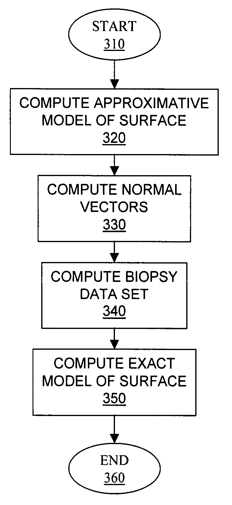
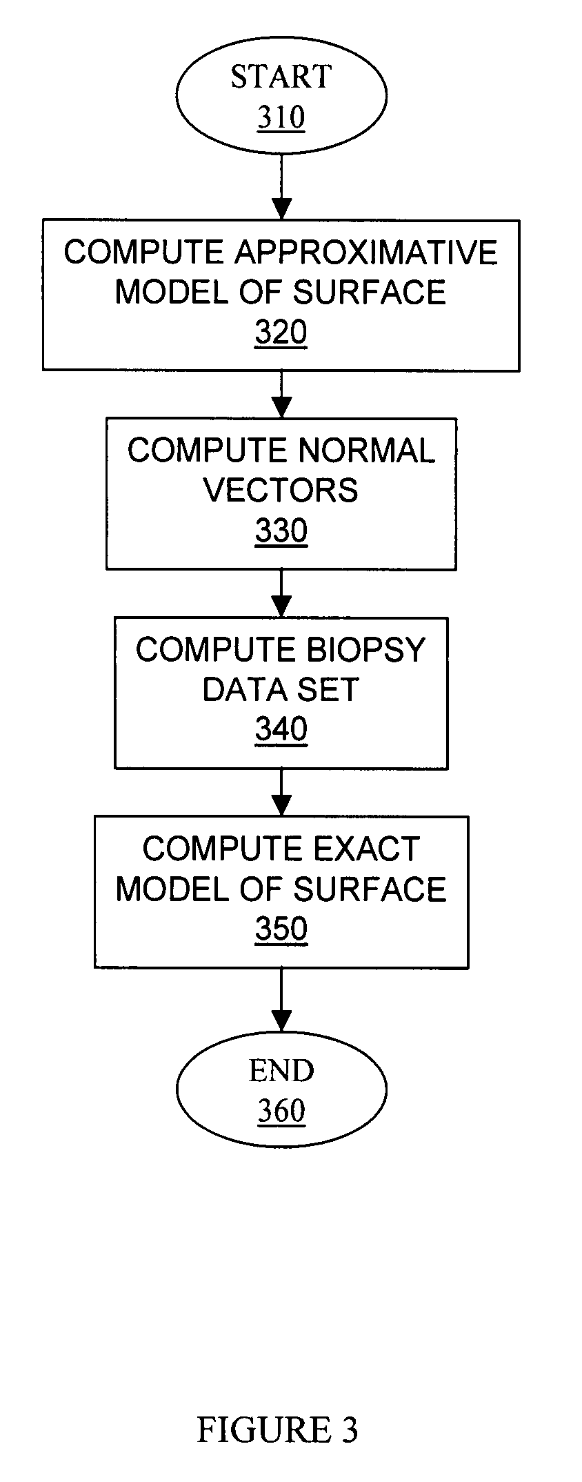
[0144] A specific embodiment of the above-described method for assessing the contraction synchronization of a heart of a patient is described hereinbelow and compared to prior art methods.

[0145] Materials and Methods

[0146] Study Population

[0147] The study population consisted of consecutive patients referred to the nuclear medicine department of the Montreal Heart Institute for assessment of left ventricular systolic function between June 2004 and June 2006. Patients hospitalized for an acute event or with decompensated heart failure were excluded. Data regarding baseline variables and past medical history were recorded and both planar and GBP-SPECT acquisition performed. The protocol was approved by the Montreal Heart Institute's research and ethic committee.

[0148] Labeling Technique

[0149] Labeling of autologous red-blood cells was performed using the usual in vivo technique with intravenous administration of 1110 MBq of 99mtechnetium. Cold stannous pyrophosphate (Amerscan™, A...


PUM


Abstract

A method for assessing the contraction synchronization of a heart of a patient using a set of tridimensional images of the heart, the heart having a contraction cycle, the set of tridimensional images including tridimensional images of the heart acquired at successive phases in the contraction cycle, the heart defining a cardiac chamber, the cardiac chamber defining a chamber surface, the method comprising: segmenting the sequence of tridimensional images to extract a model of an evolution over time of the configuration of the chamber surface; computing a synchronization index for the cardiac chamber using the model of the evolution over time of the configuration of the chamber surface, the synchronization index being indicative of the contraction synchronization of the movements of the chamber surface during the contraction cycle; and assessing the contraction synchronization of the heart based on the contraction index.

Description

[0001] This application claims priority from and is a continuation-in-part of U.S. patent application Ser. No. 11 / 052,077, filed on Feb. 8, 2005. This application further claims the benefit of U.S. Provisional Patent Application Ser. No. 60 / 809,335, filed on May 31, 2006. These US Patent Applications are hereby incorporated by reference in their entirety.FIELD OF THE INVENTION [0002] The present invention relates to the art of tomography data sets processing. More specifically, the present invention is concerned with a method for assessing the contraction synchronization of a heart. BACKGROUND OF THE INVENTION [0003] Many geometric parameters related to cardiac chambers of a patient are of interest to clinicians. Specific examples of such geometric parameters include surface coordinates and cavity volume at specific moments in the cardiac cycle, ejection fraction and phase differences between data relating to two distinct cardiac chambers or to different parts of a specific cardiac ...

Claims


Application Information

Patent Timeline
Patent Type & Authority Applications(United States)
IPC IPC(8): A61B5/02A61B5/05A61B6/03G06F19/00G06K9/00G06T7/00G06T7/60
CPCG06T7/0012G06T2207/30048G06T7/602G06T7/62Y10S378/901
Inventor HAREL, FRANCOISFINNERTY, VINCENT
Owner INST DE CARDIOLOGIE DE MONTREAL
Why Eureka
Try Eureka