Ultrasound method for enhanced visualization of thermal lesions and other features of biological tissues

Inactive Publication Date: 2007-10-11
RIVERSIDE RES INST
View PDF4 Cites 32 Cited by
  • Summary
  • Abstract
  • Description
  • Claims
  • Application Information

AI Technical Summary

Benefits of technology

[0008] In an exemplary ultrasound imaging system, spectrum analysis of the backscattered signals is performed separately on the frequency ranges centered about the fundamental and the second harmonic or other harmonics. Midband-fit (integrated backscatter) values are converted into pixel values in grayscale images. Thus, thr

Problems solved by technology

However, the HIFU induced lesions can b

Method used


Image

  • Ultrasound method for enhanced visualization of thermal lesions and other features of biological tissues
  • Ultrasound method for enhanced visualization of thermal lesions and other features of biological tissues
  • Ultrasound method for enhanced visualization of thermal lesions and other features of biological tissues

Examples


Embodiment Construction

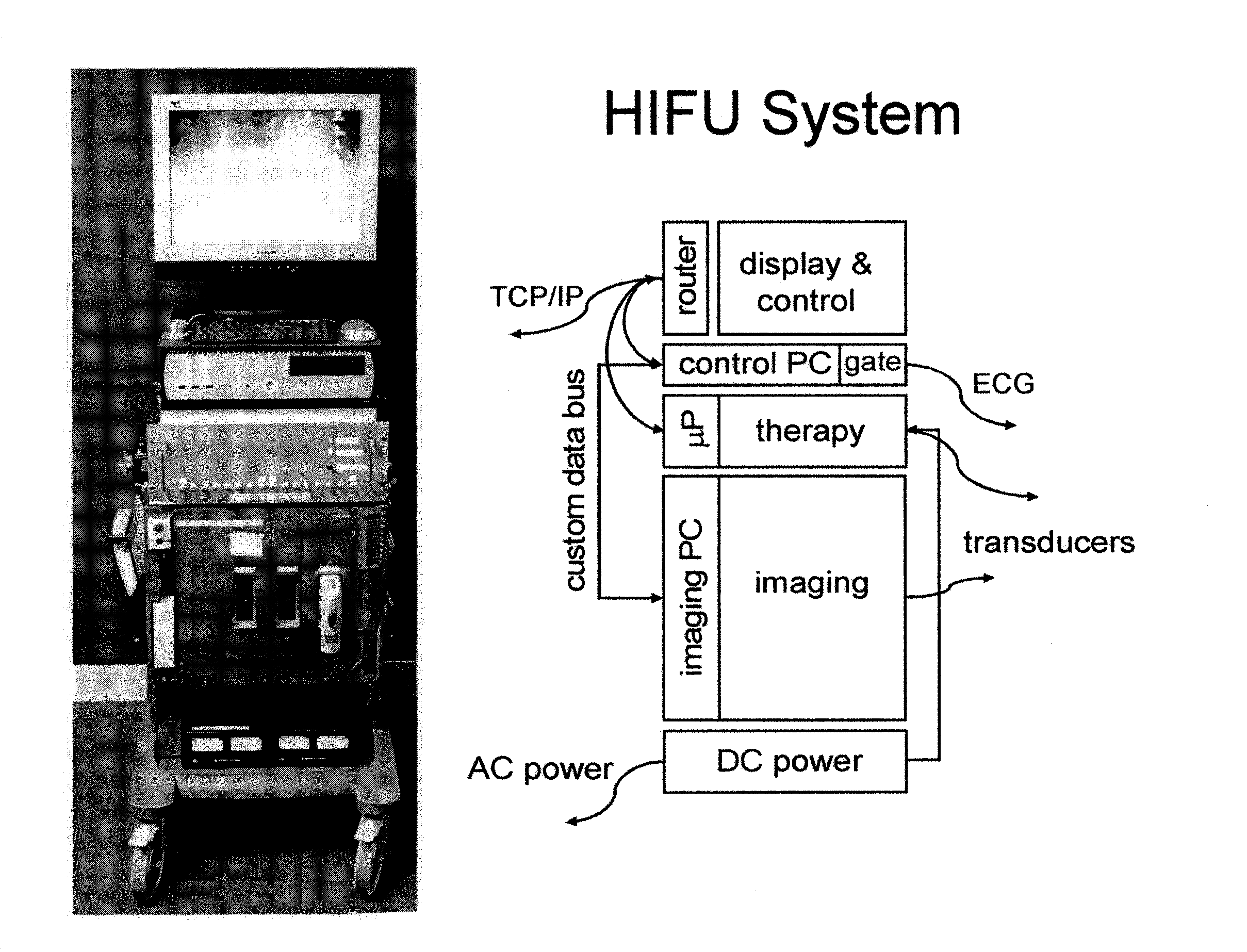
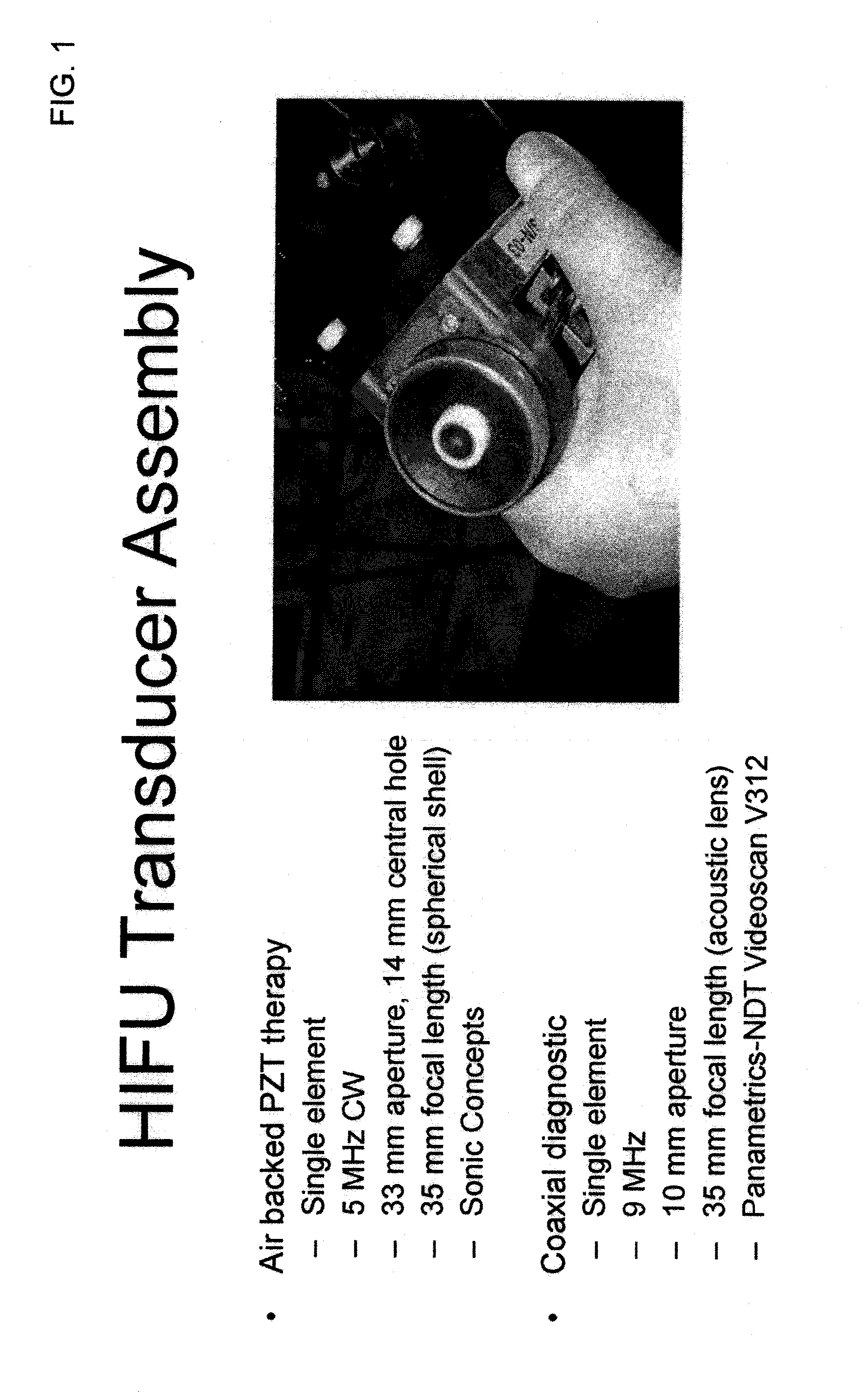
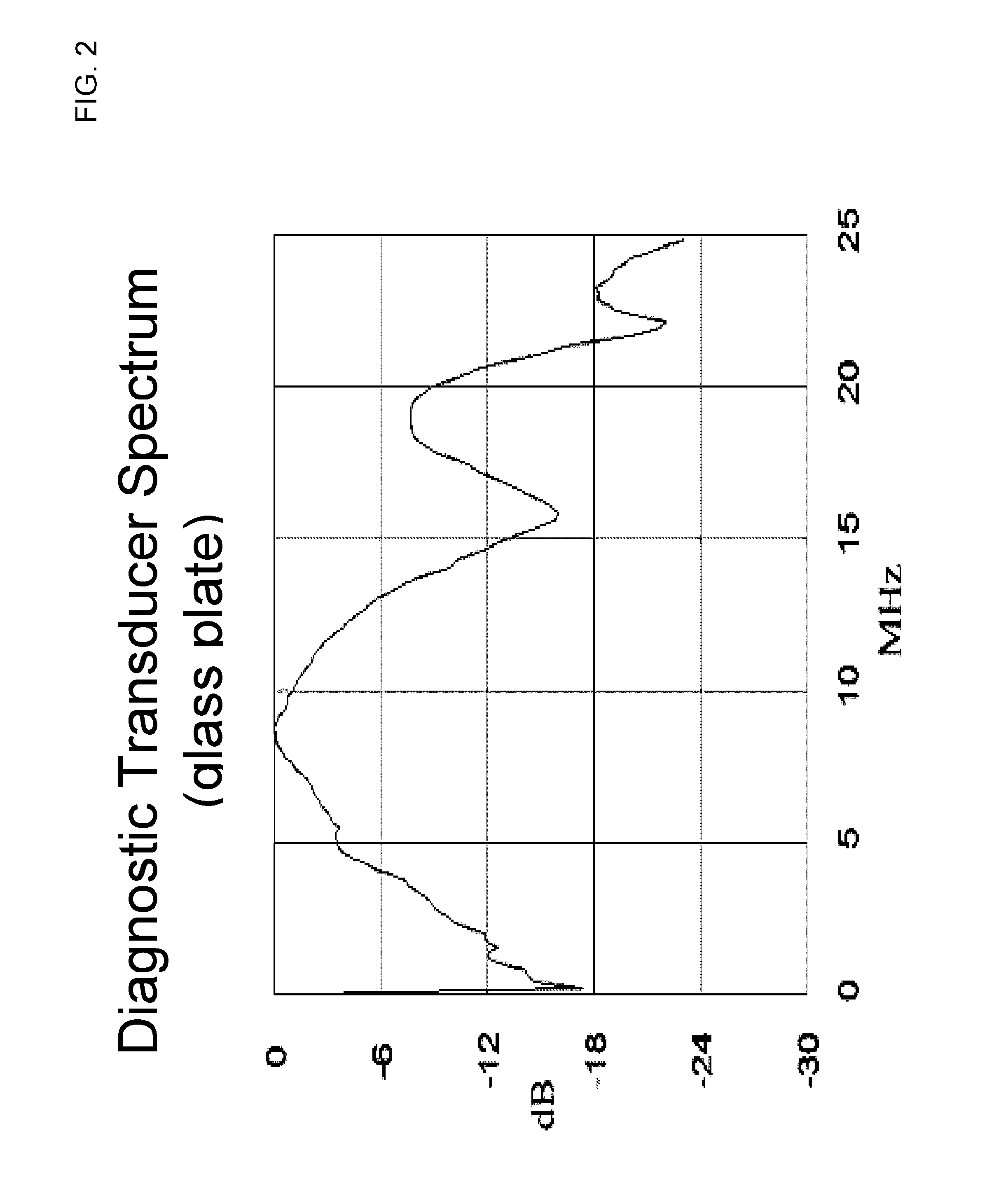
[0024] Imaging systems and visualization techniques are provided for distinguishing lesioned from normal tissue. The imaging systems and visualization techniques are based on spectrum analysis of the harmonics in diagnostic ultrasound waves that are differentially backscattered from lesioned and normal tissues. The imaging systems use suitable diagnostic transducers for the receiving backscattered ultrasound waves. The imaging may be conducted in brightness mode (B-Mode), pitch-catch mode, or any other appropriate mode suitable for defining lesion geometry, location and size.

[0025] The tissue lesions of interest may be the result of natural processes or may be clinically induced, for example, as part of treatments of cardiac disease and cancer. Such treatments may, for example, use HIFU to ablate subsurface structures without injuring intervening tissues. Ultrasonic energy can be applied in a target volume to induce tissue lesions. The imaging systems and lesion visualization techn...


PUM


Abstract

Systems and methods for visualizing lesions in tissue are provided. The systems and the system and methods exploit the differential scattering of ultrasound waves by normal and lesioned tissues to image the lesions. Spectrum analysis of the harmonics in diagnostic ultrasound waves that are differentially backscattered from lesioned and normal tissues provides image contrast and resolution.

Description

CROSS-REFERENCE TO RELATED APPLICATIONS [0001] This application claims the benefit of U.S. provisional patent application No. 60 / 779,348 filed on Mar. 2, 2006, which application is hereby incorporated by reference herein in its entirety. STATEMENT REGARDING FEDERALLY SPONSORED RESEARCH OR DEVELOPMENT [0002] The United States government has certain rights in the present invention pursuant to research grant No. RO1 CA84588 awarded by the National Cancer Institute and the National Heart, Lung, and Blood Institute.FIELD OF THE INVENTION [0003] The present invention relates to systems and methods of imaging and visualization of biological materials (e.g., medical imaging). In particular, the invention relates to visualization of thermal lesions and other structural features of biological tissues. BACKGROUND OF THE INVENTION [0004] High-intensity focused ultrasound (HIFU) technique is a noninvasive extracorporeal technique capable of ablating subsurface structures without injuring interve...

Claims


Application Information

Patent Timeline
IPC IPC(8): A61B8/00
CPCA61B8/08A61N7/02G01S15/899G01S15/8977G01S7/52038
Inventor SILVERMAN, RONALD H.MURATORE, ROBERT
Owner RIVERSIDE RES INST
Why Eureka
Try Eureka