Ultrasonic diagnostic apparatus and ultrasonic diagnostic method

Inactive Publication Date: 2007-10-11
TOSHIBA MEDICAL SYST CORP
View PDF5 Cites 12 Cited by
  • Summary
  • Abstract
  • Description
  • Claims
  • Application Information

AI Technical Summary

Benefits of technology

[0025]The present invention has taken into consideration the above-described problems, and it is an object of the present invention to provide an ultrasonic diagnostic apparatus and an ultrasonic diagnostic metho

Problems solved by technology

However, according to the method using the alternate scanning technology, since the packet size is not large, it is not possible to increase the number of the power signals of the blood flow added to determine the presence/absence of the blood flow.
Therefore, there is such a problem that the above-mentioned advantage to improve the S/N ratio cannot be obtained.
In addition, there is such a problem to reduce a return velocity because the sending period of the ultrasonic waves in the same direction is long.
If the return velocity is low as mentioned above, there is such a da

Method used


Image

  • Ultrasonic diagnostic apparatus and ultrasonic diagnostic method
  • Ultrasonic diagnostic apparatus and ultrasonic diagnostic method
  • Ultrasonic diagnostic apparatus and ultrasonic diagnostic method

Examples


Embodiment Construction

[0038]Hereinbelow, a description will be given of an ultrasonic diagnostic apparatus and an ultrasonic diagnostic method according to an embodiment of the present invention with reference to the drawings.

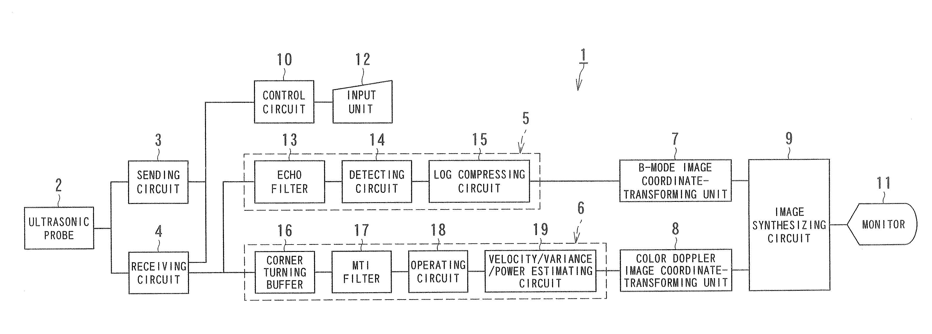
[0039]FIG. 1 is a block diagram showing the structure of the ultrasonic diagnostic apparatus according to the embodiment of the present invention.

[0040]An ultrasonic diagnostic apparatus 1 has an ultrasonic probe 2, a sending circuit 3, a receiving circuit 4, a B-mode processing system 5, a color Doppler processing system 6, a B-mode image coordinate-transforming unit 7, a color Doppler image coordinate-transforming unit 8, an image synthesizing circuit 9, a control circuit 10, a monitor 11, and an input unit 12.

[0041]The ultrasonic probe 2 has a plurality of piezoelectric vibrators containing ceramic in parallel at the end thereof. The piezoelectric vibrators of the ultrasonic probe 2 have a function for generating ultrasonic waves (ultrasounds) on the basis of a voltage pulse appl...


PUM


Abstract

An ultrasonic diagnostic apparatus has a scanning executing unit, a Doppler signal obtaining unit, a storage unit, a removing unit, a blood flow information obtaining unit, and a B-mode image generating unit. The scanning executing unit executes a first scanning that is iteratively performed by a predetermined period so as to obtain a B-mode image of an object and executes a second scanning that is iteratively performed a predetermined number of times by a first period between the first scanning so as to obtain a two-dimensional distribution of a blood flow information and that is iteratively performed by a second period while inserting the first scanning. The Doppler signal obtaining unit sequentially obtains a Doppler signal from a reflection signal obtained by the second scanning. The storage unit stores, for each raster direction, a Doppler signal string at unequal time intervals collected for the first period and the second period by the Doppler signal obtaining unit. The removing unit obtains a blood flow signal by reading the Doppler signal string at the unequal time intervals from the storage unit and removes a signal component with a small motion. The blood flow information obtaining unit obtains the blood flow information based on the blood flow signal. The B-mode image generating unit generates the B-mode image based on the reflection signal obtained by the first scanning.

Description

BACKGROUND OF THE INVENTION[0001]1. Field of the Invention[0002]The present invention relates to an ultrasonic diagnostic apparatus and an ultrasonic diagnostic method that two-dimensionally visualizes a blood flow with a Doppler signal obtained by receiving and sending ultrasonic waves.[0003]2. Description of the Related Art[0004]In diagnosis using an ultrasonic diagnostic apparatus, a method for detecting a blood flow velocity by using a Doppler effect of ultrasonic waves and displaying a blood flow with colors is called a color Doppler method. In a conventional color Doppler ultrasonic diagnostic apparatus having a blood flow display function according to the color Doppler method, ultrasonic waves are received and sent in the same direction plural times. A data string of the ultrasonic waves received and sent in the same direction is called a packet.[0005]By increasing the packet size of the data string of the ultrasonic waves, the detection sensitivity of the blood flow can be i...

Claims


Application Information

Patent Timeline
IPC IPC(8): A61B8/00
CPCG01S15/8979G01S7/52085
Inventor SATO, TAKESHI
Owner TOSHIBA MEDICAL SYST CORP
Why Eureka
Try Eureka