Systems and methods for magnetic resonance imaging

a magnetic resonance imaging and magnetic resonance imaging technology, applied in the field of magnetic resonance imaging, can solve the problems of small neural structures such as spinal cord or optic nerve, and difficult to achieve in vivo high resolution dti of brain regions near temporal bone or sinuses, and achieve the effect of high resolution dti of brain regions near the temporal bone or sinuses, and increase the duration of data

Inactive Publication Date: 2007-10-25
UNIV OF UTAH RES FOUND
View PDF20 Cites 90 Cited by
  • Summary
  • Abstract
  • Description
  • Claims
  • Application Information

AI Technical Summary

Benefits of technology

[0013] In certain embodiments, the first and second inversion RF signals include first and second inversion RF pulses. In certain embodiments, the first inversion RF pulse is applied substantially immediately after the excitation RF signal. In certain embodiments, the first and second inversion RF pulses are separated by a time approximately 5 ms or larger. In certain embodiments, the method further includes providing slice-selective gradients that are selected such that magnetization within the FOV is substantially preserved while magnetization external to the FOV is substantially suppressed, thereby allowing magnetization in each of a plurality of slices to be substantially maintained in its equilibrium state while exciting and imaging one or more others of the plurality of slices.
[0020] In certain embodiments, the first and second inversion RF signals include first and second inversion RF pulses. In certain embodiments, the first inversion RF pulse is applied substantially immediately after the excitation RF signal. In certain embodiments, the first and second inversion RF pulses are separated by a time approximately 5 ms or larger. In certain embodiments, the one or more instructions further includes an instruction for providing slice-selective gradients that are selected such that magnetization within the FOV is substantially preserved while magnetization external to the FOV is substantially suppressed, thereby allowing magnetization in each of a plurality of slices to be substantially maintained in its equilibrium state while exciting and imaging one or more others of the plurality of slices.

Problems solved by technology

Although DTI can provide useful information about white-matter diseases in the brain, high resolution DTI of brain regions near the temporal bone or sinuses, of small neural structures such as the spinal cord or optic nerve, or of extracranial organs in vivo has been difficult to achieve using conventional two-dimensional (2D) singleshot diffusion-weighted EPI techniques (2D ss-DWEPI).
There are strong non-uniform local magnetic fields created by the magnetic susceptibility changes at tissue / bone or tissue / air interfaces, which typically induce severe distortion on the resultant ss-DWEPI images.
Typically, increasing spatial resolution requires an increase in the duration of the data acquisition window, which in turn increases the distortion from off-resonance effects.
As a result, the spatial resolution obtained using conventional 2D ss-EPI is generally much lower than that obtainable with conventional, multi-shot MRI, giving decreased resolution for measurements of interest, such as white matter tract anatomy and nerve fiber anatomy.
However most multishot DWI acquisition techniques suffer from the instability of phase errors among shots due to global or localized motions during application of the large diffusion gradients.
Because most of these are 2D acquisition techniques, they produce relatively poor resolution along the slice direction.

Method used


Image

  • Systems and methods for magnetic resonance imaging
  • Systems and methods for magnetic resonance imaging
  • Systems and methods for magnetic resonance imaging

Examples


Embodiment Construction

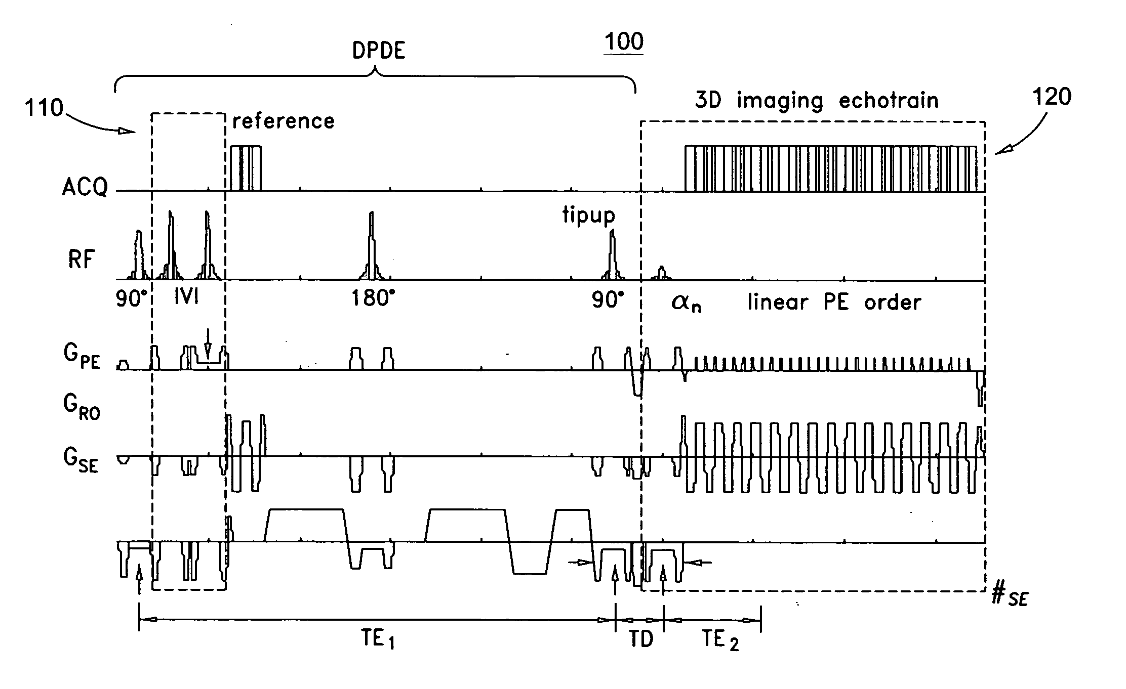
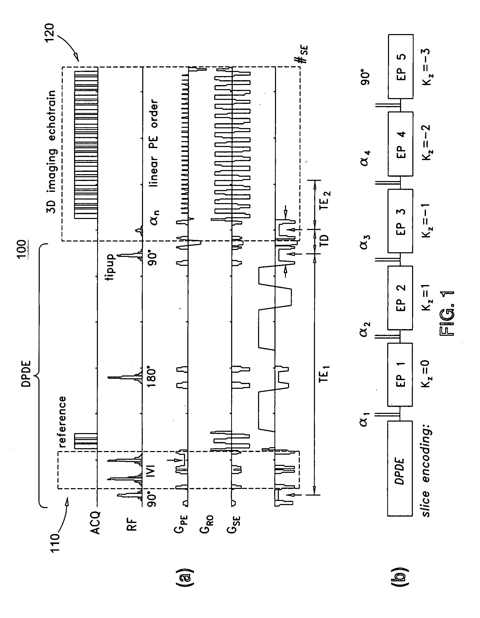
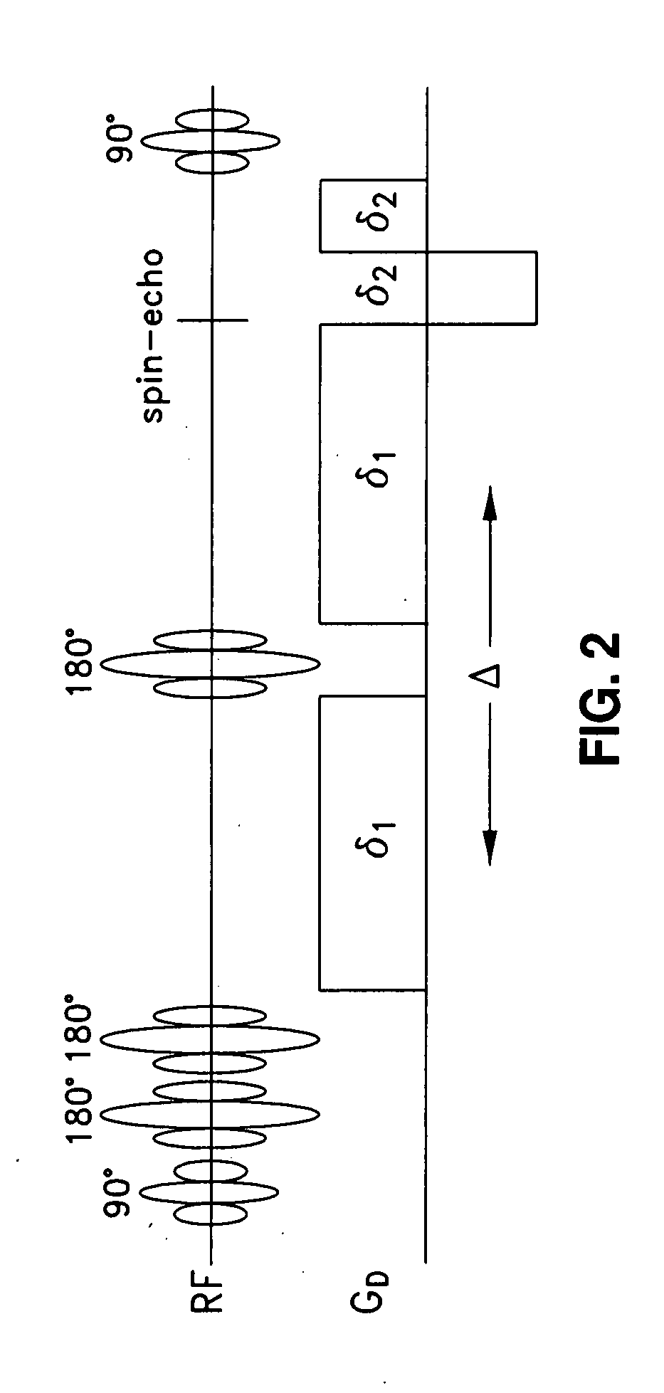
[0047] An improved three-dimensional (3D) singleshot stimulated echo planar imaging (3D ss-DWSTEPI or ss-STEPI or STEPI) is presented which includes a novel technique to perform 3D singleshot DWI and DTI of a restricted 3D volume. In certain embodiments, 3D ss-DWSTEPI acquires 3D raw data from a limited 3D volume after a single diffusion-prepared driven-equilibrium (DPDE) preparation by short EPI readouts of several stimulated echoes. In certain embodiments, the raw data includes k-space data. In certain embodiments, the raw data includes data that has not undergone any transformation. The EPI readout time is preferably shortened by using an inner volume imaging (IVI) technique along the phase-encoding direction.

[0048] In certain embodiments, 3D ss-STEPI may be used to image any localized anatomical volume within a body without aliasing artifact and with high-resolution. The results from 3D ss-STEPI imaging studies of phantoms, an excised animal heart, and in vivo results from huma...


PUM


Abstract

Methods and apparatus for operating an MRI system is provided. The disclosure provides a diffusion-prepared driven-equilibrium preparation for an imaging volume and acquiring 3-dimensional k-space data from said prepared volume by a plurality of echoplanar readouts of stimulated echoes. An excitation radio-frequency signal and first and second inversion RF signals are provided to define a field-of-view (FOV).

Description

CROSS-REFERENCE TO RELATED APPLICATIONS [0001] This application claims the benefit under 35 U.S.C. §119(e) of U.S. Provisional Patent Application No. 60 / 788,533, filed on Mar. 31, 2006, and titled “SYSTEMS AND METHODS FOR MAGNETIC RESONANCE IMAGING,” the entirety of which is hereby incorporated by reference.GOVERNMENT LICENSE RIGHTS [0002] The U.S. Government has a paid-up license in this invention and the right in limited circumstances to require the patent owner to license others on reasonable terms as provided for by the terms of Grant Nos. R21 NS052424, R21 EB005705, and R01 HL57990 awarded by the National Institutes of Health.BACKGROUND OF THE INVENTION [0003] 1. Field of the Invention [0004] This application relates to magnetic resonance imaging. [0005] 2. Description of the Related Art [0006] Diffusion-weighted magnetic resonance (MR) imaging (DWI) is a known tool for detecting abnormal water diffusion in the brain (e.g., ischemic stroke). The directional information obtained...

Claims


Application Information

Patent Timeline
Patent Type & Authority Applications(United States)
IPC IPC(8): A61B5/05
CPCG01R33/4806G01R33/5615G01R33/5616A61B5/055G01R33/56509G01R33/56536G01R33/5676G01R33/56341
Inventor JEONG, EUN-KEEPARKER, DENNIS L.CHOI, KIM SEONG-EUNKHOLMOVSKI, EVGUENI G.
Owner UNIV OF UTAH RES FOUND
Why Eureka
Try Eureka