

Compound subretinal prostheses with extra-ocular parts and surgical technique therefore
a subretinal prosthesis and surgical technique technology, applied in the field of compound subretinal prosthesis with extraocular parts and surgical technique, can solve the problems of inability to fully address and solve surgical and technical problems, and the inability to use subretinal prostheses with permanent extra-ocular connections to supply additional energy, etc., to achieve a highly controlled introduction and minimal damage to the retina and the retinal pigment epithelium
- Summary
- Abstract
- Description
- Claims
- Application Information
AI Technical Summary
Benefits of technology
Problems solved by technology
Method used
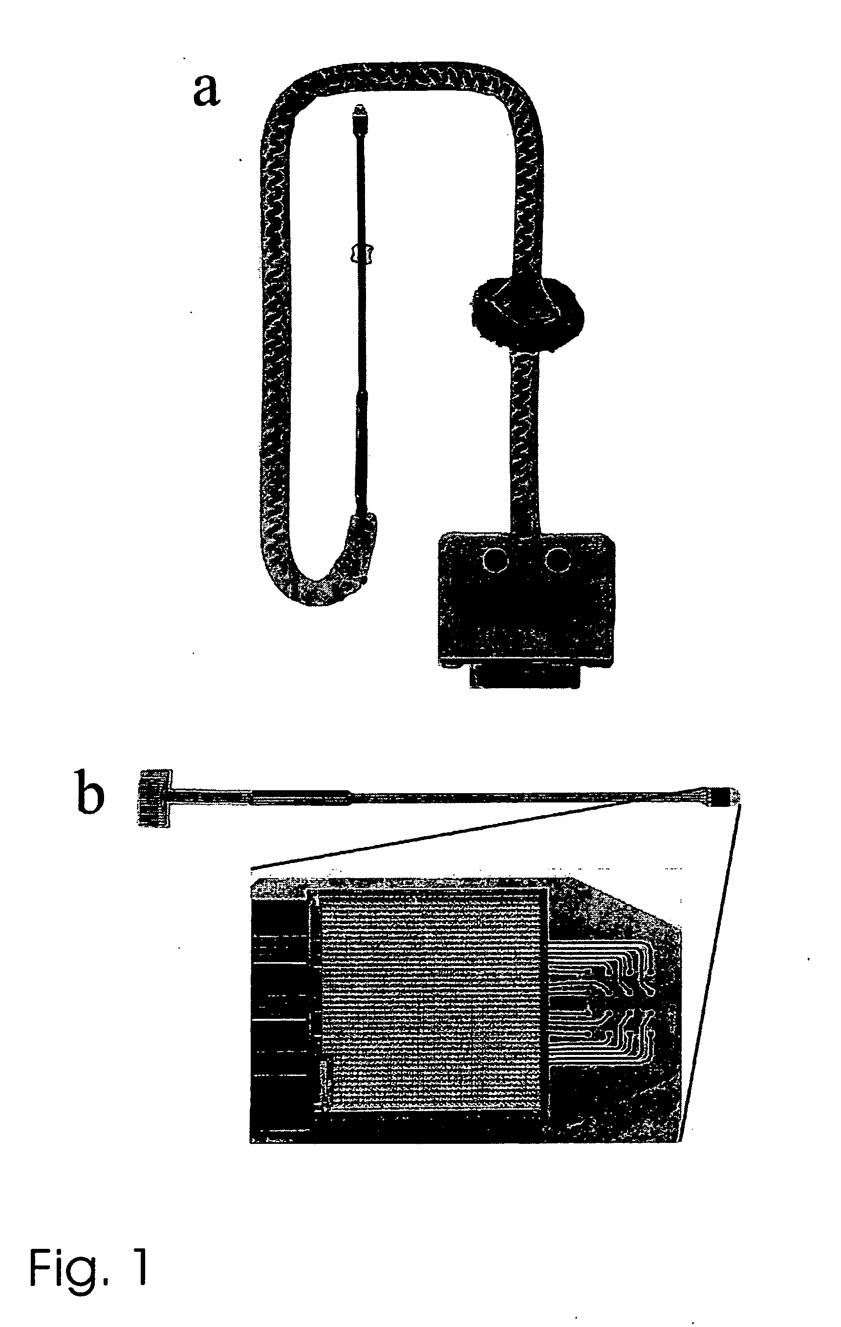
Image

Examples
embodiment 1
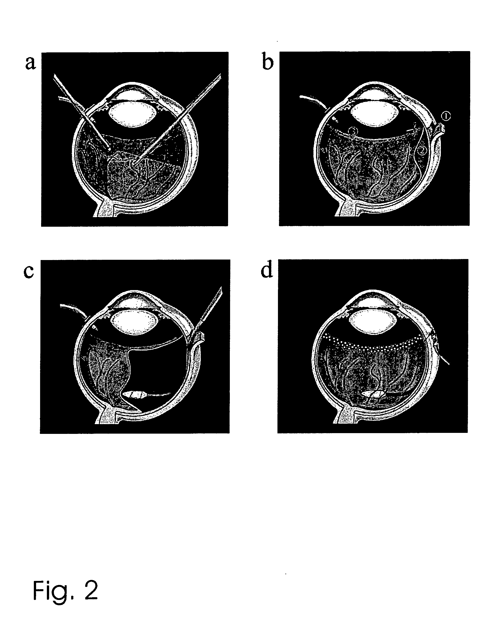
2. The method of embodiment 1, wherein in step b) a subretinal cannula is used to create said localized bubble.
3. The method of embodiment 1 or 2, wherein in step c) the retinotomy is performed around 180° and approximately half of the retina is detached.
[0071] 4. The method of any one of embodiments 1 to 3, wherein in step c), during choroidal penetration, localized application of diathermia, localized application of laser energy, systemic reduction of blood pressure, localized application of cryotherapy, localized application of vasoconstrictory agents, and the like is used to avoid choroidal hemorrhage.
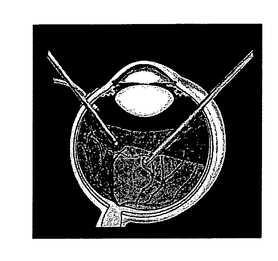
5. The method of any one of embodiments 1 to 4, wherein in step d) subretinal forceps under fundus visualization are used for positioning the implant.
6. The method of any one of embodiments 1 to 5, wherein in step e) a physiologically acceptable solution, preferably a heavy fluid such as Perfluorodecaline is instilled to flatten the retina, prior to closing the sclera flap.
embodiment 6
7. The method of embodiment 6, wherein in step e), after the retina has been flattened, the eye is filled with silicone oil and a circumferential peripheral laser photocoagulation is applied near the ora serrata.
embodiment 7
8. The method of embodiment 7, wherein in step e), after the sclera flap has been closed, at least one fixating patch provided at said connecting portion of the implant is attached to the sclera before the conjunctiva is closed.
PUM
 Abstract
Description
Claims
Application Information