[0012]In one aspect, the present invention provides a method of creating an incision in a body structure. An electrosurgical probe is positioned adjacent the target tissue so that one or more active electrode(s) are brought into at least partial contact or close proximity with the target tissue.
High frequency voltage is then applied between the active electrode(s) and one or more return electrode(s) and the active electrode(s) are moved, translated, reciprocated, or otherwise manipulated to
cut through a portion of the tissue. In some embodiments, an
electrically conductive fluid, e.g., isotonic
saline or conductive gel, is delivered or applied to the
target site to substantially surround the active electrode(s) with the fluid. In other embodiments, the active electrode(s) are immersed within the
electrically conductive fluid. In both embodiments, the
high frequency voltage may be selected to effect a controlled depth of
hemostasis of severed blood vessels within the tissue, which greatly improves the surgeon's view of the
surgical site.
[0013]In one aspect, tissue is
cut, dissected, and / or transected by molecular dissociation or disintegration processes. (In contrast, in conventional
electrosurgery tissue is
cut by rapidly heating the tissue until cellular fluids explode, producing a
cutting effect along the pathway of localized heating.) The present invention volumetrically removes the tissue along the
cutting pathway in a cool
ablation process that minimizes
thermal damage to surrounding tissue. In these embodiments, the high frequency voltage applied to the active electrode(s) is sufficient to vaporize the electrically
conductive fluid (e.g., gel or
saline) between the active electrode(s) and the tissue. Within the vaporized fluid, a
plasma is formed and charged particles (e.g., electrons) cause the molecular breakdown or disintegration of the tissue, perhaps to a depth of several
cell layers. This molecular dissociation is accompanied by the volumetric removal of the tissue, e.g., along the incision of the tissue. This process can be precisely controlled to effect the volumetric removal of tissue as thin as 10 microns to 150 microns with minimal heating of, or damage to, surrounding or underlying tissue structures. A more complete description of this phenomenon is described in commonly assigned U.S. Pat. No. 5,683,366, the complete disclosure of which is incorporated herein by reference.
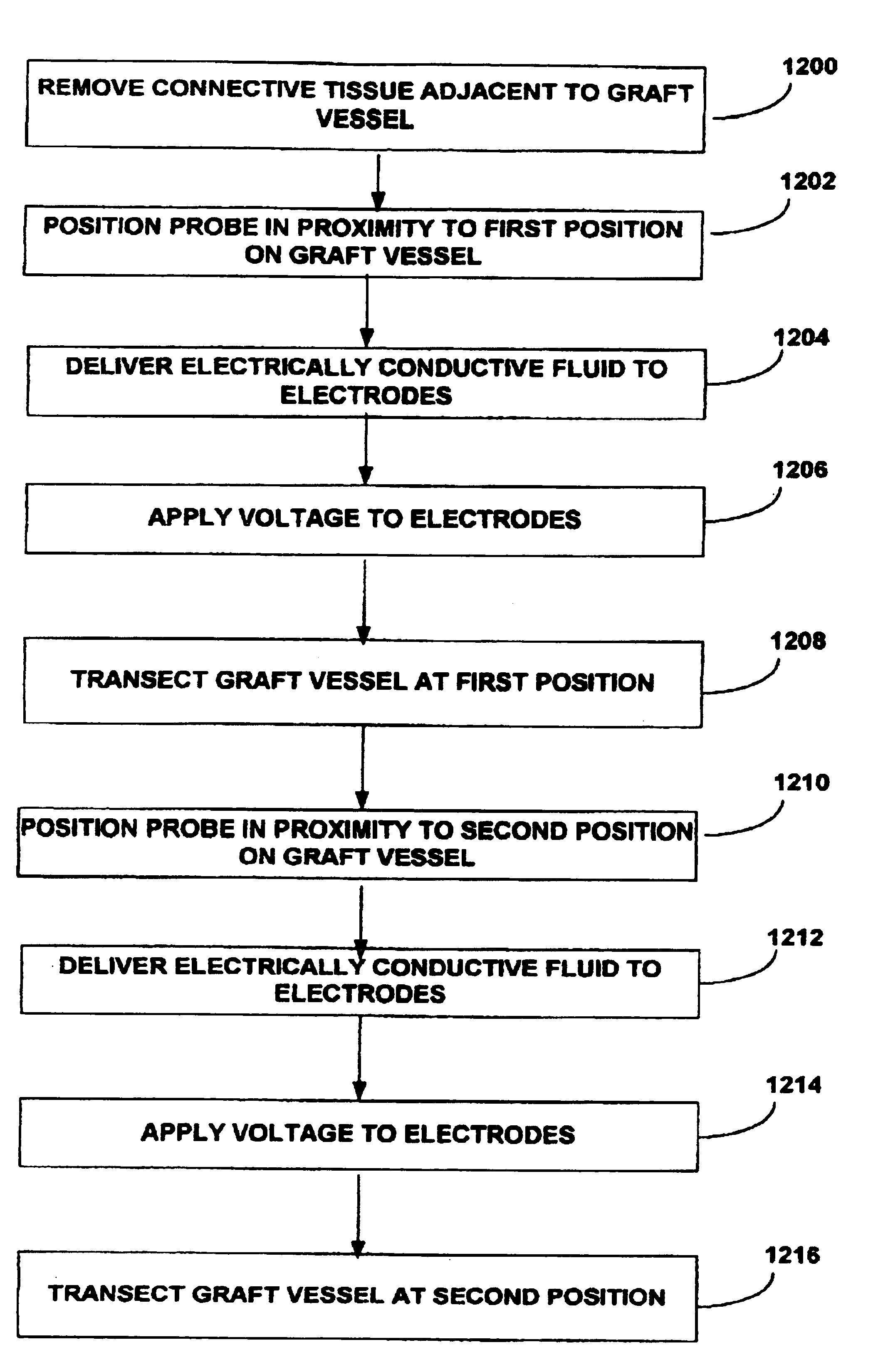
[0015]In another exemplary embodiment, the present invention provides a method for harvesting the IMA from a patient. The electrosurgical probe is positioned either by direct
visualization and access of a sternotomy or indirectly via endoscopic access adjacent the IMA and high frequency electrical energy is applied between one or more active electrode(s) and one or more return electrode(s). The probe is then moved so that the active electrode(s) volumetrically removes
connective tissue adjacent to the IMA so that the IMA is free from
connective tissue along a portion of its length. In an exemplary embodiment, the probe is positioned adjacent to the IMA, and advanced along the length of the IMA while high frequency electrical energy is applied between the active electrode(s) and a return electrode to remove or cut the
connective tissue or other structures surrounding the IMA. The residual heat from the electrical energy may also provides simultaneous
hemostasis of severed blood vessels, which increases
visualization and improves
recovery time for the patient. In addition, the ability to simultaneously cut through tissue on either side of the IMA decreases the length of the procedure, which further improves patient
recovery time. After a suitable length of the IMA has been dissected, it may be transected, and anastomosed to a diseased coronary
artery using known methods. In some embodiments, an electrically
conductive fluid (liquid, gas, or gel) is placed at the
target site adjacent to the IMA so as to provide a current flow path between the return electrode and the active electrode.
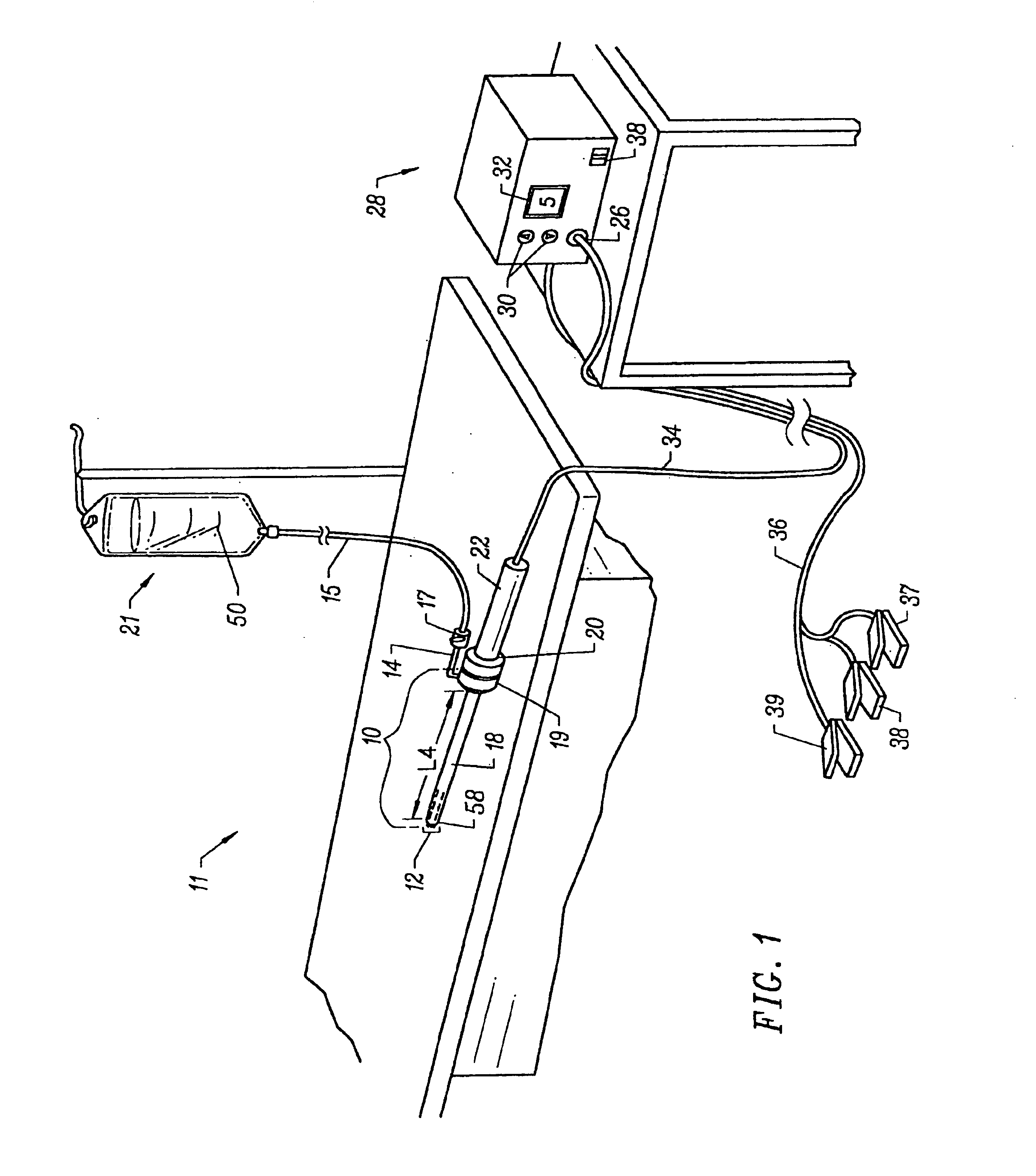
[0017]The apparatus can further include a fluid delivery element for delivering electrically
conductive fluid to the active electrode(s) and the target site. The fluid delivery element may be located on the probe, e.g., a fluid lumen or tube, or it may be part of a separate instrument. Alternatively, an electrically conductive gel or spray, such as a
saline electrolyte or other conductive gel, may be applied the target site. In this embodiment, the apparatus may not have a fluid delivery element. In both embodiments, the electrically conductive fluid preferably provides a current flow path between the active electrode(s) and one or more return electrode(s). In an exemplary embodiment, the return electrode is located on the probe and spaced a sufficient distance from the active electrode(s) to substantially avoid or minimize current shorting therebetween and to shield the return electrode from tissue at the target site.
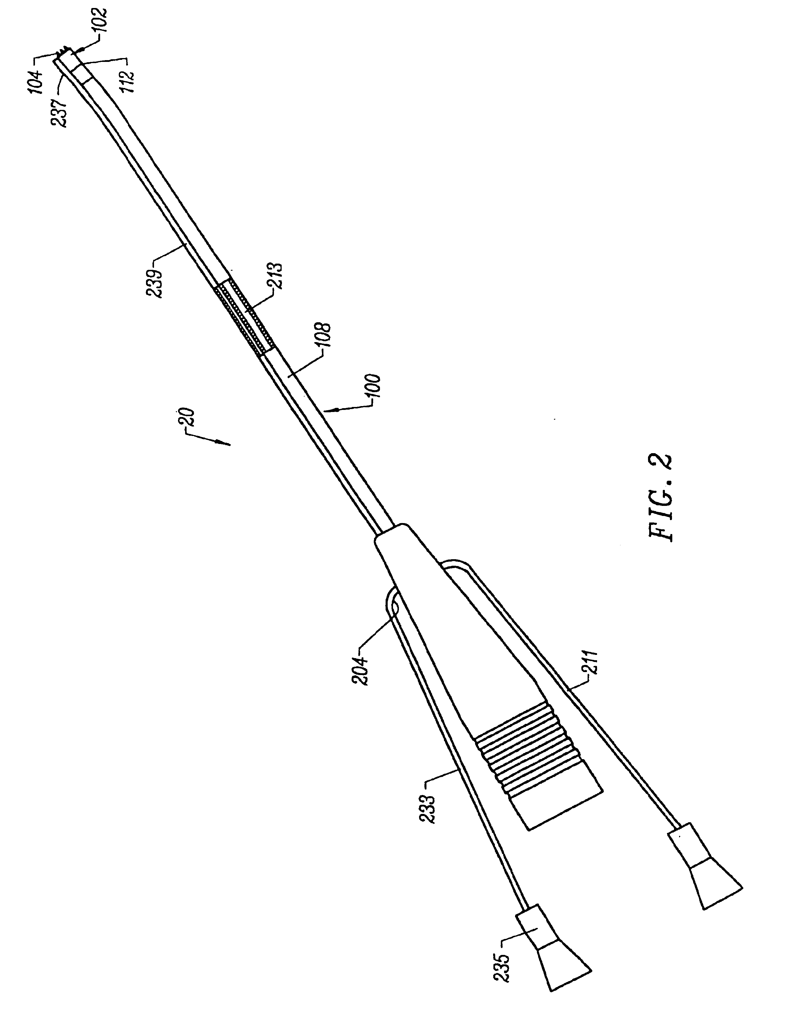
[0020]In another aspect of the invention, there is provided an electrosurgical probe having a blade-like active electrode affixed to an electrically insulating electrode support on the distal end of a shaft. In a specific configuration, the active electrode is in the form of a substantially flat
metal blade having an
active edge and first and second blade sides. The return electrode is typically located on the shaft distal end proximal to the electrode support. In use, the active electrode and the return electrode are coupled to opposite poles of a
high frequency power supply. The
active edge may have a variety of shapes, and is adapted for generating
high current densities thereon, and for precisely severing or ablating tissue or an organ in a highly controlled manner via molecular dissociation of tissue components. The first and second blade sides are adapted for engaging with tissue, such as tissue severed by the
active edge, and for coagulating tissue engaged therewith.
[0022]In another aspect of the invention, there is provided a method of harvesting a tissue or organ using an electrosurgical probe having at least one active electrode disposed on the distal end of the probe. In one embodiment, the active electrode is in the form of a single blade including an active edge and first and second blade sides. In situations where the tissue to be harvested is concealed by an overlying tissue, the tissue to be harvested must first be accessed by incising or removing the overlying tissue. Removal of the overlying tissue may be performed in various ways, including: 1) mechanically, e.g. using a scalpel, rongeur,
surgical saw or
drill, etc. or a combination thereof; 2) via conventional
electrosurgery, e.g., a Bovie; or 3) using an electrosurgical probe of the instant invention adapted for severing tissue in a cool
ablation process involving molecular dissociation of tissue components. Once the tissue or organ to be harvested is accessible, the tissue or organ to be harvested may be dissected by juxtaposing the active edge of the blade electrode against the surrounding connective tissue, and applying a high frequency voltage difference between the active and return electrodes sufficient to cause molecular dissociation of connective tissue components. In this way, the connective tissue adjacent to the active electrode is ablated at a temperature in the range of 40° C. to 70° C., with no, or minimal,
thermal damage to the tissue to be harvested.


