Electroporation to interrupt blood flow

a technology of electroporation and blood flow, applied in the field of electroporation devices, can solve the problems of undesirable irreversible electroporation of tissue, and achieve the effect of minimizing cellular damage to surrounding cells and tissue and high control level

Inactive Publication Date: 2005-08-04
RGT UNIV OF CALIFORNIA
View PDF7 Cites 289 Cited by
  • Summary
  • Abstract
  • Description
  • Claims
  • Application Information

AI Technical Summary

Benefits of technology

[0026] The invention provides the simultaneous irreversible electroporation of multitudes of cells providing a direct indication of the actual occurrence of electroporation and an indication of the degree of electroporation averaged over the multitude sufficient to disrupt blood flow. The discovery is likewise useful in the irreversible electroporation of cells which make-up vessels and of cells in blood. The benefits of this process include a high level of control over the beginning point of irreversible electroporation and the control of blood flow to and out of very specific areas.
[0027] A feature of the invention is t

Problems solved by technology

Others have generally considered irreversible electroporation of tissue to be undesirable due to the possibility of ins

Method used


Image

  • Electroporation to interrupt blood flow
  • Electroporation to interrupt blood flow
  • Electroporation to interrupt blood flow

Examples


example 1

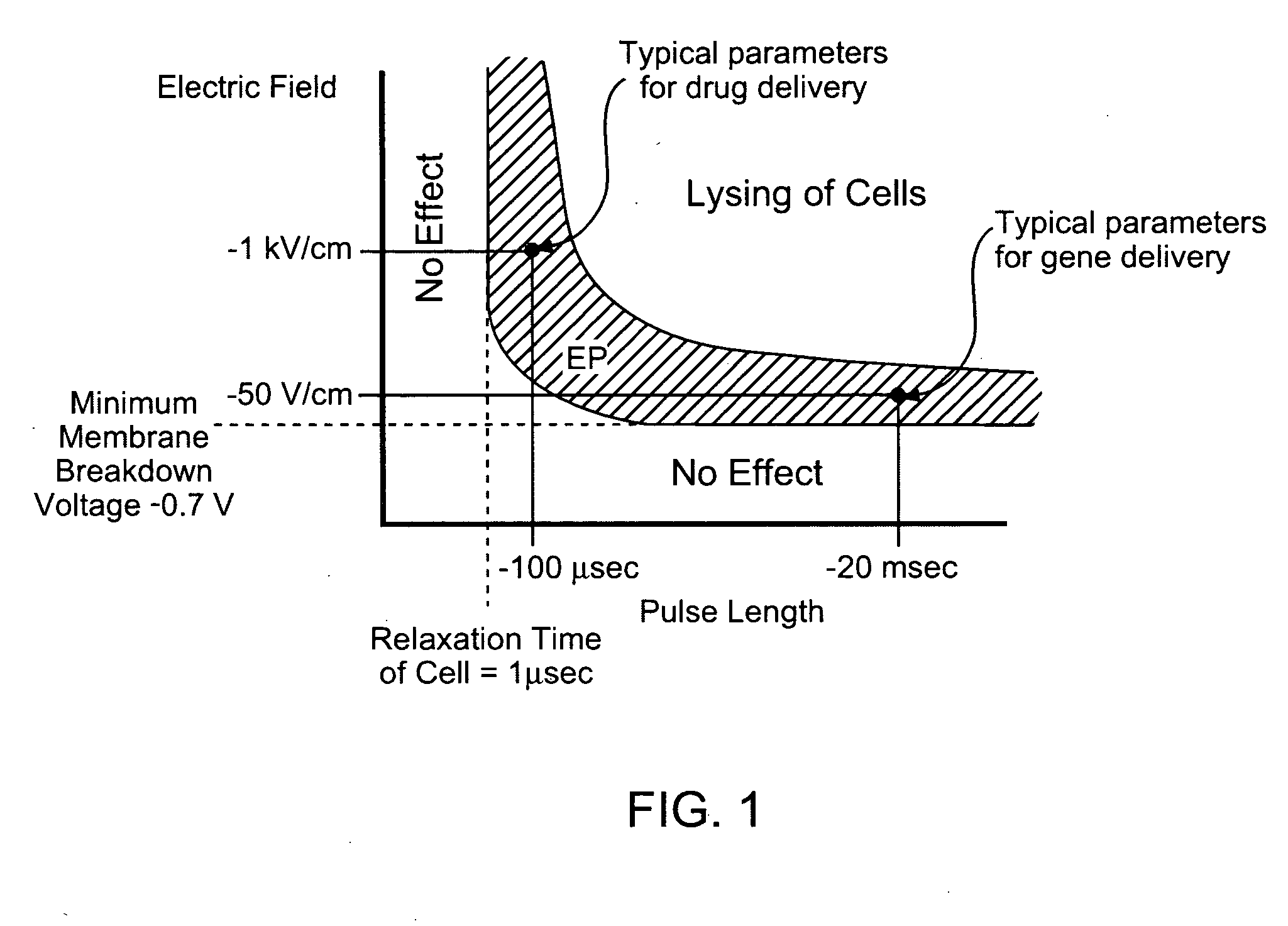
[0090] The mathematical model provided here shows that irreversible tissue ablation can affect substantial volumes of tissue, without inducing damaging thermal effects. To this end, the present invention uses the Laplace equation to calculate the electrical potential distribution in tissue during typical electroporation pulses and a modified Pennes (bioheat), (Pennes, H. H., Analysis of tissue and arterial blood flow temperatures in the resting forearm. J of Appl. Physiology., 1948. 1: p. 93-122), equation to calculate the resulting temperature distribution. It is important to note that there are several forms of the bioheat equation which have been reviewed (Carney, C. K., Mathematical models of bioheat transfer, in Bioengineering heat transfer, Y. I. Choi, Editor. 1992, Academic Press, Inc: Boston. p. 19-152; Eto, T. K. and B. Rubinsky, Bioheat transfer, in Introduction to bioengineering, S. A. Berger, W. Goldsmith, and E. R. Lewis, Editors. 1996, Oxford press). While the Pennes e...

example 2

[0113] This example was developed to produce a correlation between electroporation pulses and thermal effects. The system analyzed is an infinitesimally small control volume of tissue exposed to an electroporation voltage gradient of V (Volts / cm). The entire electrical energy is dissipated as heat and there is no conduction of heat from the system. The calculations produce the increase in temperature with time during the application of the pulse and the results are a safe lower limit for how long a certain electroporation pulse can be administered until a certain temperature is reached. To generate the correlation an energy balance is made on a control volume between the Joule heating produced from the dissipation of heat of the V (volt / cm) electrical potential dissipating through tissue with an electrical conductivity of σ (ohm-cm) and the raise in temperature of the control volume made of tissue with a density ρ (g / cc) and specific heat, c, (J / g K). the calculation produces the fo...

example 3

[0120] The goal of this experiment was to verify the ability of irreversible electroporation pulses to produce substantial tissue ablation in the non-thermal regime. To this end we have performed experiments on the liver of Spraque-Dawley male rats (250 g to 350 g) under an approved animal use and care protocol. After the animals were anesthetized by injection of Nembutal Sodium Solution (50 mg / ml Pentobarbital) the liver was exposed via a midline incisions and one lobed clamped between two cylindrical electrodes of Ag / AgCl, with a diameter of 10 mm (In Vivo Metric, Healdsburg, Calif.). The electrodes had their flat surface parallel; they were concentric and the liver between the electrodes was compressed so that the lobes were separated by 4 mm. A schematic of the electrodes and the liver is shown in FIG. 9. The liver was exposed to a single electroporation pulse of 40 milliseconds. One electrode was set to 400 V and the other grounded. The rest of the liver was not in contact with...


PUM


Abstract

A method for disrupting blood flow to undesirable tissue such as cells of a cancerous or non-cancerous tumor is disclosed. It involves the placement of electrodes into or near the vicinity of vessels supplying blood to the undesirable tissue and through the application of electrical pulses causing blood flow disruption. The electric pulses irreversibly permeate the cell membranes, thereby invoking cell death. The irreversibly permeabilized cells are left in situ and are removed by the body immune system. The process may further comprise monitoring blood flow and/or infusion of a material such as a chemotherapeutic agent or marker into the blood.

Description

CROSS-REFERENCE [0001] This application claims the benefit of U.S. Provisional Application No. 60 / 532,588, filed Dec. 24, 2003, which application is incorporated herein by reference.FIELD OF THE INVENTION [0002] This invention relates to the field of electrode devices useful to disrupt blood flow in order to carry out treatments. BACKGROUND OF THE INVENTION [0003] In many medical procedures, such as the treatment of benign or malignant tumors, it is important to be able to ablate the undesirable tissue in a controlled and focused way without affecting the surrounding desirable tissue. Over the years, a large number of minimally invasive methods have been developed to selectively destroy specific areas of undesirable tissues as an alternative to resection surgery. There are a variety of techniques with specific advantages and disadvantages, which are indicated and contraindicated for various applications. For example, cryosurgery is a low temperature minimally invasive technique in w...

Claims


Application Information

Patent Timeline
IPC IPC(8): A61B18/04A61B18/12A61B18/18A61N1/00A61N1/04A61N1/05A61N1/32
CPCA61B18/12A61B18/1477A61B2018/1425A61N1/327A61N1/0472A61N1/05A61N1/0412A61B18/1233A61B2018/00577A61B2018/00613A61B2018/00714A61B2018/00761A61B2018/00827
Inventor RUBINSKY, BORISEDD, JONHOROWITZ, LIANA
Owner RGT UNIV OF CALIFORNIA
Why Eureka
Try Eureka