
Acoustical-based tissue resuscitation

a tissue resuscitation and acoustic technology, applied in the field of acoustic vibrational based methods, can solve the problems of invasive and associated with various complications, rapid damage or death of affected tissue, and approved uses, and achieve the effects of convenient use, low cost of devices, and easy adaptability to a wide variety of situations

Active Publication Date: 2012-06-12
VIRGINIA COMMONWEALTH UNIV
View PDF30 Cites 2 Cited by
  • Summary
  • Abstract
  • Description
  • Claims
  • Application Information

AI Technical Summary

Benefits of technology

[0019]Another advantage of the invention is that the devices used to carry out the methods are portable, convenient for use, and readily adaptable to a wide variety of situations. For example, the technology may be utilized in emergency situations, in pre-hospital settings (e.g., by paramedics), in emergency rooms, operating rooms, intensive care units, schools, airports, in the home, etc. The devices are also suitable for use in remote locations where it would be difficult or impossible to provide equipment or highly specialized personnel such as surgical equipment and surgeons to carry out more invasive techniques. Further, the methodology requires minimal or no participation of the subject being treated. Thus, even an unconscious patient in a supine position may be treated with relative ease. Also, the cost of the devices is relatively low, making them fiscally accessible to health care practitioners in less affluent settings.
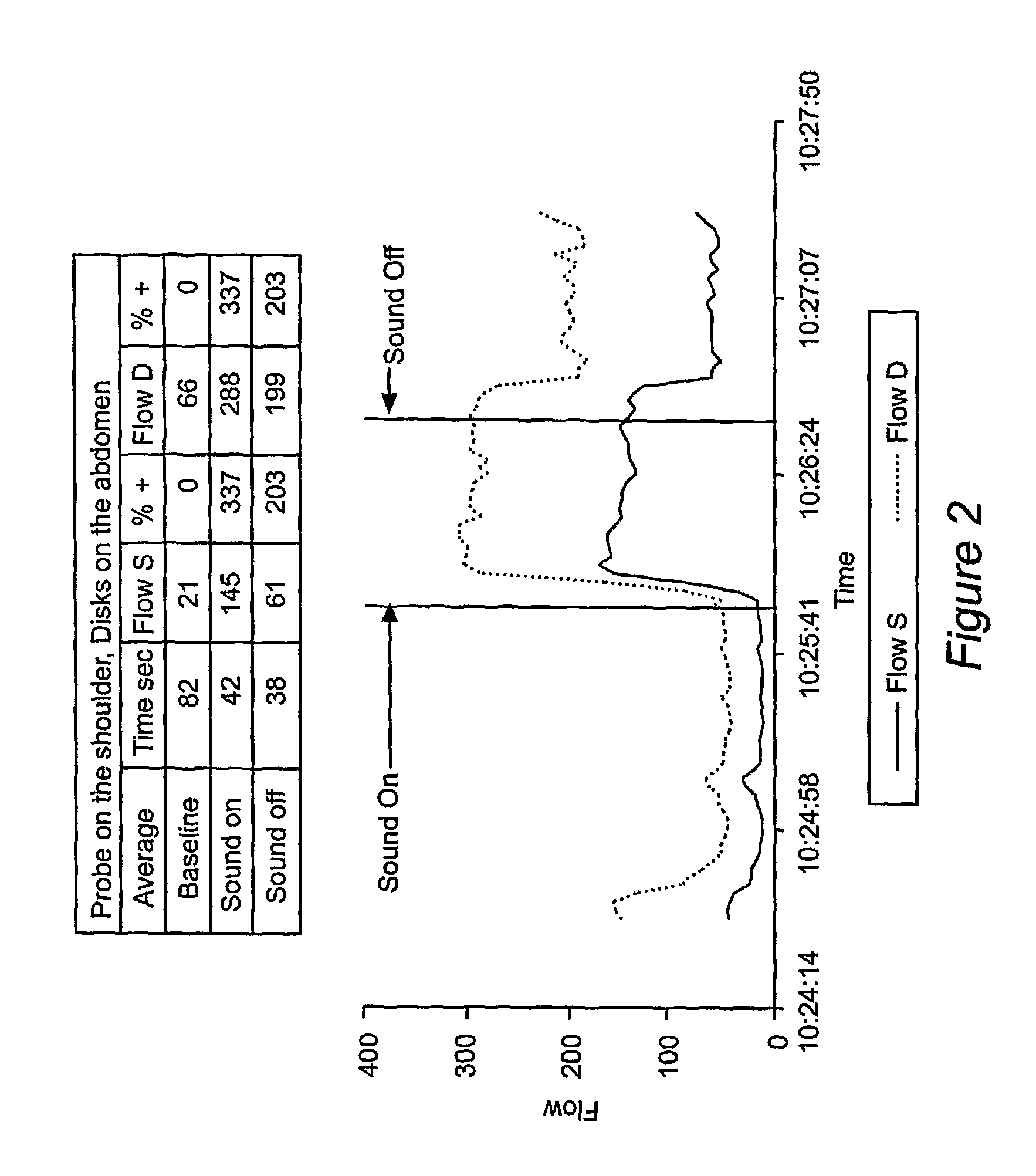
[0020]An additional advantage of the method is that it is subject to a high level of control. The effects of sound and / or vibrational transmission on blood flow are rapid, with increases occurring within seconds of the application of a source of low frequency acoustical energy. Likewise, after withdrawal of the source of the acoustical energy, the effects diminish within minutes. Thus, the effect of sound and / or vibrational transmission on blood flow in the targeted tissue can be closely regulated and accurately titrated.
[0021]According to the methods of the invention, a source of low frequency sound and / or vibration is typically placed directly on a protective layer or material that is external to the tissue in which blood flow is to be increased. In a preferred embodiment, this protective layer is skin, and the sound or vibrational source is placed directly on the skin. However, those of skill in the art will recognize that there may be instances where the protective layer is a substance other than skin, for example, a synthetic bandage or bioartificial covering such as those used to cover wounds (e.g. of burn patients); or some other type of prosthetic covering or device.
[0022]Further, circumstances may arise which preclude direct contact with skin, so that the source is instead placed on material that covers the skin (e.g. clothing), for example, in an emergency situation where there is not sufficient time to remove a patient's clothing, or when removal might be inadvisable (patient or patients limbs cannot be moved without causing possible further trauma), or in which direct exposure of the skin might otherwise have untoward effects (e.g. treatment being carried out in conditions of severe cold, exposure to chemicals, etc.).
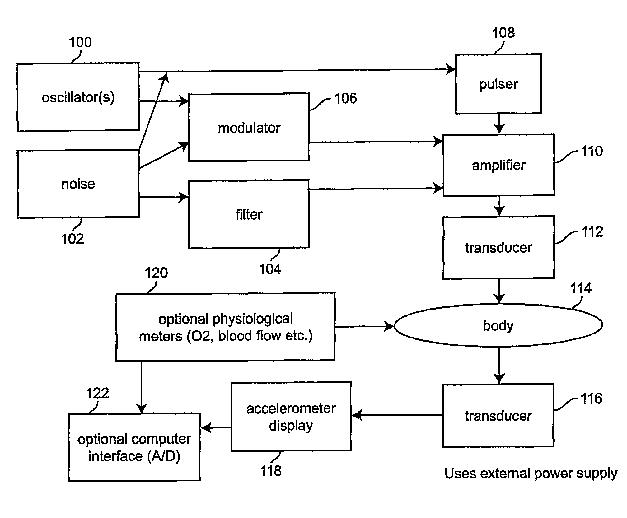
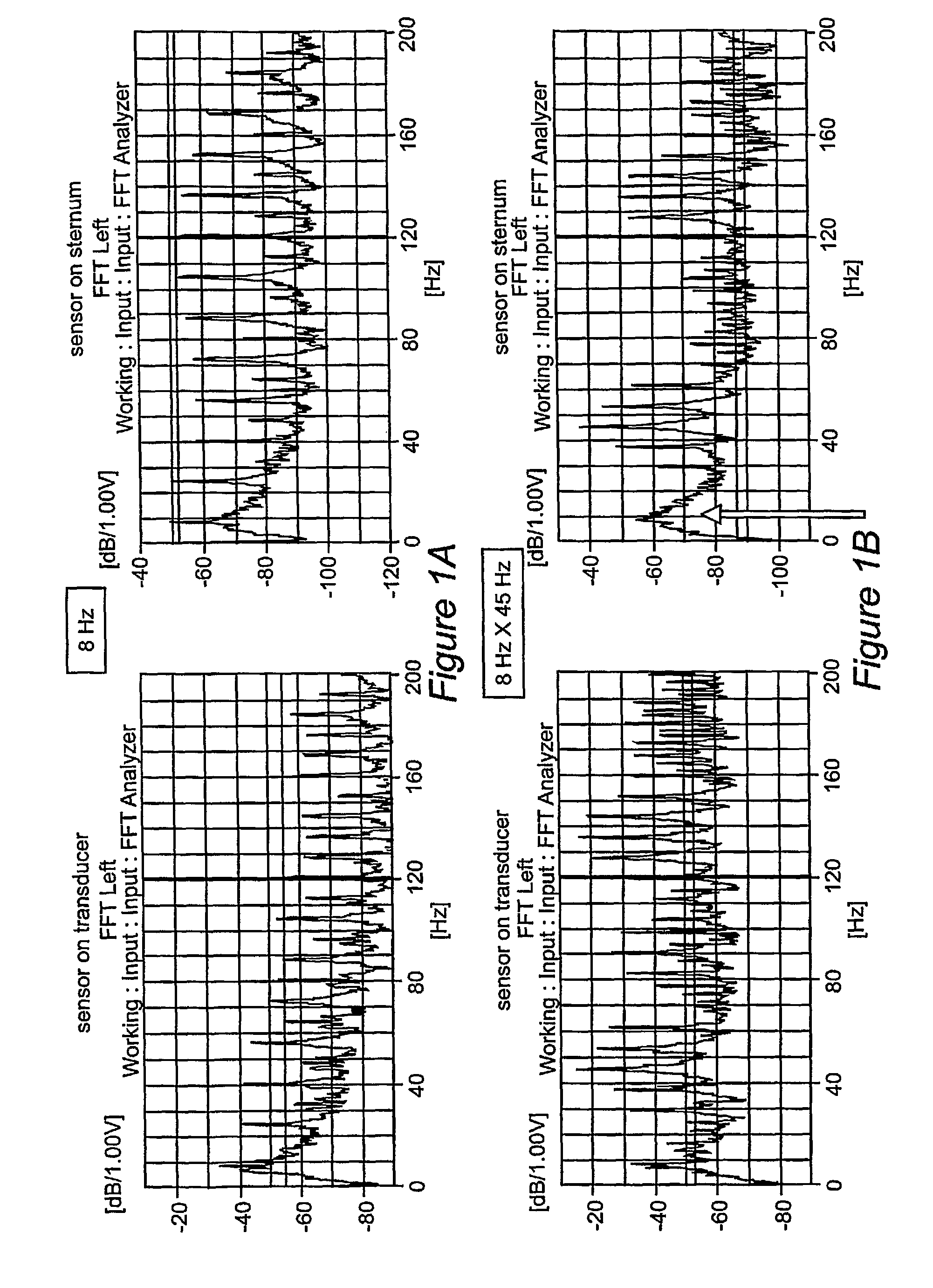
[0023]The acoustical or vibrational energy that is used in the practice of the invention is low frequency sound (acoustical energy) or vibration (mechanical energy). In preferred embodiments of the invention, the sound or vibration that is delivered is comparable to the resonant frequencies of the internal organs (e.g., in the range from less than 1 Hz to about 50 Hz, and more preferably from about 5 Hz to about 20 Hz). For example, the frequency may be about 8 Hz in some applications (e.g., 8 Hz may be preferred for the chest, but the frequency would be different for different organs based on their acoustic, i.e., resonant, properties and geometries); however, depending on the application the frequency may be different (e.g., 0.1-1000 Hz). These frequencies can be modulated (modulating frequencies) by carriers up to 100 kHz. The result of multiplying one frequency by another (Amplitude Modulation [AM]) is the carrier plus or minus the modulators. In one embodiment of the invention, in order to produce such low frequencies with relatively small, conveniently sized transducers, the present invention takes advantage of the fact that the body is a nonlinear medium in which modulated frequencies are demodulated. Thus, in order to attain an 8 Hz frequency within the targeted tissue, a modulated combination of, for example, a 40 or 45 Hz carrier frequency modulated by 8 Hz is generated and administered. Outside the body, the result would be the carrier (40 Hz)+ / −8 Hz. However, within the body (a nonlinear medium), the signal is demodulated to the independent components: 40 Hz carrier plus the separate 8 Hz modulating frequency. As a result, the targeted tissue receives sound energy at a frequency of 8 Hz via non parametric demodulation. Addition combinations of carrier and modulator frequencies may also be used. For example, the carrier frequency will generally be in the range of about 10 to about 100,000 Hz, and preferably in the range of about 50 to 250 Hz. In a preferred embodiment of the invention, the frequency that is employed is about 8 Hz modulated by about 40-45 Hz for the chest application.
[0024]It should be understood that “sound” is actually vibration being applied to the body. The vibration is measured as acceleration in reference to one meter per second squared. This is the second derivative of vibration displacing the skin. It some applications, it is preferable to provide exposure to sound or vibration for a period sufficient to increase oxygen levels by greater than 10 m / s2 (which is about one gravity unit root mean square (1 g rms)). Safety concerns may arise for levels of more than 2 g rms depending upon frequency and plane of simulation. The vibration needed may be “perceptual”, but it is safe and not annoying.

Problems solved by technology

In these cases, ischemia can cause rapid, irreversible damage or death of the affected tissue.
These methods are invasive and associated with various complications.
Recent interest has been demonstrated in modulating blood flow pharmacologically using the nitric oxide pathway but there are currently no approved uses for such with the exception of inhaled nitric oxide for severe pulmonary hypertension.
However, this device is not portable, and cannot be readily adapted to emergency situations.
The prior art has thus far failed to provide non-invasive methodology for the prevention or treatment of low blood flow / ischemia, particularly methodology that can be readily employed in emergency situations.

Method used


Image

  • Acoustical-based tissue resuscitation
  • Acoustical-based tissue resuscitation
  • Acoustical-based tissue resuscitation

Examples


example 1

Demonstration of Transmission of Acoustical Energy Through Skin and into Underlying Tissue

[0045]In order to overcome this problem, a modulated signal was used. According to the mathematics of vibration, amplitude modulation is the multiplication of one frequency by another. For example, if a frequency of 8 Hz is modulated by a 40 Hz frequency, the resulting frequency, when propagated through a liner medium, will be the carrier (40 Hz)±8 Hz. However, if the frequencies are propagated through a non-linear acoustic system (e.g. water, or biological tissue such as skin) the combined signal demodulates into the component frequencies of 40 Hz and 8 Hz. Theoretically, this provides a way to introduce an 8 Hz signal into tissues within the body in a facile manner.

[0046]This can be illustrated as follows:

Modulation=8 by 45 Hz

AM=(1=cos Ωt×cos ωt

[0047]where Ω=2πF [modulating ˜] and ω=πf [carriers˜]

Demodulation=8.45 Hz

Demodulation Ω=ΩaFΩ[f2(t)], where a is an exponent=1.85.

[0048]In order to te...

example 2

Acoustical Pulsing at Various Locations of the Body

[0052]Experimental results were extended by applying acoustical signals to various parts of the body of a human subject. The acoustical vibration source was placed, in succession, on the feet, the chest, the back of the head, and the back. Changes in O2 levels in response to the changing position of the vibrational energy source were measured using a near infrared spectroscopy sensor located at the shoulder of the subject. This measures the aggregate tissue hemoglobin saturation level of the deltoid muscle at a depth of about 4 cm. As can be seen in FIG. 3, rapid increases in the level of tissue hemoglobin oxygen saturation occurs indicating that blood flow is improved to a point where there is more left over oxygen in the venous blood.

[0053]This example demonstrates again that improvements in blood flow and tissue oxygenation can be produced at locations which are removed from the area of blood flow and oxygenation monitoring meani...

example 3

Acoustically Induced Blood Flow in an Animal Model

[0054]Similar experiments were carried out using a swine animal model. In this case, blood flow Doppler sensors were placed in muscle tissue of the pig. An acoustical signal of 8 Hz modulated by 45 hz was applied across the chest similar to that shown in FIGS. 6 and 7 and the results are presented in FIG. 4. As can be seen in the Figure, blood flow is increased.

[0055]In addition, the effect of acoustical stimulation on blood flow under conditions of low blood pressure were examined. One liter of blood was removed from the pig, and the experiment was repeated as above. The results are presented in FIG. 5, where it can be seen that blood flow to the region increased despite the fact that there was an overall reduction in blood volume. This provides evidence that the technique may be useful in the treatment of hemorrhagic shock.

[0056]This example demonstrates that even with a stopped heart, blood was forced from the body using vibration...


PUM


Abstract

Acoustical-based methods that increase tissue oxygenation, and equipment for carrying out the methods, are provided. The methods involve exposing tissue to low frequency sound in order to increase blood flow in the tissue, and hence oxygenation of the tissue. The methods may be used to treat or prevent disorders related to ischemia and low blood flow, such as shock, stroke and congestive heart failure.

Description

BACKGROUND OF THE INVENTION[0001]1. Field of the Invention[0002]The invention generally relates to acoustical and / or mechanical vibrational based methods that increase tissue blood flow and oxygenation. In particular, the invention provides methods and equipment for the use of low frequency sound or mechanical vibration to increase blood flow and hence tissue oxygenation. The methods are useful for the treatment or prevention of disorders related to ischemia and low blood flow.[0003]2. Background of the Invention[0004]Pathological conditions associated with decreased or low blood flow (ischemia) kill or incapacitate millions of people annually. Examples of such conditions include hemorrhagic shock, cardiogenic shock, congestive heart failure, cardiac arrest, septic shock, intestinal ischemia, myocardial ischemia, stroke, traumatic brain injury, sickle cell vaso-occlusive crisis, burns, compartment syndrome, and acute and chronic wounds, among others. In these cases, ischemia can cau...

Claims


Application Information

Patent Timeline
Patent Type & Authority Patents(United States)
IPC IPC(8): A61H1/00
CPCA61H23/0236A61H2201/5074A61H2230/207A61H2230/25A61H2201/5005
Inventor WARD, KEVIN R.LENHARDT, MARTIN L.
Owner VIRGINIA COMMONWEALTH UNIV
Learn More