
Method and apparatus for solid organ tissue approximation

a solid organ and approximation technology, applied in the field of medical devices and methods for trauma and general surgery, combat medicine, emergency medical services, etc., can solve the problems of not being designed for use in parenchymal tissue sutures, the optimal solution of open visceral wound repair, and the use of sutures in the medical practice of using sutures, etc., to improve the apposition of haemostatic tissue, and improve the effect of quick

Inactive Publication Date: 2007-11-01
DAMAGE CONTROL SURGICAL TECH
View PDF48 Cites 12 Cited by
  • Summary
  • Abstract
  • Description
  • Claims
  • Application Information

AI Technical Summary

Benefits of technology

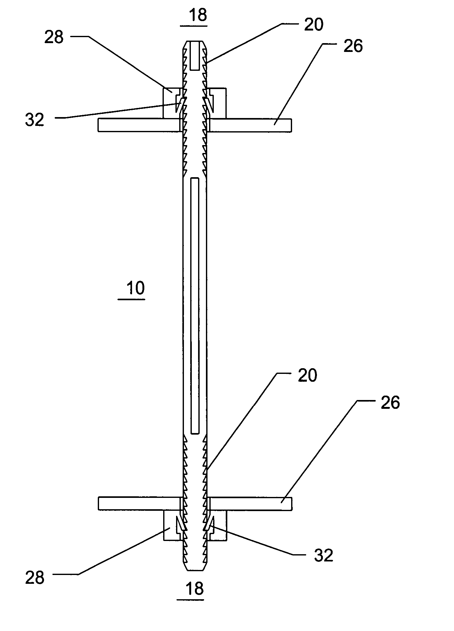
[0009] This invention relates to an improved haemostatic tissue apposition device for use in trauma care. The present invention is a transfixing trans-parenchymal bolt. Key features of the bolt include column strength, adjustable depth of penetration, flexibility, tissue non-reactivity, quick and simple application, and adjustment of the pressure plates. The trans-parenchymal bolt uses pressure plates that are affixed to the ends of the bolt to distribute the pressure over a wide area of tissue and compress the tissue. Key features of the pressure plates include one-way ratcheting with quick release or a friction lock, ability to quickly and cleanly remove the pressure plate, and the ability to adjust the pressure plate to ensure optimum tissue apposition and compression. The trans-parenchymal bolt generates pressure tamponade to provide for wound hemostasis. The pressure plates are atraumatic structures such as tabs, leafs, solids, meshes, or other structures that distribute force over a wide area of tissue. By contrast, traumatic structures include pointed projections or small thin wires or whiskers that could rip through parenchymal, or other, tissue. The trans-parenchymal bolt may be placed through an open surgical access site or through a laparoscopic access and manipulation system. The trans-parenchymal bolt, or bolt, can act as, perform the function of, or be equivalent to a soft-tissue rivet. The bolt can also be placed blindly into tissue wherein the distal end of the bolt expands to form a pressure plate thus eliminating the need to access the distal end of the bolt to apply the pressure plate. In this embodiment, the distal pressure plate is activated or expanded by control energy or force applied at the proximal end of the bolt, said control energy being transmitted along the length of the bolt by a linkage, coupling, electronic cabling, or the like. The control energy, or force, then expands the distal pressure plate. Release, or re-collapse, of the distal pressure plate can also be accomplished using the same mechanism at the proximal end of the bolt. The distal and proximal pressure plates are not sharp but are blunted and atraumatic and apply distributed pressure to the tissue.
[0010] Once the bolt has been placed, it remains in place either temporarily or permanently. Temporary placement necessitates removal of the bolt. The bolt may be made from materials that permit long-term implantation or it may be fabricated from resorbable materials that obviate the need to remove the bolt in a subsequent surgical procedure. Both the bolt and the pressure plates are fabricated from materials with smooth outer surfaces that do not encourage tissue or clot ingrowth. The bolts and pressure plates are radiopaque and can be visualized on fluoroscopy or X-ray. Thus, the bolts and pressure plates may be removed with minimal re-bleeding.

Problems solved by technology

The current medical practice of utilizing sutures is not an optimized solution to open visceral wound repair.
Sutures were not designed for use in parenchymal tissue.

Method used


Image

  • Method and apparatus for solid organ tissue approximation
  • Method and apparatus for solid organ tissue approximation
  • Method and apparatus for solid organ tissue approximation

Examples


Embodiment Construction

[0034] The present invention may be embodied in other specific forms without departing from its spirit or essential characteristics. The described embodiments are to be considered in all respects only as illustrative and not restrictive. The scope of the invention is therefore indicated by the appended claims rather than the foregoing description. All changes that come within the meaning and range of equivalency of the claims are to be embraced within their scope.

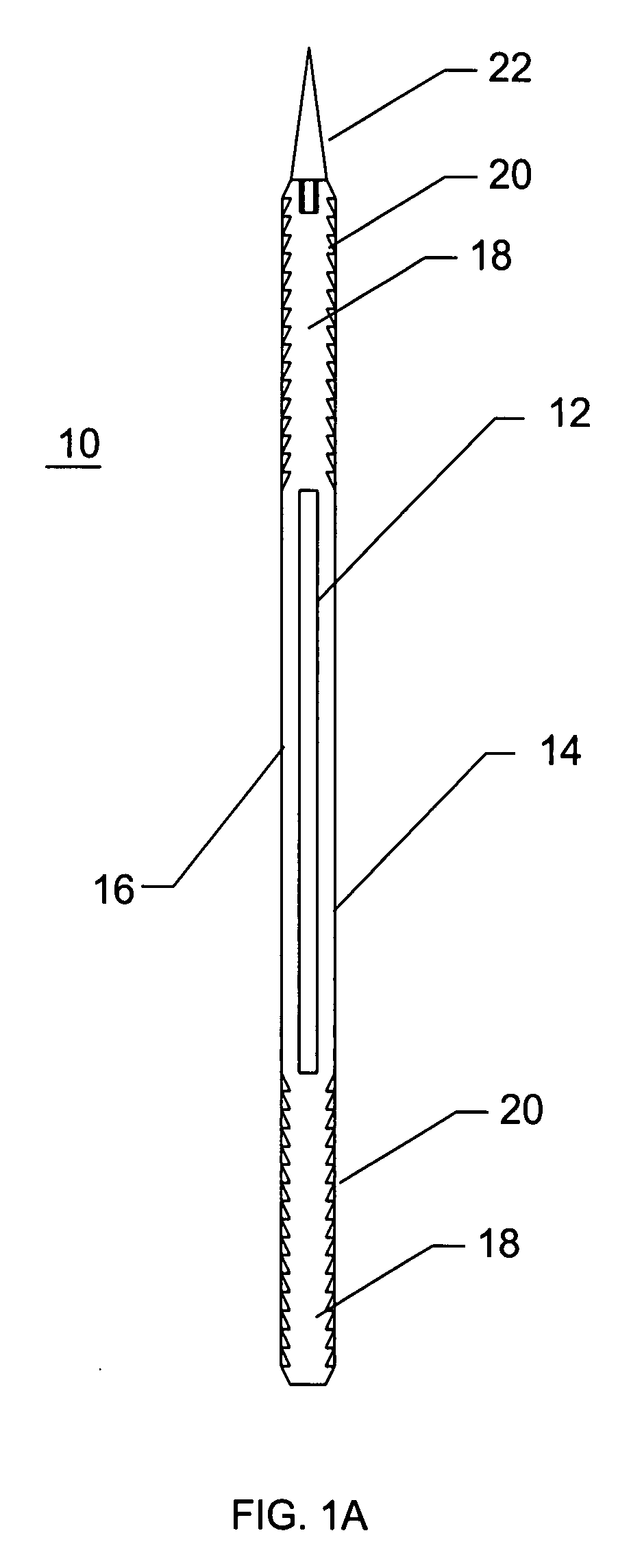
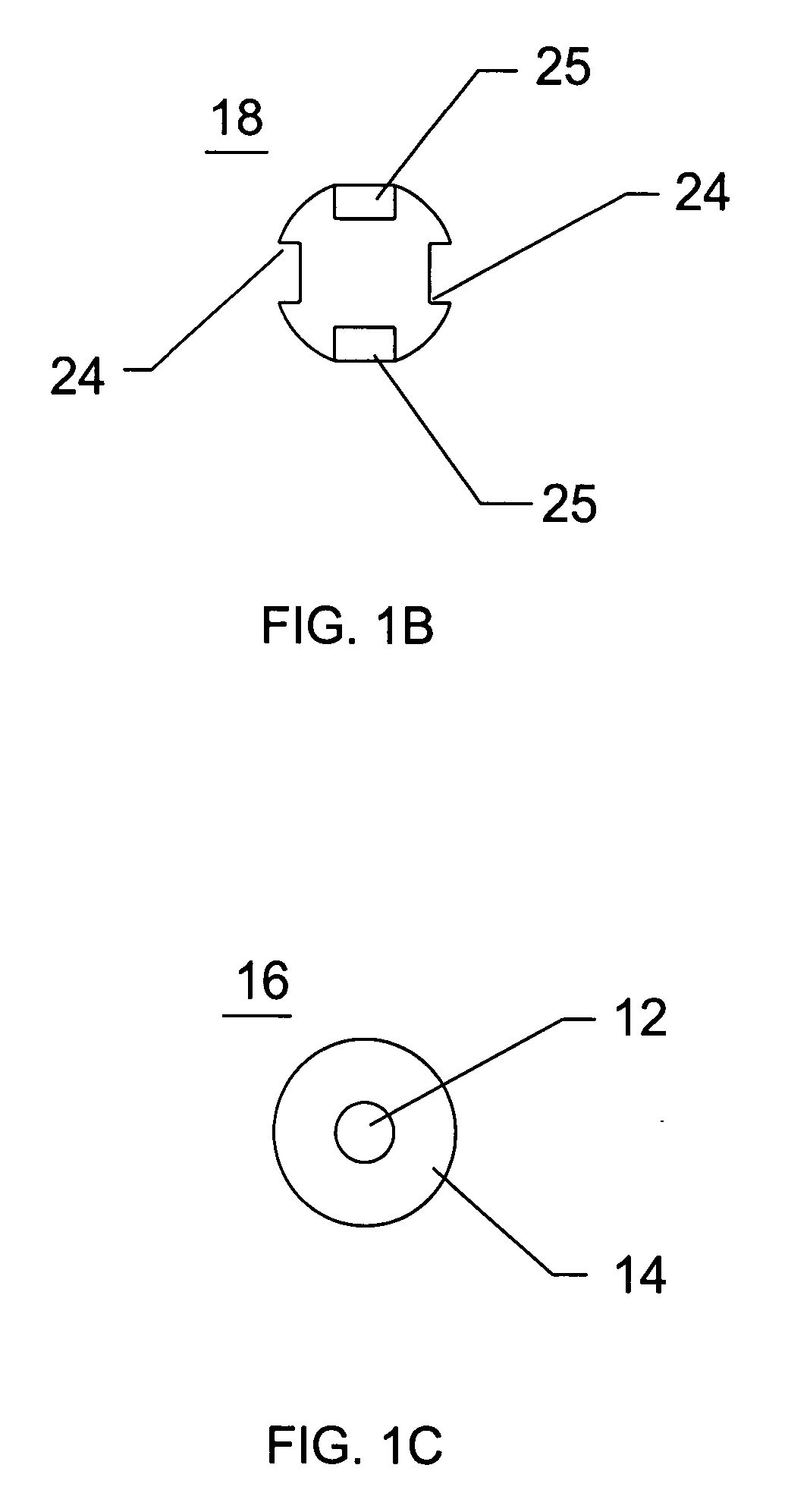
[0035]FIG. 1A illustrates a longitudinal cross-sectional view of a parenchymal bolt 10 of the present invention. The parenchymal bolt 10 comprises an inner core 12, an outer coating 14, a central region 16, a plurality of ends 18, and a plurality of serrations 20 on one or both ends 18. The parenchymal bolt 10 further comprises an optional pointed tip or trocar 22.

[0036] Referring to FIG. 1A, inner core 12 of the parenchymal bolt 10 is coaxially affixed to interior of the outer coating 14. The central connecting region 16...


PUM


Abstract

Devices and methods are disclosed for achieving hemostasis in solid visceral wounds. Such devices and methods are especially useful in the emergency, trauma surgery or military setting. In such cases, the patient may have received trauma to the abdominal viscera. The devices utilize flexible, variable depth transfixing bolts that penetrate the viscera. These bolts are pulled tight to bring the tissue into apposition and hold said tissue in apposition while the wound heals. These bolts, or soft tissue rivets, overcome the limitations of sutures that are currently used for the same purposes. The bolts come in a variety of lengths and diameters. Since the bolts are flexible, the curvature can be adjusted by the surgeon. The devices are flexible, bendable, and conformable in their wet or dry state. They can be used either straight or configured in a broad range of curvatures to suit the needs of various pathologies. The bolts include pressure plates that are capable of exerting compressive pressure over broad areas of visceral wounds without causing tearing of the friable parenchyma. The bolts may be placed and removed by open surgery or laparoscopic access. The bolts can be placed into tissue where both sides of the bolt are exposed, or the bolts can be placed blindly into tissue where the bolt does not protrude out of the tissue at its distal end.

Description

RELATED APPLICATIONS [0001] This application is a continuation-in-part of, and claims priority benefit from, U.S. patent application Ser. No. 10 / 358,735 filed Feb. 4, 2003, now U.S. Pat. No. 7,235,090, entitled “Method and Apparatus for Solid Organ Tissue Approximation”, the entire contents of which are hereby incorporated herein by reference.FIELD OF THE INVENTION [0002] The field of this invention relates to devices and methods for trauma and general surgery, combat medicine, and emergency medical services. BACKGROUND OF THE INVENTION [0003] As recently as the early 1990s, surgical operations for trauma were directed at the anatomic repair of all injuries at time of the initial operation. It was observed during these exercises that many patients became hypothermic, acidotic, and coagulopathic. Patients showing these three signs often died. Death often occurred in the operating room due to exsanguination, or postoperatively, due to the complications of prolonged shock and massive t...

Claims


Application Information

Patent Timeline
Patent Type & Authority Applications(United States)
IPC IPC(8): A61B17/08
CPCA61B17/08A61B2017/081A61B6/485A61B6/12A61B2017/088
Inventor BUCKMAN, ROBERT F.LENKER, JAY A.
Owner DAMAGE CONTROL SURGICAL TECH
Learn More